Pediatric Distal Femoral Osteosarcoma: A Detailed Orthopedic Case Study
Key Takeaway
Pediatric distal femoral osteosarcoma diagnosis uses clinical presentation like knee pain and mass. Imaging includes X-rays (lytic/blastic lesions, sunburst reaction, Codman's triangle), MRI for local staging, and CT for pulmonary metastases. This guides critical orthopedic oncology treatment.
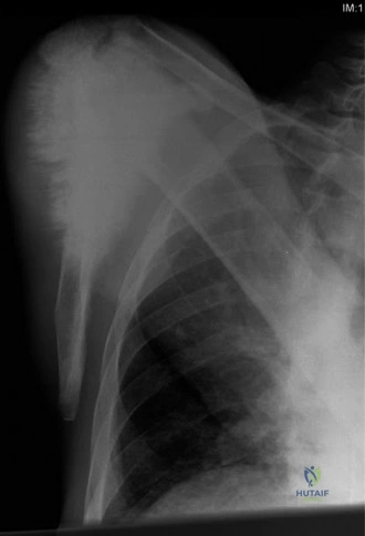
You are presented with a 14-year-old male athlete with a 5-month history of progressive left knee pain and a palpable mass in the distal thigh. Examine the provided radiograph. Describe the findings and state your primary working diagnosis.
Candidate: The radiograph shows an aggressive lesion in the metaphysis of the distal femur. There is a mixed lytic and blastic appearance with cortical destruction and a large soft tissue mass. I see a sunburst periosteal reaction and a Codman's triangle. My primary diagnosis is an osteosarcoma.
Failing to use systematic terminology. Candidates often miss describing the transition zone or the specific matrix characteristics. Simply saying "it looks like cancer" is insufficient for an elite examination; you must demonstrate recognition of the "sunburst" and "Codman's triangle" as signs of a rapidly expanding, high-grade process.
A structured response is required: 1. Location: Metaphyseal-diaphyseal lesion of the distal femur. 2. Bone/Matrix: Mixed permeative lytic and blastic (sclerotic) appearance, indicative of neoplastic osteoid production. 3. Aggression: Wide zone of transition, cortical breakthrough, and extraosseous soft tissue involvement. 4. Periosteal Reaction: Sunburst pattern and Codman's triangle, representing rapid periosteal elevation. 5. Conclusion: These findings are classic for a high-grade osteosarcoma.
The biopsy is pending. Given the age of the patient and the radiographic findings, what is your differential diagnosis, and how would you investigate this patient further?
Candidate: My differential includes Osteosarcoma, Ewing Sarcoma, and Osteomyelitis. I would investigate with an MRI of the whole femur for local staging, a CT of the chest to rule out pulmonary metastases, and a PET-CT or bone scan for systemic staging. Serum markers like ALP and LDH are also helpful.
Forgetting to mention the importance of blood tests (ALP/LDH) as prognostic markers, or failing to emphasize that the biopsy approach must be planned in conjunction with the definitive surgical team to avoid compromising limb-salvage options.
Categorize the investigation: 1. Local Staging: MRI of the entire bone to evaluate skip metastases and neurovascular relationship. 2. Systemic Staging: CT Chest (the most common site of metastasis) and PET-CT/Bone Scan for distant disease. 3. Laboratory: Serum ALP and LDH levels (prognostic). 4. Definitive Diagnosis: Core needle biopsy. I would emphasize that the biopsy tract must be placed in a location that can be excised during the definitive resection, avoiding cross-contamination of muscle compartments.
The staging is confirmed as Enneking Stage IIB. Discuss the principles of management and the role of "sandwich" chemotherapy.
Candidate: Management is multidisciplinary. We use neoadjuvant chemotherapy, surgery, and adjuvant chemotherapy. The neoadjuvant phase shrinks the tumor, treats micrometastases early, and allows us to see how well the tumor responds histologically to the drugs.
Failing to mention the prognostic value of chemotherapy. Examiners look for you to mention that histologic necrosis (Huvos grade) is the single most important predictor of survival.
Structure the rationale: 1. Systemic Control: Addressing presumed micrometastatic disease early is essential for survival. 2. Cytoreduction: Making the local tumor easier to resect with limb-salvage margins. 3. Prognostic Assessment: Assessing Huvos grade (>90% necrosis is "good response"). 4. Multidisciplinary: Mentioning the team (Oncologist, Orthopedic Oncologist, Pathologist, Radiologist) is crucial.
