Compartment Syndrome: Pathophysiology, Diagnosis, and Surgical Management
Key Takeaway
Compartment syndrome represents a critical orthopedic emergency characterized by elevated interstitial pressure within a closed osteofascial space, leading to microvascular compromise and irreversible myoneural ischemia. Prompt recognition, primarily through clinical evaluation of pain out of proportion and pain with passive stretch, is paramount. When intracompartmental pressures reach critical thresholds, emergent surgical decompression via fasciotomy is the definitive treatment to prevent devastating complications such as Volkmann ischemic contracture or limb loss.
INTRODUCTION
Compartment syndrome is a devastating orthopedic emergency defined by an elevation of interstitial pressure within a closed osteofascial compartment that results in microvascular compromise and subsequent myoneural ischemia. Because skeletal muscle is enveloped by relatively noncompliant fascial and osseous structures, any condition that either increases the volume of the compartment's contents or decreases the overall capacity of the compartment can precipitate this cascade.
While most commonly involving the anterior and deep posterior compartments of the leg and the volar compartment of the forearm, compartment syndrome can develop anywhere skeletal muscle is surrounded by substantial fascia. Documented anatomical sites include the buttock, thigh, shoulder, hand, foot, arm, and lumbar paraspinous muscles.
Compartment syndromes are broadly classified into acute and chronic forms, dictated by the etiology of the increased pressure and the temporal evolution of symptoms. Acute compartment syndrome (ACS) is a surgical emergency, whereas chronic exertional compartment syndrome (CECS) is a recurrent, exercise-induced pathology that resolves with rest but may ultimately require elective surgical intervention.
ANATOMY AND PATHOPHYSIOLOGY
The Ischemic Cascade
The pathophysiology of compartment syndrome involves a profound insult to normal local tissue homeostasis. The fundamental mechanism is the disruption of the arteriovenous (A-V) pressure gradient. Under normal physiological conditions, intracompartmental pressure (ICP) ranges from 0 to 8 mm Hg. Capillary perfusion pressure is determined by the difference between arterial inflow pressure and venous outflow pressure.
When interstitial pressure rises, it first exceeds venous pressure, leading to venous collapse and outflow obstruction. This venous congestion further increases the interstitial pressure, creating a vicious cycle. As ICP approaches the diastolic blood pressure, the local arteriovenous gradient is obliterated, resulting in decreased capillary blood flow.
Clinical Pearl: Arterial inflow continues until the intracompartmental pressure exceeds systolic blood pressure. Therefore, the presence of palpable distal pulses does not rule out compartment syndrome. Pulselessness is a late, pre-terminal sign indicating irreversible ischemic damage.
Tissue Tolerance and Necrosis
Experimental evidence and clinical observation dictate strict temporal thresholds for tissue viability:
* Muscle Ischemia: Significant muscle necrosis begins when intracompartmental pressure is increased to more than 30 mm Hg for longer than 8 hours. Irreversible damage can occur in as little as 4 to 6 hours in the presence of profound hypotension or complete arterial occlusion.
* Nerve Ischemia: Peripheral nerves are highly sensitive to ischemia. Paresthesias can develop within 30 minutes of onset. Irreversible axonal damage and nerve death typically occur after 12 to 24 hours of sustained critical pressure.
Relevant Osteofascial Anatomy
A thorough understanding of compartmental anatomy is mandatory for effective surgical decompression.
* The Leg: Comprises four distinct compartments: Anterior, Lateral, Superficial Posterior, and Deep Posterior. The deep posterior compartment (containing the tibialis posterior, flexor hallucis longus, flexor digitorum longus, and the tibial neurovascular bundle) is frequently under-released, leading to persistent ischemia.
* The Forearm: Comprises three primary compartments: Volar (flexor), Dorsal (extensor), and the Mobile Wad (brachioradialis, extensor carpi radialis longus and brevis). The volar compartment is most frequently affected following supracondylar humerus or radius/ulna fractures.
ETIOLOGY AND RISK FACTORS
Acute Compartment Syndrome (ACS)
The most common causes of acute compartment syndrome involve high-energy trauma, though iatrogenic and medical causes are also prevalent:
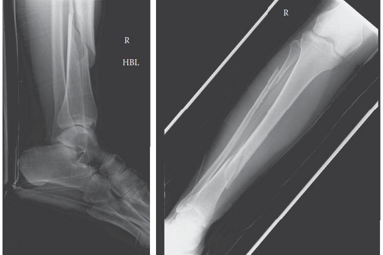
* Fractures: Tibial shaft fractures are the most common cause of ACS, accounting for up to 36% of all cases. Supracondylar humerus fractures and forearm fractures are the leading causes in the pediatric population.
* Soft Tissue Trauma: Crush injuries without fracture can cause massive muscle edema and hemorrhage.
* Vascular Injury and Reperfusion: Prolonged ischemia followed by arterial repair and reperfusion leads to massive capillary leak and cellular swelling (reperfusion injury).
* Extrinsic Compression: Tight casts, circumferential dressings, or prolonged limb compression during altered consciousness (e.g., drug overdose, prolonged surgical positioning).
* Iatrogenic/Medical: Intravenous fluid extravasation, bleeding diatheses, and anticoagulant therapy.
Chronic Exertional Compartment Syndrome (CECS)
Chronic exertional compartment syndrome is characterized by a transient, reversible increase in pressure. During strenuous exercise, muscle volume can increase by up to 20% due to increased blood flow and capillary filtration. In a noncompliant compartment, this physiological engorgement causes a pathological rise in pressure.
CECS of the lower extremity is most common in long-distance runners and military recruits pushed past normal limits of functional tolerance. It has also been reported in the forearms of weightlifters, rowers, and welders.
Surgical Warning: Fascial hernias have a definite association with the development of exertional compartment symptoms. Approximately 15-40% of patients treated for CECS exhibit fascial defects. These hernias represent a focal weakness where muscle bellies protrude, often impinging superficial nerves (e.g., the superficial peroneal nerve in the lateral leg).
CLINICAL PRESENTATION AND DIAGNOSIS
Clinical Evaluation: The "6 Ps"
The diagnosis of acute compartment syndrome is primarily clinical. The classic "6 Ps" are taught universally, but their diagnostic utility varies significantly:
1. Pain out of proportion: The earliest and most reliable indicator. The pain is typically described as deep, unremitting, and poorly localized.
2. Pain with passive stretch: Highly sensitive. Stretching the muscles within the affected compartment elicits severe pain (e.g., passive toe extension causing severe pain in the deep posterior compartment of the leg).
3. Paresthesia: An early sign of nerve ischemia in the distribution of the nerve traversing the compartment.
4. Pallor: A late sign indicating severe arterial compromise.
5. Paralysis: A late sign indicating irreversible muscle and nerve damage.
6. Pulselessness: The final, pre-terminal sign.
Objective Intracompartmental Pressure (ICP) Monitoring
In obtunded, intubated, or polytrauma patients where clinical examination is unreliable, objective ICP measurement is mandatory. This is typically performed using a solid-state transducer (e.g., Stryker needle) or an arterial line setup.
The Delta P Concept:
Absolute pressure readings are less reliable than the differential pressure (Delta P).
* Delta P = Diastolic Blood Pressure - Intracompartmental Pressure
* A Delta P of less than 30 mm Hg is the universally accepted threshold for emergent fasciotomy. For example, if a patient's diastolic BP is 70 mm Hg and the ICP is 45 mm Hg, the Delta P is 25 mm Hg, necessitating immediate surgical decompression.
SURGICAL INDICATIONS
Absolute Indications:
* Unequivocal clinical signs of acute compartment syndrome (pain out of proportion, pain on passive stretch) in an alert, cooperative patient.
* Delta P < 30 mm Hg in an obtunded, polytrauma, or pediatric patient.
* Arterial injury requiring repair with a prolonged ischemic time (> 4-6 hours), necessitating prophylactic fasciotomy.
Relative Indications:
* Chronic exertional compartment syndrome that has failed conservative management (rest, orthotics, gait retraining), confirmed by elevated pre- and post-exercise ICP measurements.
OPERATIVE MANAGEMENT: STEP-BY-STEP SURGICAL TECHNIQUES
The definitive treatment for acute compartment syndrome is emergent, complete surgical fasciotomy of all affected compartments. Minimalist or endoscopic approaches have no role in the acute setting.
Lower Extremity: Double-Incision Four-Compartment Fasciotomy
The Mubarak and Owen double-incision technique is the gold standard for decompressing the leg, as it safely accesses all four compartments while preserving a wide skin bridge.
1. Preoperative Preparation and Positioning:
* Place the patient supine on a radiolucent table.
* Do NOT use a tourniquet, as it exacerbates ischemia and prevents the assessment of muscle viability post-decompression.
* Prep and drape the entire lower extremity free.
2. Anterolateral Incision (Decompressing Anterior and Lateral Compartments):
* Make a 15 to 20 cm longitudinal incision centered exactly halfway between the tibial crest and the fibular shaft.
* Dissect through the subcutaneous tissue to expose the crural fascia.
* Identify the anterior intermuscular septum, which separates the anterior and lateral compartments.
* Anterior Compartment Release: Make a longitudinal incision in the fascia over the anterior compartment, approximately 2 cm anterior to the septum. Extend the release proximally to the tibial tubercle and distally to the extensor retinaculum using fasciotomy scissors.
* Lateral Compartment Release: Make a parallel longitudinal incision in the fascia over the lateral compartment, 2 cm posterior to the septum. Extend proximally toward the fibular head and distally toward the lateral malleolus.
* Pitfall: Beware of the superficial peroneal nerve, which exits the lateral compartment fascia in the distal third of the leg.
3. Posteromedial Incision (Decompressing Superficial and Deep Posterior Compartments):
* Make a 15 to 20 cm longitudinal incision 2 cm posterior to the posteromedial border of the tibia.
* Warning: Carefully identify and retract the saphenous vein and nerve anteriorly.
* Superficial Posterior Compartment Release: Incise the fascia over the gastrocnemius-soleus complex. Extend the release fully from the proximal calf to the musculotendinous junction distally.
* Deep Posterior Compartment Release: Retract the superficial posterior compartment posteriorly. Detach the soleus bridge from the posterior tibia to expose the deep transverse fascia. Incise this fascia longitudinally to release the tibialis posterior, FDL, and FHL.
* Clinical Pearl: The deep posterior compartment is the most frequently missed release. Ensure you visualize the posterior tibial neurovascular bundle to confirm adequate decompression.
Upper Extremity: Volar Forearm Fasciotomy
The volar compartment is the most critical space in the upper extremity. An extended Henry approach is utilized to decompress the superficial and deep flexors.
1. Incision and Approach:
* Begin the incision proximal to the antecubital fossa, medial to the biceps tendon.
* Extend the incision distally across the antecubital fossa in a curvilinear fashion, continuing down the volar forearm over the path of the radial artery.
* At the wrist, cross the flexion crease obliquely toward the ulnar side to avoid contracture, extending into the palm to release the carpal tunnel.
2. Fascial Release:
* Divide the lacertus fibrosus (bicipital aponeurosis) proximally to decompress the brachial artery and median nerve.
* Incise the superficial fascia of the forearm over the flexor carpi radialis (FCR).
* Retract the FCR ulnarly and the brachioradialis radially to expose the deep flexor compartment (Flexor Pollicis Longus and Flexor Digitorum Profundus).
* Incise the fascia over the deep muscles. If individual muscle bellies remain tense, perform epimysiotomies (incising the epimysium of individual muscles).
* Complete the release by dividing the transverse carpal ligament to decompress the median nerve in the carpal tunnel. Guyon's canal may also be released if ulnar nerve compromise is suspected.
POSTOPERATIVE PROTOCOL AND WOUND MANAGEMENT
Following successful fasciotomy, the management of the open wounds is critical to prevent infection and facilitate eventual closure.
-
Immediate Postoperative Care:
- The wounds must be left open. Primary closure is strictly contraindicated as it will recreate the compartment syndrome.
- Assess muscle viability. Non-viable, necrotic muscle (which does not bleed when cut, does not contract with electrocautery, and appears dusky/gray) must be aggressively debrided.
- Apply Negative Pressure Wound Therapy (NPWT / Wound VAC) at 75 to 125 mm Hg continuous pressure. NPWT reduces tissue edema, manages exudate, and promotes angiogenesis.
-
Second-Look Operation:
- Return the patient to the operating room at 48 to 72 hours for a "second look."
- Perform further debridement of any evolving necrotic tissue.
-
Definitive Closure:
- Once edema has subsided (typically 5 to 10 days post-injury), attempt delayed primary closure. Techniques such as the "shoelace" vessel loop technique or dynamic wound closure devices can assist in gradual skin approximation.
- If the skin cannot be approximated without tension, a Split-Thickness Skin Graft (STSG) must be applied to cover the defect.
COMPLICATIONS
Failure to recognize and promptly treat compartment syndrome leads to catastrophic outcomes:
* Volkmann Ischemic Contracture: The classic sequela of untreated compartment syndrome. Necrotic muscle is replaced by dense, inelastic fibrotic scar tissue, leading to severe joint contractures, clawing of the digits, and a functionless, insensate limb.
* Infection: Open fasciotomy wounds are highly susceptible to nosocomial infections. Deep infection may necessitate radical debridement.
* Rhabdomyolysis and Acute Kidney Injury (AKI): Massive muscle necrosis releases myoglobin into the systemic circulation, which precipitates in the renal tubules. Aggressive intravenous hydration and urine alkalinization are required to prevent renal failure.
* Amputation: In cases of delayed presentation (> 12-24 hours) with complete, irreversible myoneural necrosis, primary amputation is often the safest and most functional option to prevent life-threatening sepsis and crush syndrome.
You Might Also Like