Geriatric Complex Acetabular Fractures: A Detailed Clinical Case Study
Key Takeaway
Geriatric complex acetabular fractures, frequently from low-energy falls in osteoporotic patients, require meticulous diagnosis. This involves a thorough clinical exam followed by advanced imaging like CT with 3D reconstructions. Imaging delineates comminution, articular impaction, and column involvement, which are crucial for effective treatment planning in this challenging patient population.
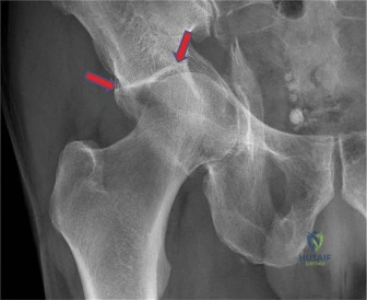
A 78-year-old female presents following a ground-level fall. She is on Apixaban. Initial radiographs and CT scan show a complex acetabular fracture with significant comminution of the quadrilateral plate, superomedial dome impaction, and medial subluxation of the femoral head. Describe your systematic approach to classifying this injury and how the "gull sign" influences your management strategy.
Candidate: I would classify this as an Anterior Column Posterior Hemitransverse (ACPHT) fracture using the Letournel system. The "gull sign" indicates superomedial dome impaction. In an elderly patient with significant bone loss, this suggests poor articular prognosis, likely requiring a 'fix and replace' approach (ORIF of the columns followed by acute THA) rather than isolated ORIF.
Failing to mention the "Fix and Replace" concept, or attempting to justify ORIF alone despite evidence of severe cartilage damage and osteopenia. Candidates also often forget to address the systemic urgency, such as managing the anticoagulation (Apixaban) before surgery.
A structured response is required: 1. Classification: Identify it as an ACPHT fracture; highlight the medial displacement of the quadrilateral plate. 2. Prognostic Marker: Explicitly define the 'gull sign' as subchondral articular impaction. 3. Surgical Rationale: Argue for 'Fix and Replace'. Pure ORIF in this demographic carries high failure rates due to osteoporosis (lack of hardware purchase) and the high probability of post-traumatic OA from the impaction. 4. Medical Optimization: Acknowledge the need for multidisciplinary input to manage Apixaban reversal or delay to minimize hemorrhage risk.
You have decided to proceed with an Anterior Intrapelvic (Modified Stoppa) approach. What is your primary concern regarding neurovascular structures in the pelvic basin, and how do you mitigate this?
Candidate: My main concern is the 'corona mortis', an anastomosis between the external iliac/inferior epigastric and the obturator vessels. I would identify it during the exposure of the superior pubic ramus, ligate it safely, and then retract the external iliac vessels and femoral nerve gently to access the quadrilateral plate.
Ignoring the corona mortis or failing to mention that it is a common site for potentially life-threatening hemorrhage. Also, failing to mention the protection of the obturator nerve as the dissection proceeds posteriorly.
The candidate must demonstrate anatomical vigilance: 1. Corona Mortis: Explicitly discuss its identification, clipping, and division to prevent retraction-related avulsion. 2. Vascular Protection: Describe protecting the external iliac vessels and the femoral nerve (using specialized retractors like the Hohmann or blunt pelvic retractors). 3. Obturator Nerve: Mention its identification to prevent neuropraxia during posterior quadrilateral plate reduction.
Following pelvic fixation, you convert to a posterior approach for THA. Why is a cemented, polished, double-tapered femoral stem preferred over an uncemented press-fit implant in this specific patient?
Candidate: In an elderly patient with Dorr Type C bone, an uncemented stem risks intraoperative periprosthetic fracture due to the thin, osteoporotic cortex. A cemented stem provides immediate fixation, better stress distribution, and allows for immediate full weight-bearing, which is essential for this patient's rehabilitation.
Arguing that uncemented stems are "more modern" or "better." Failing to link the implant choice to the patient's specific bone quality (Dorr Type C) and the functional goal of immediate weight-bearing.
The answer must synthesize bone quality and mechanical goals: 1. Bone Biology: Recognize Dorr Type C anatomy (thin cortices, widened canal). 2. Fracture Risk: Explain that press-fit implants in poor bone increase the risk of hoop-stress-induced fracture. 3. Immediate Stability: Highlight that cemented implants (specifically polished tapered designs) provide reliable "immediate" stability, facilitating early mobilization and reducing post-operative complications like pneumonia and DVT.
