
العربية
Comprehensive Master Guide · Medically Reviewed

Comprehensive Guide to AC Joint Injuries: Epidemiology, Surgical Anatomy & Biomechanics

Discover the ultimate guide to AC joint injuries. Learn about key symptoms, epidemiology, surgical anatomy, biomechanics, and Rockwood classification.

15 Detailed Chapters
26 min read
Updated: Jun 2026
Dr. Mohammed Hutaif Clinic
Medically Reviewed by
Prof. Dr. Mohammed Hutaif Clinic
Verified Content Expert Reviewed

Quick Medical Answer

AC joint injuries affect the articulation between the clavicle and scapula, commonly due to direct shoulder blows in athletes. They involve disruption of the AC ligaments (horizontal stability) and the critical coracoclavicular ligaments (vertical stability: trapezoid, conoid). The Rockwood classification categorizes injuries based on displacement and ligament damage, guiding management from conservative care to complex surgical reconstruction, emphasizing precise anatomical understanding.

Illustration of ac joint injuries - Dr. Mohammed Hutaif
🎓

FRCS Masterclass: Clinical Viva

Interactive Examiner Scenario • Test your knowledge before revealing the answers.

👨‍⚕️ Examiner Scenario

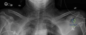
A 28-year-old rugby player presents to the Emergency Department following a direct tackle to the right shoulder. He is holding his arm in adduction. Clinical examination reveals a prominent distal clavicle and significant tenderness at the AC joint. You are presented with the following radiograph.

Clinical Image
Anteroposterior radiograph of the shoulder

Describe your findings and classify the injury according to the Rockwood system.

Candidate: The radiograph shows a superior displacement of the distal clavicle relative to the acromion. The coracoclavicular (CC) interval is significantly increased compared to the contralateral side. This represents a complete disruption of both the AC and CC ligaments. Based on the Rockwood classification, this is a Type III injury.

❌ Common Pitfall (Poor Answer)

Candidates often stop at "Type III." A high-scoring candidate must differentiate between Type III and Type V. Failing to comment on the degree of displacement relative to the clavicular shaft diameter or failing to assess the soft tissue envelope (deltoid/trapezius) often leads to a lower score.

⭐ The Gold Standard (Perfect Answer)

Start with a systematic description: "The radiograph demonstrates a superior dislocation of the distal clavicle. The CC interval is increased by [X]%. This indicates a complete tear of both the AC and CC ligaments. To classify, I would measure the displacement; if it is 25-100%, it is a Rockwood Type III. If it exceeds 100% of the normal CC interval, or the clavicle is displaced by more than one shaft diameter, it represents a Type V injury, which implies extensive stripping of the deltoid and trapezius origin."

👨‍⚕️ Examiner Scenario

The patient in the previous scenario is a professional athlete. He wants to know your management plan. What are the key decision-making factors for surgical versus non-operative treatment in high-grade AC joint injuries?

Candidate: For Type III injuries, non-operative management is often successful for the general population. However, for a professional athlete, I would consider operative stabilization due to their high functional demands, overhead requirements, and desire to return to sport without pain or deformity.

❌ Common Pitfall (Poor Answer)

Suggesting surgery for every Type III injury is incorrect. Candidates must demonstrate an understanding of the literature showing that long-term functional outcomes for non-operative management are often equal to operative management, thereby framing surgery as a decision based on "patient-specific demands" rather than a blanket rule.

⭐ The Gold Standard (Perfect Answer)

Structure the answer by categories: 1. Injury Type: Types IV, V, and VI are absolute indications for surgery due to mechanical instability or soft tissue incarceration. 2. Functional Demand: In an overhead athlete or heavy laborer, the persistent weakness and scapular dyskinesis associated with Type III can be debilitating; therefore, anatomical reconstruction is often preferred. 3. Evidence Base: Acknowledge that while radiographic reduction is better with surgery, meta-analyses show no significant difference in long-term functional scores between non-operative and operative cohorts for Type III. The decision for the athlete is therefore a risk/benefit discussion regarding return-to-play speed vs. surgical morbidity (e.g., hardware failure, infection).

👨‍⚕️ Examiner Scenario

You decide to proceed with a surgical repair using a suture-button construct. What are the main risks associated with this technique, and how do you minimize the risk of neurovascular injury?

Candidate: Suture-button risks include tunnel widening, infection, or hardware breakage. To protect neurovascular structures, I ensure careful dissection around the coracoid and use fluoroscopy to guide the drill, keeping the trajectory away from the brachial plexus and subclavian vessels.

❌ Common Pitfall (Poor Answer)

Being vague about the anatomy. An examiner expects you to know exactly where the danger lies: the brachial plexus and subclavian vessels lie medial and posterior to the coracoid. A "perfect" candidate will explicitly mention the use of a protective retractor or a "finger-palpation" technique under the coracoid base.

⭐ The Gold Standard (Perfect Answer)

Start with the technical risks: Suture-button constructs carry risks of tunnel osteolysis, hardware failure, and loss of reduction. Neurovascular Safety: 1. Anatomy: The brachial plexus and subclavian vessels are at risk, specifically medial and posterior to the coracoid. 2. Technique: I would utilize a blunt dissection to expose the base of the coracoid. 3. Protection: I would place a small, malleable retractor or a Freer elevator between the inferior surface of the coracoid and the underlying neurovascular structures to act as a 'backstop' during drilling and suture passage. 4. Fluoroscopy: Intraoperative imaging is mandatory to confirm drill trajectory through the base of the coracoid and the midpoint of the clavicle.

Detailed Chapters & Topics

Dive deeper into specialized chapters regarding ac-joint-injuries

15 Chapters
01
Chapter 1 21 min

Arthroscopic AC Joint Reduction and Coracoclavicular Stabilization: A TightRope Masterclass

Master arthroscopic AC joint reduction and coracoclavicular stabilization using the TightRope system for acute Rockwood…

02
Chapter 2 15 min

Arthroscopic Distal Clavicle Resection: An Intraoperative Masterclass for AC Joint Pathology

Master arthroscopic distal clavicle resection for AC joint pathology. Learn key anatomy and surgical steps to effective…

03
Chapter 3 16 min

Masterclass: Comprehensive Surgical Reconstruction of Acromioclavicular Joint Injuries

Master AC joint reconstruction in this surgical masterclass. Learn to restore complex biomechanics and stabilize ligame…

04
Chapter 4 24 min

Acromioclavicular (AC) Joint Dislocation: Epidemiology, Anatomy, Biomechanics, and Clinical Evaluation

Discover the essentials of AC joint dislocation. This guide covers epidemiology, surgical anatomy, biomechanics, and cl…

05
Chapter 5 86 min

Precise Joint Dislocation Examination: AC Joint Diagnosis Demystified

KEY POINTS Acromioclavicular joint dislocation Examination corner Rockwood classification (1984)4 Or more simply the “S…

06
Chapter 6 14 min

Choosing mm Nonabsorbable Radiolucent for AC Joint Repair

Repair and Reconstruction of Acromioclavicular Injuriey DEFINITION About 9% of shoulder girdle injuries involve damage …

07
Chapter 7 14 min

Is distal clavicle resection the fix for your AC joint pain?

Repair and Reconstruction of Acromioclavicular Injuries DEFINITION About 9% of shoulder girdle injuries involve damage …

08
Chapter 8 20 min

Solving Elbow Cases & Acromioclavicular Arthritis Pain

A 55-year-old, right-hand-dominant female presents with right shoulder pain for 6 months. She localizes the pain over t…

09
Chapter 9 24 min

Acromioclavicular (AC) Joint Separation: Epidemiology, Classification & Biomechanics

Discover AC joint separation epidemiology and biomechanics. Learn the Rockwood classification system to ensure accurate…

10
Chapter 10 27 min

AC Joint Injury? Effective Healing with a Clavicle Strap Sling

ACROMIOCLAVICULAR AND STERNOCLAVICULAR JOINT INJURIES ACROMIOCLAVICULAR JOINT INJURY Epidemiology It is most common in …

11
Chapter 11 15 min

Acromioclavicular (AC) Joint Injuries: Epidemiology, Anatomy, Rockwood Classification & Management

Explore the epidemiology, anatomy, and management of AC joint injuries. Learn the six types of the Rockwood classificat…

12
Chapter 12 16 min

Heal Your AC Joint Injury: Causes, Symptoms & Treatment Guide

Acromioclavicular (AC) joint injury is a common shoulder injury that occurs when the ligaments that connect the collarb…

13
Chapter 13 10 min

Acromioclavicular Joint Excision & Management: Surgical Guide

Master acromioclavicular joint pathology management with this expert surgical guide. Explore conservative treatments an…

14
Chapter 14 19 min

Acromioclavicular Joint Injuries: Comprehensive Surgical Management

Explore the surgical management of acromioclavicular joint injuries. Learn about AC joint etiology, biomechanics, and c…

15
Chapter 15 20 min

Acute Shoulder Dislocation & AC Joint Reconstruction

Explore the evolving management of acute anterior shoulder dislocations. Learn about irreducible cases, ORIF surgery, a…

Dr. Mohammed Hutaif Clinic
Medically Verified Content by
Prof. Dr. Mohammed Hutaif Clinic
Consultant Orthopedic & Spine Surgeon
Guide Overview