Full Question & Answer Text (for Search Engines)
Question 1:
A 48-year-old tennis player presents with chronic lateral elbow pain, exacerbated by gripping and wrist extension. On examination, maximal tenderness is consistently localized to an area just distal and anterior to the lateral epicondyle. Which specific structure is most likely the primary source of pathology?
Options:
- Common extensor origin at the supracondylar ridge
- Anconeus muscle belly
- Origin of the Extensor Carpi Radialis Brevis (ECRB) tendon
- Lateral collateral ligament complex
- Radial nerve proper as it crosses the elbow joint
Correct Answer: Origin of the Extensor Carpi Radialis Brevis (ECRB) tendon
Explanation:
The most common site of pathology in lateral epicondylitis (tennis elbow) is the origin of the Extensor Carpi Radialis Brevis (ECRB) tendon, specifically its deep fibers, just distal and anterior to the lateral epicondyle. While the common extensor origin is affected, the ECRB is the primary culprit. The anconeus muscle is more posterior and not typically the primary pain generator. The lateral collateral ligament complex is associated with elbow instability. The radial nerve proper is rarely the direct source of pain but can be entrapped in radial tunnel syndrome, which is a differential diagnosis, but the precise localization points strongly to the ECRB.
Question 2:
During your physical examination for suspected lateral epicondylitis, you perform Cozen's test. Which maneuver constitutes a positive Cozen's test?
Options:
- Pain with resisted elbow flexion while the forearm is supinated.
- Pain with resisted wrist extension while the elbow is flexed at 90 degrees.
- Pain with resisted wrist extension while the elbow is extended, the forearm is pronated, and the wrist is radially deviated.
- Pain with passive wrist flexion and forearm pronation while the elbow is extended.
- Pain with resisted middle finger extension while the forearm is pronated.
Correct Answer: Pain with resisted wrist extension while the elbow is extended, the forearm is pronated, and the wrist is radially deviated.
Explanation:
Cozen's test involves the examiner palpating the lateral epicondyle while the patient makes a fist, pronates the forearm, radially deviates the wrist, and then extends the wrist against resistance. A positive test is reproduction of pain at the lateral epicondyle. Option C accurately describes this maneuver. Option B describes a component but misses the critical elbow extension and forearm pronation. Option D describes Mill's test, which is passive. Option E describes Maudsley's test.
Question 3:
A 55-year-old accountant presents with lateral elbow pain that started insidiously. He denies any acute trauma but notes pain with typing and lifting objects, especially with his palm down. Which of the following findings on examination would be MOST specific for lateral epicondylitis rather than a radial tunnel syndrome?
Options:
- Pain with resisted forearm supination.
- Tenderness over the supinator muscle.
- Pain elicited by passive wrist flexion with the elbow extended.
- Normal sensation in the distribution of the superficial radial nerve.
- Pain with resisted long finger extension (Maudsley's test).
Correct Answer: Pain elicited by passive wrist flexion with the elbow extended.
Explanation:
Pain elicited by passive wrist flexion with the elbow extended (Mill's test) is a classic maneuver that stretches the common extensor origin, particularly the ECRB, and is highly suggestive of lateral epicondylitis. While Maudsley's test (resisted long finger extension) is also positive in lateral epicondylitis, it can sometimes be positive in radial tunnel syndrome due to irritation of the nerve passing beneath the ECRB. Tenderness over the supinator muscle and pain with resisted forearm supination are more indicative of radial tunnel syndrome. Normal sensation in the superficial radial nerve distribution is common in both, as PIN entrapment is a motor neuropathy. Therefore, Mill's test specifically targets the common extensor origin's stretch sensitivity.
Question 4:
Which histological finding is most consistently associated with chronic lateral epicondylitis specimens obtained surgically?
Options:
- Acute inflammatory cell infiltration (e.g., neutrophils, macrophages)
- Vascular proliferation and disorganized collagen with fibroblasts (angiofibroblastic hyperplasia)
- Extensive calcification within the tendon substance
- Complete rupture of the ECRB tendon
- Focal areas of bacterial infection
Correct Answer: Vascular proliferation and disorganized collagen with fibroblasts (angiofibroblastic hyperplasia)
Explanation:
Chronic lateral epicondylitis is primarily a degenerative tendinopathy, not an inflammatory process. Histologically, it is characterized by angiofibroblastic hyperplasia, which involves disordered collagen fibers, increased fibroblasts, and neovascularization, rather than acute inflammatory cells. While some minor inflammation may be present, it's not the hallmark. Calcification can occur but is less consistent. Complete rupture is rare. Infection is not part of the pathology.
Question 5:
A patient presents with lateral elbow pain that radiates distally to the dorsal aspect of the forearm and hand. They report weakness, particularly with gripping, and exquisite tenderness over the extensor muscle mass, approximately 3-5 cm distal to the lateral epicondyle, specifically in the arcade of Frohse region. Pain is exacerbated by repetitive forearm rotation. Which condition should be prioritized in your differential diagnosis?
Options:
- Lateral epicondylitis
- Radiohumeral joint osteoarthritis
- Posterior interosseous nerve (PIN) entrapment syndrome
- Capitellar osteochondritis dissecans
- Cervical radiculopathy (C6-C7)
Correct Answer: Posterior interosseous nerve (PIN) entrapment syndrome
Explanation:
The description of pain radiating distally to the dorsal forearm/hand, weakness with gripping, and exquisite tenderness 3-5 cm distal to the lateral epicondyle (over the arcade of Frohse where the PIN can be entrapped), especially exacerbated by repetitive forearm rotation, is classic for posterior interosseous nerve (PIN) entrapment syndrome, a form of radial tunnel syndrome. While lateral epicondylitis is a differential, the specific tenderness location and nerve-like radiation strongly favor PIN entrapment. Radiohumeral OA typically presents with pain with rotation and sometimes catching, but less nerve-like radiation. Capitellar OCD affects younger patients and usually involves mechanical symptoms. Cervical radiculopathy would have more widespread neurological deficits and often neck pain. PIN entrapment affects motor function, leading to weakness without sensory changes, which aligns with the presentation of grip weakness.
Question 6:
Which of the following imaging modalities is considered most useful in confirming the diagnosis of lateral epicondylitis and assessing its severity in cases where the clinical diagnosis is equivocal or non-operative treatment has failed?
Options:
- Plain radiographs of the elbow
- CT scan of the elbow
- Electromyography (EMG) and Nerve Conduction Studies (NCS)
- Magnetic Resonance Imaging (MRI) or Musculoskeletal Ultrasound
- Bone scintigraphy
Correct Answer: Magnetic Resonance Imaging (MRI) or Musculoskeletal Ultrasound
Explanation:
Plain radiographs are typically normal in lateral epicondylitis and are mainly used to rule out bony pathology. CT scans offer excellent bony detail but are less effective for soft tissue. EMG/NCS are useful for differentiating nerve entrapment syndromes (like radial tunnel) but not for diagnosing lateral epicondylitis directly. MRI and high-resolution musculoskeletal ultrasound are the most useful imaging modalities. Ultrasound can show hypoechogenicity, tendon thickening, tears, and neovascularization. MRI can detect signal changes within the ECRB tendon, edema, and tendinosis/tears. These modalities help confirm the diagnosis, assess the extent of degenerative changes, and rule out other soft tissue pathologies. Bone scintigraphy is rarely indicated for this condition.
Question 7:
A patient with suspected lateral epicondylitis has undergone a corticosteroid injection at the common extensor origin. They return three months later with recurrent, slightly worse pain. What is the MOST appropriate next step in management, assuming initial non-operative treatment (PT, NSAIDs) was also attempted without success?
Options:
- Repeat corticosteroid injection at the same site.
- Proceed directly to surgical debridement.
- Initiate a trial of Platelet-Rich Plasma (PRP) injection.
- Order an EMG/NCS to rule out radial tunnel syndrome.
- Prescribe a stronger oral NSAID regimen.
Correct Answer: Initiate a trial of Platelet-Rich Plasma (PRP) injection.
Explanation:
Repeat corticosteroid injections are generally discouraged due to evidence suggesting potential long-term adverse effects on tendon integrity and often diminished efficacy after initial failure. While surgery is an option for recalcitrant cases, a trial of biologic injections like PRP or autologous blood is often considered before surgery, especially after a failed corticosteroid injection, as they aim to promote healing. Ordering an EMG/NCS is a reasonable diagnostic step if nerve entrapment is suspected as a differential or co-morbidity, but given the recurrence after a targeted injection, biological augmentation is a strong consideration before resorting to surgery. A stronger NSAID regimen is unlikely to succeed if initial NSAIDs failed and the condition is chronic. Therefore, PRP offers a rehabilitative option prior to surgery.
Question 8:
What is the primary rationale for recommending a counterforce brace (tennis elbow strap) in the management of lateral epicondylitis?
Options:
- To restrict elbow range of motion and prevent overuse.
- To improve blood flow to the extensor tendon origin.
- To provide proprioceptive feedback and improve neuromuscular control.
- To alter the angle of pull of the extensor muscles, thereby reducing tension at their origin.
- To provide direct compression over the area of maximal tenderness to reduce pain.
Correct Answer: To alter the angle of pull of the extensor muscles, thereby reducing tension at their origin.
Explanation:
The primary rationale for a counterforce brace is to alter the angle of pull of the extensor muscles distal to their origin, effectively lengthening the muscle-tendon unit and reducing the tensile load and strain at the common extensor origin, particularly the ECRB, during gripping and wrist extension activities. This mechanism offloads the injured area. While some proprioceptive feedback may occur, it's not the primary effect. It does not restrict elbow ROM, improve blood flow directly, or primarily act via direct compression for pain reduction, although comfort may be a side effect.
Question 9:
A 32-year-old active construction worker presents with typical symptoms of lateral epicondylitis. He reports that his pain is worse when performing tasks requiring sustained grip and repetitive hammering. Which of the following statements regarding the prognosis of lateral epicondylitis is most accurate?
Options:
- Surgical intervention is required in the majority of patients to achieve lasting relief.
- Most cases resolve spontaneously within 6 weeks, regardless of intervention.
- Approximately 80-95% of patients achieve satisfactory relief with non-operative management.
- Patients with workers' compensation claims have a significantly better prognosis due to access to extensive rehabilitation.
- The duration of symptoms before presentation is the most critical factor determining the success of treatment.
Correct Answer: Approximately 80-95% of patients achieve satisfactory relief with non-operative management.
Explanation:
Lateral epicondylitis has a generally favorable prognosis with non-operative management. Approximately 80-95% of patients achieve satisfactory relief with a combination of rest, activity modification, physical therapy, NSAIDs, and sometimes injections. While the course can be protracted (up to 12-18 months), surgical intervention is only required in a small percentage (5-10%) of recalcitrant cases. Spontaneous resolution within 6 weeks is optimistic; it often takes longer. Workers' compensation claims are often associated with a poorer prognosis, not a better one. While symptom duration can influence treatment response, it's not the single 'most critical factor' for overall success, which is primarily driven by the high success rate of conservative measures.
Question 10:
Which muscle is most commonly implicated in the pathology of lateral epicondylitis?
Options:
- Extensor Digitorum Communis (EDC)
- Extensor Carpi Ulnaris (ECU)
- Extensor Carpi Radialis Longus (ECRL)
- Extensor Carpi Radialis Brevis (ECRB)
- Supinator
Correct Answer: Extensor Carpi Radialis Brevis (ECRB)
Explanation:
The Extensor Carpi Radialis Brevis (ECRB) is almost universally accepted as the primary muscle/tendon involved in lateral epicondylitis. Its origin on the lateral epicondyle is the most common site of tendinopathic changes. While other extensors (ECRL, EDC, ECU) also originate from the common extensor tendon, the ECRB is most consistently implicated due to its anatomical position and biomechanical loading characteristics, especially with wrist extension and radial deviation combined with gripping.
Question 11:
When performing Maudsley's test, which specific finding indicates a positive result and points towards lateral epicondylitis?
Options:
- Pain over the medial epicondyle with resisted wrist flexion.
- Pain over the lateral epicondyle with resisted supination.
- Pain over the lateral epicondyle with resisted extension of the third digit (middle finger).
- Pain over the dorsal aspect of the wrist with resisted wrist flexion.
- Pain in the biceps groove with resisted forearm supination.
Correct Answer: Pain over the lateral epicondyle with resisted extension of the third digit (middle finger).
Explanation:
Maudsley's test, also known as the 'middle finger extension test,' specifically assesses the extensor digitorum communis (EDC) which has a common origin with the ECRB. A positive test involves pain over the lateral epicondyle with resisted extension of the third digit (middle finger). This test places direct stress on the common extensor origin. Options A, B, D, and E describe tests for medial epicondylitis, radial tunnel, wrist pain, or biceps pathology respectively.
Question 12:
A 40-year-old administrative assistant complains of insidious onset lateral elbow pain. She states her pain is worse when lifting a coffee cup or using a computer mouse. On physical exam, you find tenderness over the lateral epicondyle and a positive Mill's test. She has full, pain-free elbow range of motion. What is the most appropriate initial management strategy?
Options:
- Immediate surgical debridement of the ECRB origin.
- Prescription of oral corticosteroids for 6 weeks.
- Referral for an MRI to confirm tendon tear.
- Activity modification, NSAIDs, and physical therapy focused on eccentric strengthening.
- Corticosteroid injection into the common extensor origin.
Correct Answer: Activity modification, NSAIDs, and physical therapy focused on eccentric strengthening.
Explanation:
For initial presentation of lateral epicondylitis, conservative management is almost always indicated. This includes activity modification (avoiding aggravating activities), NSAIDs for pain relief, and physical therapy focusing on pain-free eccentric strengthening of the wrist extensors. Surgery is reserved for chronic, recalcitrant cases (typically >6-12 months of failed non-operative treatment). Oral corticosteroids are generally not used due to systemic side effects and limited long-term efficacy. An MRI is not necessary for initial diagnosis in a classic presentation. While corticosteroid injections can provide short-term relief, they are often considered after an initial trial of activity modification, NSAIDs, and PT, and their long-term benefit is debated compared to other non-operative treatments.
Question 13:
Which of the following conditions is LEAST likely to be confused with lateral epicondylitis based on clinical presentation and physical examination?
Options:
- Radial tunnel syndrome
- Cervical radiculopathy (C6/C7)
- Posterior interosseous nerve (PIN) entrapment
- Ulnar neuropathy at the elbow (cubital tunnel syndrome)
- Radiohumeral osteoarthritis
Correct Answer: Ulnar neuropathy at the elbow (cubital tunnel syndrome)
Explanation:
Ulnar neuropathy at the elbow (cubital tunnel syndrome) primarily causes pain and paresthesias in the medial forearm and little/ring fingers, with motor weakness in ulnar-innervated intrinsic hand muscles. Its location and symptom distribution are distinct from lateral elbow pain, making it the least likely to be confused with lateral epicondylitis. Radial tunnel syndrome, PIN entrapment, cervical radiculopathy (which can refer pain to the lateral elbow/forearm), and radiohumeral osteoarthritis (with lateral elbow pain and mechanical symptoms) are all important differential diagnoses for lateral epicondylitis.
Question 14:
A 28-year-old overhead athlete presents with chronic lateral elbow pain and occasional clicking, particularly with pronation and supination. Examination reveals tenderness over the radial head and capitellum, along with some crepitus during elbow rotation. Resisted wrist extension is mildly painful. What is the most important differential diagnosis to consider in this patient?
Options:
- Lateral epicondylitis
- Radial tunnel syndrome
- Capitellar osteochondritis dissecans (OCD)
- Posterior interosseous nerve entrapment
- Triceps tendinopathy
Correct Answer: Capitellar osteochondritis dissecans (OCD)
Explanation:
Given the patient's age (28, though OCD is more common in adolescents), overhead athlete status, chronic lateral elbow pain, clicking, tenderness over the radial head/capitellum, and crepitus with rotation, Capitellar Osteochondritis Dissecans (OCD) is a critical differential. While lateral epicondylitis can cause lateral elbow pain, the mechanical symptoms (clicking, crepitus, tenderness specifically over the joint line) in an athlete should raise suspicion for intra-articular pathology like OCD or early radiohumeral arthritis. Radial tunnel and PIN entrapment are less likely to cause mechanical joint symptoms. Triceps tendinopathy causes posterior elbow pain.
Question 15:
Which factor has been shown to be a positive prognostic indicator for successful non-operative treatment of lateral epicondylitis?
Options:
- Long duration of symptoms (over 6 months)
- High pain intensity at presentation
- Presence of concomitant radial tunnel syndrome
- Early initiation of physical therapy within 6 weeks of symptom onset
- Significant tears on MRI
Correct Answer: Early initiation of physical therapy within 6 weeks of symptom onset
Explanation:
Early initiation of physical therapy, especially eccentric strengthening, tends to be associated with better outcomes in non-operative management. Long duration of symptoms generally predicts a more difficult course. High pain intensity may correlate with greater pathology and potentially longer recovery. Concomitant radial tunnel syndrome complicates treatment and may require addressing both conditions. Significant tears on MRI might indicate a more severe condition that could be less responsive to non-operative treatment, though small tears can still heal conservatively.
Question 16:
Regarding the pathophysiology of lateral epicondylitis, which of the following statements is most accurate?
Options:
- It is primarily an inflammatory condition, hence the suffix '-itis'.
- It involves microscopic tearing and degenerative changes in the tendon, often referred to as tendinosis.
- It is caused by direct trauma to the lateral epicondyle.
- It is an autoimmune disorder affecting tendon integrity.
- It is a calcific deposit within the tendon, similar to calcific tendinitis of the shoulder.
Correct Answer: It involves microscopic tearing and degenerative changes in the tendon, often referred to as tendinosis.
Explanation:
Despite the historical term 'epicondylitis,' the underlying pathology of chronic lateral epicondylitis is primarily degenerative, involving microscopic tearing, collagen disorganization, and angiofibroblastic hyperplasia, a process best described as tendinosis. There is typically an absence of acute inflammatory cells. While direct trauma can initiate symptoms, it's not the primary underlying cause of the chronic degenerative changes. It is not an autoimmune disorder, nor is calcification the primary pathology.
Question 17:
What is the typical sensory deficit, if any, associated with a true lateral epicondylitis?
Options:
- Numbness in the little finger and ulnar half of the ring finger.
- Hypesthesia in the dorsal web space of the thumb and index finger.
- Decreased sensation along the lateral forearm.
- No specific sensory deficit.
- Paresthesias in the thumb, index, and middle fingers.
Correct Answer: No specific sensory deficit.
Explanation:
Lateral epicondylitis is a tendinopathy and does not directly cause specific sensory nerve deficits. If sensory changes are present, they point towards a differential diagnosis such as cervical radiculopathy (C6-C7), radial tunnel syndrome (though primarily motor), or less commonly, superficial radial nerve entrapment (which would cause sensory changes on the dorsal thumb/index finger). Therefore, a pure lateral epicondylitis, without nerve involvement, should present with no specific sensory deficits.
Question 18:
A 60-year-old patient with lateral epicondylitis reports persistent pain despite physical therapy, activity modification, and two corticosteroid injections over 9 months. An MRI shows diffuse tendinosis with a partial-thickness tear of the ECRB origin. Which surgical approach is most commonly employed for recalcitrant lateral epicondylitis?
Options:
- Open release and debridement of the common extensor origin.
- Endoscopic repair of the ECRB tendon tear.
- Ulnar nerve transposition.
- Radial head excision.
- Lateral collateral ligament repair.
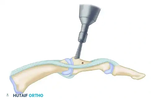
Correct Answer: Open release and debridement of the common extensor origin.
Explanation:
For recalcitrant lateral epicondylitis, the most commonly performed surgical procedure is an open (or increasingly, arthroscopic) release and debridement of the common extensor origin, specifically addressing the pathologic portion of the ECRB tendon. This involves excising the diseased, degenerative tissue. Endoscopic repair of a partial tear is not the standard. Ulnar nerve transposition is for cubital tunnel syndrome. Radial head excision is for conditions like severe radial head fractures or arthritis. Lateral collateral ligament repair is for instability.
Question 19:
When evaluating a patient with suspected lateral epicondylitis, which observation, if present, would most strongly suggest an alternative diagnosis such as radiohumeral osteoarthritis?
Options:
- Pain with resisted wrist extension.
- Point tenderness over the lateral epicondyle.
- Crepitus and pain with forearm pronation/supination.
- Pain relief with rest.
- Weak grip strength.
Correct Answer: Crepitus and pain with forearm pronation/supination.
Explanation:
While pain with resisted wrist extension and point tenderness are hallmarks of lateral epicondylitis, crepitus and pain specifically with forearm pronation and supination are highly indicative of intra-articular pathology, such as radiohumeral osteoarthritis, or possibly a plica. These mechanical symptoms are less typical for isolated tendinopathy. Pain relief with rest and weak grip strength can be present in both conditions to varying degrees.
Question 20:
A patient is referred to you for chronic lateral elbow pain. You suspect radial tunnel syndrome as a differential. Which physical examination maneuver would be most helpful in differentiating radial tunnel syndrome from lateral epicondylitis?
Options:
- Pain with resisted wrist extension (Cozen's test).
- Pain with resisted middle finger extension (Maudsley's test).
- Tenderness over the lateral epicondyle.
- Pain with resisted supination of the forearm.
- Pain with passive wrist flexion with the elbow extended (Mill's test).
Correct Answer: Pain with resisted supination of the forearm.
Explanation:
While there can be overlap, pain with resisted supination of the forearm, especially when the elbow is extended, specifically stresses the supinator muscle, under which the posterior interosseous nerve (PIN) passes, making it a key maneuver for diagnosing radial tunnel syndrome. The other tests (Cozen's, Maudsley's, Mill's, and lateral epicondyle tenderness) are classic signs of lateral epicondylitis, though some can be mildly positive in radial tunnel due to proximity or associated inflammation.
Question 21:
Which of the following describes the most common anatomical site of compression for the posterior interosseous nerve (PIN) in radial tunnel syndrome?
Options:
- Arcade of Struthers
- Fibrous arch of the supinator muscle (Arcade of Frohse)
- Ligament of Struthers
- Medial intermuscular septum
- Between the two heads of the pronator teres
Correct Answer: Fibrous arch of the supinator muscle (Arcade of Frohse)
Explanation:
The most common anatomical site of compression for the posterior interosseous nerve (PIN) in radial tunnel syndrome is the fibrous arch of the supinator muscle, known as the Arcade of Frohse. The Arcade of Struthers and Ligament of Struthers are associated with high median nerve compression. The medial intermuscular septum is relevant to the ulnar nerve. Compression between the two heads of the pronator teres is a site for median nerve entrapment (pronator syndrome).
Question 22:
Which intrinsic muscle of the hand is innervated by the ulnar nerve and commonly tested for weakness in cases of suspected ulnar neuropathy, a condition distinct from lateral epicondylitis?
Options:
- Abductor pollicis brevis
- Flexor pollicis longus
- First dorsal interosseous
- Opponens pollicis
- Flexor digitorum profundus to the index finger
Correct Answer: First dorsal interosseous
Explanation:
The first dorsal interosseous muscle is a key intrinsic hand muscle innervated by the ulnar nerve. Weakness here, along with other ulnar-innervated intrinsic muscles, is a hallmark of ulnar neuropathy (e.g., cubital tunnel syndrome). The abductor pollicis brevis, opponens pollicis, and flexor pollicis longus are primarily innervated by the median nerve. Flexor digitorum profundus is median and ulnar nerve-innervated, but the specific finger innervation varies.
Question 23:
A 50-year-old patient presents with lateral epicondylitis. An occupational therapist recommends an eccentric exercise program. What is the primary theoretical benefit of eccentric exercises in tendinopathy rehabilitation?
Options:
- To increase tendon length and flexibility.
- To reduce inflammation within the tendon.
- To induce collagen remodeling and strengthen the tendon.
- To increase concentric muscle strength for improved performance.
- To improve joint proprioception.
Correct Answer: To induce collagen remodeling and strengthen the tendon.
Explanation:
The primary theoretical benefit of eccentric exercises in tendinopathy rehabilitation is to induce collagen remodeling, strengthen the tendon, and improve its load-bearing capacity. While they may contribute to flexibility and improved performance (by strengthening the entire muscle-tendon unit), the specific effect on tendon structure and resistance to injury is the key. They do not directly reduce inflammation, and proprioception is a secondary benefit. The progressive loading during the lengthening phase of muscle contraction is thought to stimulate fibroblast activity and collagen synthesis in a more organized fashion.
Question 24:
Which of the following laboratory tests is most helpful in the routine diagnosis and workup of lateral epicondylitis?
Options:
- Erythrocyte Sedimentation Rate (ESR)
- C-Reactive Protein (CRP)
- Rheumatoid Factor (RF)
- Complete Blood Count (CBC)
- None of the above
Correct Answer: None of the above
Explanation:
Lateral epicondylitis is a clinical diagnosis based on history and physical examination. There are no specific laboratory tests that diagnose or are routinely helpful in the workup of uncomplicated lateral epicondylitis. ESR, CRP, and RF might be considered if an inflammatory arthropathy is suspected as a differential, but not for typical lateral epicondylitis. CBC is a general health screen. Therefore, none of the listed tests are routinely indicated.
Question 25:
When advising a patient on activity modification for lateral epicondylitis, which type of activity should be MOST emphasized to reduce strain on the ECRB?
Options:
- Avoiding heavy lifting with the elbow flexed.
- Minimizing repetitive elbow flexion and extension.
- Reducing activities that involve forceful wrist extension or gripping with the wrist extended.
- Limiting shoulder abduction beyond 90 degrees.
- Avoiding direct pressure on the ulnar nerve at the elbow.
Correct Answer: Reducing activities that involve forceful wrist extension or gripping with the wrist extended.
Explanation:
The ECRB is a primary wrist extensor. Activities involving forceful wrist extension, especially combined with gripping (e.g., hammering, tennis backhand, using a screwdriver), significantly load the ECRB origin and are the main aggravators of lateral epicondylitis. Reducing these activities is paramount for activity modification. The other options pertain to different elbow/shoulder pathologies or nerve entrapments.
Question 26:
What is the typical age range for patients presenting with lateral epicondylitis?
Options:
- Adolescence (10-18 years old)
- Young adults (19-25 years old)
- Middle-aged adults (30-60 years old)
- Elderly (over 70 years old)
- Infants and toddlers
Correct Answer: Middle-aged adults (30-60 years old)
Explanation:
Lateral epicondylitis is most prevalent in middle-aged adults, typically between 30 and 60 years old, with a peak incidence in the 4th and 5th decades of life. It is less common in younger individuals and generally not seen in infants or toddlers. While elderly individuals can develop it, the peak incidence is earlier.
Question 27:
Following surgical debridement for chronic lateral epicondylitis, which of the following is an expected post-operative rehabilitation goal during the early phase (first 2-4 weeks)?
Options:
- Initiating heavy eccentric wrist strengthening exercises.
- Immediate return to sport-specific activities.
- Restoration of full, pain-free elbow range of motion.
- Aggressive passive stretching into wrist flexion.
- Removal of external sutures/staples and wound care.
Correct Answer: Restoration of full, pain-free elbow range of motion.
Explanation:
In the early post-operative phase (2-4 weeks), the primary goals are pain control, wound healing (suture/staple removal is often around 10-14 days, not the sole goal for 2-4 weeks), and gradual restoration of pain-free elbow range of motion. Heavy eccentric wrist strengthening and immediate return to sport are delayed until the later stages of rehabilitation to allow for adequate healing and tissue maturation. Aggressive passive stretching can be counterproductive and potentially re-injure the healing tissue.
Question 28:
Which nerve is at greatest risk of iatrogenic injury during surgical intervention for lateral epicondylitis?
Options:
- Median nerve
- Ulnar nerve
- Musculocutaneous nerve
- Superficial radial nerve
- Posterior interosseous nerve
Correct Answer: Posterior interosseous nerve
Explanation:
During surgical intervention for lateral epicondylitis, especially with deeper dissection, the posterior interosseous nerve (PIN), a branch of the radial nerve, is at greatest risk. It winds around the radial neck and passes through the supinator muscle (Arcade of Frohse), near the surgical field for the common extensor origin. The superficial radial nerve is also a risk, but typically more distal and subcutaneous. Median, ulnar, and musculocutaneous nerves are more distant from the lateral epicondyle.
Question 29:
When considering the use of Platelet-Rich Plasma (PRP) for lateral epicondylitis, what is the primary proposed mechanism of action?
Options:
- Direct analgesic effect from the injected plasma.
- Anti-inflammatory properties of concentrated platelets.
- Delivery of growth factors to stimulate tendon healing and regeneration.
- Mechanical disruption of calcific deposits.
- Neurolysis of peripheral nerves at the elbow.
Correct Answer: Delivery of growth factors to stimulate tendon healing and regeneration.
Explanation:
The primary proposed mechanism of action for Platelet-Rich Plasma (PRP) in tendinopathy is the delivery of concentrated growth factors (e.g., PDGF, TGF-β, VEGF, IGF-1) released from activated platelets. These growth factors are believed to stimulate cellular proliferation, collagen synthesis, and neovascularization, thereby promoting tissue healing and regeneration rather than just providing analgesia or anti-inflammatory effects (which are secondary or debated).
Question 30:
A patient with lateral epicondylitis symptoms also describes numbness and tingling in the thumb and index finger. Which additional diagnostic consideration becomes critical?
Options:
- Ulnar nerve entrapment
- Median nerve entrapment (e.g., carpal tunnel syndrome or pronator syndrome)
- Cervical radiculopathy
- Thoracic outlet syndrome
- All of the above
Correct Answer: All of the above
Explanation:
Numbness and tingling in the thumb and index finger suggest involvement of the median nerve (e.g., carpal tunnel, pronator syndrome) or cervical radiculopathy at C6 or C7. While ulnar nerve entrapment affects the little and ring fingers, and thoracic outlet syndrome can affect various nerves, the specific distribution (thumb and index finger) makes median nerve and cervical radiculopathy particularly critical to consider alongside lateral epicondylitis. Therefore, to ensure a comprehensive differential, 'All of the above' encompasses the potential for multiple etiologies or concomitant conditions.
Question 31:
Which component of the lateral collateral ligament complex is most important for resisting varus stress at the elbow?
Options:
- Annular ligament
- Accessory collateral ligament
- Lateral ulnar collateral ligament (LUCL)
- Radial collateral ligament
- Oblique ligament
Correct Answer: Radial collateral ligament
Explanation:
The Radial Collateral Ligament (RCL) is the primary static stabilizer against varus stress at the elbow. The Annular Ligament stabilizes the radial head. The Lateral Ulnar Collateral Ligament (LUCL) is critical for posterolateral rotatory stability. The accessory collateral ligament provides additional support. While the question asks about lateral epicondylitis, a thorough examiner will know surrounding anatomy and potential differential diagnoses involving instability. The Radial Collateral Ligament originates from the lateral epicondyle, making it relevant to the region, though LUCL is more important for posterolateral instability.
Question 32:
A high-resolution musculoskeletal ultrasound for a patient with chronic lateral epicondylitis is most likely to reveal which of the following findings?
Options:
- Joint effusion within the radiohumeral joint.
- Hypoechoic thickening of the common extensor tendon, often with neovascularization.
- Complete rupture of the ECRB tendon with retraction.
- Anterior dislocation of the radial head.
- Calcification within the ulnar collateral ligament.
Correct Answer: Hypoechoic thickening of the common extensor tendon, often with neovascularization.
Explanation:
Musculoskeletal ultrasound in chronic lateral epicondylitis commonly shows hypoechoic (darker) thickening and disorganization of the common extensor tendon, particularly at the ECRB origin. Doppler ultrasound can also reveal neovascularization (increased blood flow), which is thought to be associated with pain. Joint effusion and radial head dislocation are unrelated. Complete rupture is rare. Calcification in the ulnar collateral ligament indicates medial elbow pathology.
Question 33:
What is the primary goal of initial rest and activity modification for a patient diagnosed with acute lateral epicondylitis?
Options:
- To prevent progression to surgery.
- To completely immobilize the elbow joint.
- To reduce the mechanical load on the healing tendon and allow symptomatic relief.
- To strengthen the surrounding musculature to compensate for the injured tendon.
- To stretch the affected tendon to improve flexibility.
Correct Answer: To reduce the mechanical load on the healing tendon and allow symptomatic relief.
Explanation:
The primary goal of initial rest and activity modification is to reduce the mechanical load and repetitive strain on the inflamed/degenerative common extensor tendon origin. This aims to decrease pain, prevent further microtrauma, and create an environment conducive to healing. Complete immobilization is rarely indicated and can lead to stiffness. Strengthening and stretching are part of later-stage rehabilitation, not the immediate goal of rest.
Question 34:
Which occupational factor is most strongly associated with an increased risk of developing lateral epicondylitis?
Options:
- Prolonged static posture.
- Repetitive, forceful gripping combined with wrist extension/pronation.
- Frequent overhead reaching.
- Exposure to cold temperatures.
- Minimal use of hand tools.
Correct Answer: Repetitive, forceful gripping combined with wrist extension/pronation.
Explanation:
Occupational factors involving repetitive, forceful gripping, especially when combined with wrist extension and/or forearm pronation (e.g., using heavy hand tools, assembly line work), are strongly associated with an increased risk of lateral epicondylitis due to the excessive strain placed on the common extensor origin, particularly the ECRB. Prolonged static posture and cold exposure are less directly implicated. Frequent overhead reaching can contribute to shoulder issues but less directly to lateral epicondylitis. Minimal tool use would decrease risk.
Question 35:
A patient with lateral epicondylitis also presents with significant weakness in wrist extension and finger extension, with minimal pain. The most likely concomitant diagnosis is:
Options:
- Radial tunnel syndrome
- Cervical disc herniation (C7 radiculopathy)
- Posterior interosseous nerve (PIN) entrapment
- Medial epicondylitis
- Elbow fracture
Correct Answer: Posterior interosseous nerve (PIN) entrapment
Explanation:
Significant weakness in wrist extension and finger extension, especially with minimal pain, is a hallmark of Posterior Interosseous Nerve (PIN) entrapment. The PIN is a purely motor nerve, and its compression leads to weakness in the muscles it innervates (wrist extensors, finger extensors) without sensory deficits. While radial tunnel syndrome encompasses PIN entrapment, PIN entrapment specifically highlights the motor weakness. Cervical radiculopathy could cause weakness but usually involves more widespread neurological symptoms and pain. Medial epicondylitis is on the opposite side. An elbow fracture would have acute pain and swelling.
Question 36:
Which injection type has been shown in some studies to have superior long-term outcomes compared to corticosteroid injections for chronic lateral epicondylitis?
Options:
- Local anesthetic (lidocaine) alone.
- Hyaluronic acid injection.
- Botulinum toxin injection.
- Platelet-Rich Plasma (PRP) injection.
- Prolotherapy (dextrose solution) injection.
Correct Answer: Platelet-Rich Plasma (PRP) injection.
Explanation:
While corticosteroid injections can provide short-term pain relief, several studies have demonstrated that Platelet-Rich Plasma (PRP) injections may offer superior long-term outcomes for chronic lateral epicondylitis, likely due to their role in stimulating tendon healing and regeneration. Local anesthetics provide only temporary relief. Hyaluronic acid and prolotherapy have less robust evidence for superiority in lateral epicondylitis compared to PRP. Botulinum toxin can reduce muscle activity but its long-term efficacy over corticosteroids is not clearly established as superior and side effects like temporary weakness are common.
Question 37:
When performing a 'grip strength' test in a patient with lateral epicondylitis, which observation is typically expected?
Options:
- Symmetrically reduced grip strength bilaterally.
- Increased grip strength due to compensatory muscle activation.
- Significantly reduced grip strength on the affected side, particularly with the elbow extended.
- Normal grip strength on the affected side.
- Reduced grip strength primarily in the little finger.
Correct Answer: Significantly reduced grip strength on the affected side, particularly with the elbow extended.
Explanation:
Patients with lateral epicondylitis typically experience significantly reduced grip strength on the affected side, especially when the elbow is extended, as this position places more tension on the common extensor origin. This is often due to pain inhibition rather than true muscle weakness. Symmetrically reduced strength or normal strength would be atypical for an affected unilateral condition. Reduced strength only in the little finger would suggest ulnar nerve involvement.
Question 38:
What is the typical timeframe for considering surgical intervention for lateral epicondylitis after exhausting non-operative treatments?
Options:
- Within 3 months of diagnosis.
- After 6-12 weeks of symptoms.
- After 6-12 months of failed non-operative management.
- Only if imaging shows a complete tendon rupture.
- Immediately after diagnosis in athletes.
Correct Answer: After 6-12 months of failed non-operative management.
Explanation:
Surgical intervention for lateral epicondylitis is typically considered for chronic, recalcitrant cases that have failed a comprehensive trial of non-operative management for at least 6 to 12 months. This allows sufficient time for various conservative treatments to have an effect. Surgery is not indicated within the first few months, nor is it reserved only for complete ruptures (which are rare), or performed immediately, even in athletes, unless it's a very specific, rare acute injury.
Question 39:
A 45-year-old patient presents with pain at the lateral epicondyle. During examination, you note that resisted wrist extension causes pain, but resisted long finger extension (Maudsley's test) is negative. What is the most likely implication of this finding?
Options:
- The patient likely has radial tunnel syndrome rather than lateral epicondylitis.
- The ECRB is unaffected, and another wrist extensor is the primary pathology.
- It suggests a less severe form of lateral epicondylitis, or atypical involvement.
- The patient is malingering.
- This pattern indicates primary involvement of the extensor digiti minimi.
Correct Answer: It suggests a less severe form of lateral epicondylitis, or atypical involvement.
Explanation:
While both Cozen's (resisted wrist extension) and Maudsley's (resisted long finger extension) tests target the common extensor origin, a positive Cozen's and negative Maudsley's suggests that the primary pathology is more specifically affecting the ECRB or ECRL rather than the Extensor Digitorum Communis (EDC), which is specifically stressed by Maudsley's test. It doesn't necessarily rule out lateral epicondylitis but might indicate a less widespread or atypical involvement of the common extensor origin. It does not directly point to radial tunnel syndrome, nor does it imply malingering. The ECRB is part of the common extensor origin, so it's not unaffected. EDC and ECRB are distinct but share a common origin.
Question 40:
Which of the following physical therapy modalities has the strongest evidence for long-term efficacy in the treatment of chronic lateral epicondylitis?
Options:
- Therapeutic ultrasound (pulsed mode).
- Hot packs and cold packs.
- Transcutaneous Electrical Nerve Stimulation (TENS).
- Eccentric strengthening exercises of the wrist extensors.
- Deep tissue massage to the forearm extensors.
Correct Answer: Eccentric strengthening exercises of the wrist extensors.
Explanation:
Of the listed modalities, eccentric strengthening exercises of the wrist extensors have the strongest evidence for long-term efficacy in the treatment of chronic tendinopathies, including lateral epicondylitis. These exercises are thought to promote collagen remodeling and increase the tendon's load-bearing capacity. The evidence for therapeutic ultrasound, TENS, and passive modalities like hot/cold packs and massage for long-term benefit is less robust or primarily for short-term pain relief.
Question 41:
A patient undergoing physical therapy for lateral epicondylitis is struggling with pain during daily activities despite an appropriate exercise regimen. The therapist notes that the patient's job involves frequent use of a heavy stapler. What would be the most important adjustment to recommend?
Options:
- Increase the intensity of eccentric exercises.
- Switch to a heavier stapler to build strength.
- Suggest using the unaffected hand or modifying the stapler for reduced force/different grip.
- Recommend immediate corticosteroid injection.
- Advise complete immobilization of the wrist and elbow.
Correct Answer: Suggest using the unaffected hand or modifying the stapler for reduced force/different grip.
Explanation:
If a specific activity like using a heavy stapler consistently aggravates symptoms, the most important adjustment is activity modification. This could involve using the unaffected hand, finding an alternative tool that requires less force, or modifying the grip/technique. Increasing exercise intensity when pain is already problematic would be counterproductive. Immediate injection is often considered after failed conservative measures, but activity modification is a primary conservative step. Complete immobilization is generally not indicated and can lead to stiffness.
Question 42:
Which statement best describes the role of surgical management for lateral epicondylitis?
Options:
- It is the first-line treatment for most patients.
- It is reserved for patients with severe pain and functional limitation who have failed at least 3 months of non-operative treatment.
- It primarily involves reattaching a completely ruptured ECRB tendon.
- It is typically considered for recalcitrant cases after 6-12 months of failed conservative management.
- It guarantees complete resolution of pain and full return to prior activity levels.
Correct Answer: It is typically considered for recalcitrant cases after 6-12 months of failed conservative management.
Explanation:
Surgical management for lateral epicondylitis is reserved for chronic, recalcitrant cases that have failed a prolonged course (typically 6-12 months) of comprehensive non-operative treatment, including physical therapy, activity modification, and sometimes injections. It is not first-line, and complete rupture is rare. While often successful, surgery does not guarantee complete pain resolution or full return to prior activity for all patients.
Question 43:
During your examination of a patient with lateral elbow pain, you find tenderness over the medial epicondyle in addition to the lateral epicondyle. What is the most appropriate interpretation of this finding?
Options:
- It suggests a bilateral lateral epicondylitis.
- It indicates a likely misdiagnosis of lateral epicondylitis.
- It suggests a concomitant medial epicondylitis or Golfer's elbow, or a more generalized overuse syndrome.
- It is a normal finding and should be disregarded.
- It is indicative of an underlying systemic inflammatory condition.
Correct Answer: It suggests a concomitant medial epicondylitis or Golfer's elbow, or a more generalized overuse syndrome.
Explanation:
Tenderness over both the medial and lateral epicondyles suggests either a concomitant medial epicondylitis (Golfer's elbow) or a more generalized overuse syndrome affecting both common flexor and extensor origins. It does not indicate a misdiagnosis of lateral epicondylitis (if lateral symptoms are present) but points to additional pathology. It is not a normal finding and, while systemic conditions can cause widespread tendinopathy, it's not the primary inference from focal tenderness at both epicondyles.
Question 44:
What is the primary differentiating feature between lateral epicondylitis and posterior interosseous nerve (PIN) entrapment on clinical examination?
Options:
- Location of maximal tenderness.
- Presence of pain with resisted wrist extension.
- Sensory deficits in the superficial radial nerve distribution.
- Presence of motor weakness in specific forearm/hand muscles.
- Pain with passive wrist flexion.
Correct Answer: Presence of motor weakness in specific forearm/hand muscles.
Explanation:
The primary differentiating feature is the presence of motor weakness in specific forearm/hand muscles without sensory loss in PIN entrapment (a purely motor nerve). Lateral epicondylitis is a tendinopathy, causing pain but typically no true motor weakness (though grip strength may be pain-inhibited). While tenderness and resisted movements can overlap, true, objective muscle weakness strongly points to PIN entrapment. Sensory deficits are not typical for PIN entrapment as it's a motor nerve, and radial tunnel syndrome typically involves pain but not usually overt motor weakness to the degree seen in PIN syndrome which affects more distal motor branches.
Question 45:
Which of the following findings on a plain radiograph of the elbow is LEAST likely to be associated with lateral epicondylitis?
Options:
- Normal bony anatomy.
- Small calcifications at the lateral epicondyle.
- Subtle periosteal reaction at the lateral epicondyle.
- Loose bodies within the radiohumeral joint.
- Degenerative changes of the radiohumeral joint.
Correct Answer: Loose bodies within the radiohumeral joint.
Explanation:
Loose bodies within the radiohumeral joint are indicative of an intra-articular pathology such as osteochondritis dissecans or advanced osteoarthritis, and are not typically associated with lateral epicondylitis itself. Plain radiographs are often normal in lateral epicondylitis. Occasionally, small calcifications or subtle periosteal reaction can be seen at the lateral epicondyle. Degenerative changes of the radiohumeral joint are a differential diagnosis that may present with lateral elbow pain but are not a finding of lateral epicondylitis itself.
Question 46:
Which activity would place the most significant biomechanical stress on the common extensor origin, predisposing to lateral epicondylitis?
Options:
- Forehand stroke in tennis with proper technique.
- Backhand stroke in tennis with inadequate wrist stabilization.
- Swimming laps using a freestyle stroke.
- Lifting light weights with the elbow flexed.
- Performing biceps curls with correct form.
Correct Answer: Backhand stroke in tennis with inadequate wrist stabilization.
Explanation:
A backhand stroke in tennis, especially with inadequate wrist stabilization (leading to excessive wrist extension on impact or forceful wrist extension to generate power), places significant tensile and eccentric load on the common extensor origin, particularly the ECRB. This is a classic precipitating factor for lateral epicondylitis. Forehand strokes, swimming, and most gym exercises (if done with proper form) typically load other muscle groups or distribute forces differently.
Question 47:
Which of the following statements about the efficacy of corticosteroid injections for lateral epicondylitis is most accurate?
Options:
- They provide excellent long-term pain relief and are superior to physical therapy.
- They offer reliable short-term pain relief but often have worse long-term outcomes than other non-operative treatments.
- They are contraindicated in all cases due to tendon rupture risk.
- They stimulate tendon healing and collagen repair.
- They are the only effective treatment for severe cases.
Correct Answer: They offer reliable short-term pain relief but often have worse long-term outcomes than other non-operative treatments.
Explanation:
Corticosteroid injections for lateral epicondylitis typically provide good short-term (e.g., 6-week) pain relief. However, numerous studies have shown that they often have worse long-term outcomes (e.g., recurrence rates) compared to watchful waiting, physical therapy, or even placebo. They do not stimulate tendon healing or collagen repair; in fact, they may potentially weaken tendon structure with repeated injections. They are not contraindicated in all cases but should be used judiciously, and are not the only effective treatment for severe cases (surgery or biologics are options).
Question 48:
When evaluating a patient for lateral epicondylitis, palpation of the lateral epicondyle reveals maximal tenderness just anterior to its most prominent point. This finding is most consistent with pathology of which structure?
Options:
- Common flexor origin
- Lateral ulnar collateral ligament
- Extensor Carpi Radialis Brevis (ECRB) origin
- Olecranon bursa
- Radial head articular cartilage
Correct Answer: Extensor Carpi Radialis Brevis (ECRB) origin
Explanation:
Maximal tenderness just anterior and distal to the most prominent point of the lateral epicondyle is the classic location for pathology involving the Extensor Carpi Radialis Brevis (ECRB) origin, which is the primary structure involved in lateral epicondylitis. The common flexor origin is on the medial epicondyle. The lateral ulnar collateral ligament is more posterior and inferior. The olecranon bursa is posterior. The radial head articular cartilage would cause tenderness with palpation and pain with rotation, but the specified location points more specifically to the ECRB tendon origin.
Question 49:
Which of the following is considered an absolute contraindication to a corticosteroid injection for lateral epicondylitis?
Options:
- History of diabetes mellitus.
- Current oral anticoagulant therapy.
- Pain for less than 6 weeks.
- Infection at the injection site.
- Previous failed corticosteroid injection.
Correct Answer: Infection at the injection site.
Explanation:
Infection at the injection site is an absolute contraindication to any injection, including corticosteroids, due to the risk of spreading infection into deeper tissues or the joint. Diabetes mellitus is a relative contraindication (steroids can elevate blood glucose). Oral anticoagulant therapy requires careful consideration due to bleeding risk but is not an absolute contraindication if benefits outweigh risks and precautions are taken. Pain duration less than 6 weeks might make a steroid injection less appropriate (favoring other conservative methods), but it's not an absolute contraindication. A previous failed injection often prompts consideration of alternative treatments rather than another steroid injection, but again, not an absolute contraindication from a safety perspective.
Question 50:
What is the term for the degenerative changes observed in chronic tendinopathy, such as lateral epicondylitis?
Options:
- Tendinitis
- Tenosynovitis
- Tendinosis
- Bursitis
- Arthritis
Correct Answer: Tendinosis
Explanation:
The term 'tendinosis' accurately describes the degenerative changes (collagen disorganization, angiofibroblastic hyperplasia, absence of inflammatory cells) seen in chronic tendinopathy. 'Tendinitis' implies acute inflammation, which is generally not the primary pathological process in chronic cases. 'Tenosynovitis' refers to inflammation of the tendon sheath. 'Bursitis' is inflammation of a bursa, and 'Arthritis' is joint inflammation.
Question 51:
A patient reports relief of lateral elbow pain with a trial of a counterforce brace. This response supports the hypothesis that the brace works by:
Options:
- Compressing the radial nerve to reduce pain signals.
- Preventing all wrist movement during activity.
- Reducing the tensile load and strain at the extensor origin.
- Increasing blood flow to the affected tendon.
- Providing direct heat therapy to the lateral epicondyle.
Correct Answer: Reducing the tensile load and strain at the extensor origin.
Explanation:
A counterforce brace is believed to work by applying compression distal to the epicondyle, thereby creating a new, more distal origin for the wrist extensor muscles. This effectively lengthens the muscle-tendon unit, altering the angle of pull and reducing the tensile load and strain experienced at the common extensor origin during gripping and wrist extension, which helps to alleviate pain. It does not compress the radial nerve, prevent all wrist movement, increase blood flow, or provide heat therapy.
Question 52:
In cases where cervical radiculopathy (C6/C7) mimics lateral epicondylitis, which of the following findings would be most indicative of a cervical origin?
Options:
- Pain with resisted wrist extension.
- Point tenderness over the lateral epicondyle.
- Positive Spurling's test (neck compression/rotation).
- Normal neurological exam of the upper extremity.
- Pain relief with a counterforce brace.
Correct Answer: Positive Spurling's test (neck compression/rotation).
Explanation:
A positive Spurling's test, which reproduces radicular symptoms by extending, rotating, and laterally flexing the neck while applying axial compression, is highly indicative of cervical nerve root irritation and would strongly suggest a cervical origin for the pain. While pain with resisted wrist extension and lateral epicondyle tenderness can be present with referred pain from the neck, a specific neck provocative test is key. A normal neurological exam would make radiculopathy less likely, but subtle changes may exist. Pain relief with a counterforce brace would typically point to elbow pathology.
Question 53:
What is the typical long-term outcome for most patients with lateral epicondylitis managed conservatively?
Options:
- Progressive worsening of symptoms requiring eventual surgery.
- Complete resolution of symptoms within 3 months in all patients.
- Resolution of symptoms within 1-2 years in the vast majority of patients, with or without intervention.
- Chronic, debilitating pain requiring lifelong pain management.
- Recurrent episodes requiring repeated injections indefinitely.
Correct Answer: Resolution of symptoms within 1-2 years in the vast majority of patients, with or without intervention.
Explanation:
The natural history of lateral epicondylitis is generally favorable. The vast majority of patients (80-95%) will experience resolution of symptoms within 1-2 years, even with conservative management or sometimes even watchful waiting. While symptoms can be protracted and recurrences can happen, it typically does not lead to progressive worsening requiring surgery in most, nor does it typically become a lifelong debilitating condition. Complete resolution within 3 months for all patients is overly optimistic, and indefinite repeated injections are not standard practice.
Question 54:
When performing an elbow examination for lateral epicondylitis, which nerve should be assessed for potential concurrent entrapment or irritation that might contribute to lateral elbow pain?
Options:
- Ulnar nerve
- Median nerve
- Musculocutaneous nerve
- Posterior interosseous nerve (PIN)
- Anterior interosseous nerve (AIN)
Correct Answer: Posterior interosseous nerve (PIN)
Explanation:
The Posterior Interosseous Nerve (PIN), a motor branch of the radial nerve, passes through the supinator muscle in the radial tunnel, an area anatomically close to the common extensor origin. Entrapment of the PIN (or the radial nerve proper in the radial tunnel) is a key differential diagnosis for lateral epicondylitis and can sometimes coexist or mimic it, causing lateral elbow pain and forearm symptoms. Therefore, assessing for PIN involvement (e.g., specific motor weakness) is crucial. The ulnar, median, musculocutaneous, and anterior interosseous nerves are located more medially or anteriorly and are less directly implicated in lateral epicondyle pain etiology, though all upper extremity nerves should be considered in a comprehensive exam if symptoms warrant.
Question 55:
Which specific population is at a disproportionately higher risk of developing lateral epicondylitis?
Options:
- Children involved in gymnastics.
- Adolescent baseball pitchers.
- Middle-aged manual laborers and tennis players.
- Elderly individuals with sedentary lifestyles.
- Individuals with rheumatoid arthritis.
Correct Answer: Middle-aged manual laborers and tennis players.
Explanation:
Middle-aged manual laborers (due to repetitive gripping and tool use) and tennis players (particularly due to improper backhand technique or overuse) are classic populations at higher risk for developing lateral epicondylitis. Children and adolescents are more prone to apophysitis or osteochondritis dissecans. Elderly sedentary individuals have a lower risk. While rheumatoid arthritis can cause tendinopathy, it's a systemic condition and not the specific demographic at disproportionately higher risk for this specific, typically mechanical, tendinopathy.