Surgical Management of Hallux Valgus: A Comprehensive Academic Guide
Key Takeaway
Hallux valgus is a complex triplanar deformity of the first ray requiring meticulous surgical correction. This comprehensive guide details the biomechanics, clinical evaluation, and step-by-step operative techniques for various osteotomies, including Chevron, Scarf, Ludloff, and Lapidus procedures. Designed for orthopedic residents and consultants, it provides evidence-based protocols, surgical pearls, and complication management strategies to optimize patient outcomes and restore forefoot kinematics.
Comprehensive Introduction and Patho-Epidemiology
The foundational principles and extensive bibliographic research underlying this chapter acknowledge the contributions of Dr. Brian G. Donley, who contributed significantly to this work during his tenure as a Foot Fellow at the Campbell Clinic. Dr. Donley subsequently established his practice at the Cleveland Clinic, Cleveland, Ohio, advancing the field of forefoot reconstruction. Building upon this legacy, the surgical management of hallux valgus remains one of the most intricately debated and continuously evolving domains within orthopedic foot and ankle surgery. Hallux valgus is not merely a localized prominence of the medial eminence; it is a complex, progressive, triplanar deformity of the first ray that profoundly alters the biomechanics of the entire forefoot.
Epidemiologically, hallux valgus represents the most common pathology of the forefoot, with a reported prevalence of nearly 23% in adults aged 18 to 65, escalating to over 35% in populations older than 65 years. The deformity exhibits a striking female predominance, often attributed to a combination of genetic predisposition, ligamentous laxity, and extrinsic factors such as constricting, narrow-toebox, and high-heeled footwear. However, while restrictive footwear exacerbates the symptomatology and accelerates the progression of the deformity, a strong familial inheritance pattern suggests an underlying intrinsic structural vulnerability, often linked to the morphology of the first metatarsocuneiform joint, generalized hypermobility, or variations in metatarsal length.
The patho-etiology of hallux valgus is characterized by a dynamic failure of the medial ligamentous structures and a subsequent mechanical advantage gained by the laterally based musculature. The deformity manifests in three distinct planes: transverse (lateral deviation of the hallux and medial deviation of the first metatarsal), sagittal (dorsiflexion or elevation of the first metatarsal), and coronal (pronation of the hallux and first metatarsal). Historically, surgical interventions primarily focused on the transverse plane, often neglecting the crucial coronal plane rotation. Modern academic understanding dictates that failure to recognize and correct the pronation of the first metatarsal is a primary driver of postoperative recurrence and persistent sesamoid subluxation.
As the deformity progresses, the altered alignment leads to a cascade of secondary forefoot pathologies. The incompetence of the first ray diminishes its load-bearing capacity during the terminal stance phase of gait, transferring disproportionate forces to the lesser metatarsals. This phenomenon, known as transfer metatarsalgia, frequently results in second metatarsophalangeal (MTP) joint synovitis, plantar plate attenuation, crossover toe deformities, and intractable plantar keratoses. Therefore, the surgical correction of hallux valgus must be viewed not as an isolated cosmetic procedure, but as a comprehensive biomechanical restoration of the forefoot's load-sharing architecture.
Detailed Surgical Anatomy and Biomechanics
A profound mastery of the surgical anatomy of the first ray is non-negotiable for the orthopedic surgeon attempting hallux valgus reconstruction. The first MTP joint is a highly complex diarthrodial joint stabilized by an intricate network of capsuloligamentous and musculotendinous structures. Unlike the lesser toes, the first MTP joint lacks a true deep transverse metatarsal ligament connecting the metatarsal heads directly; instead, the deep transverse metatarsal ligament tethers the sesamoid apparatus of the first ray to the second metatarsal head. This anatomical nuance is the crux of hallux valgus pathoanatomy.
The sesamoid apparatus consists of the tibial (medial) and fibular (lateral) sesamoids, which are embedded within the tendons of the flexor hallucis brevis (FHB). These sesamoids articulate with the plantar aspect of the first metatarsal head, separated by the crista (sagittal groove). In the pathogenesis of hallux valgus, it is a common misconception that the sesamoids subluxate laterally. In reality, the sesamoids remain tethered in their anatomical position relative to the second metatarsal via the deep transverse metatarsal ligament, while the first metatarsal head drifts medially and pronates off the sesamoid apparatus. As the metatarsal head deviates medially, the crista is eroded, and the fibular sesamoid appears to migrate into the first web space.
This medial drift of the first metatarsal precipitates a devastating biomechanical cascade. The abductor hallucis tendon, which normally stabilizes the medial aspect of the joint, shifts plantarward and rotates beneath the metatarsal head. In this displaced position, it loses its abductor function and paradoxically contributes to the pronation and plantarflexion of the hallux. Simultaneously, the adductor hallucis and the lateral head of the FHB gain a mechanical advantage, becoming unopposed deforming forces that tether the proximal phalanx and sesamoids laterally. This dynamic imbalance relentlessly drives the hallux into further valgus and pronation.
The disruption of this delicate anatomical balance severely compromises the windlass mechanism, originally described by Hicks. The plantar fascia, inserting into the base of the proximal phalanx via the sesamoid apparatus, relies on a stable first MTP joint to elevate the medial longitudinal arch during hallux dorsiflexion. When the first ray is deformed and destabilized by hallux valgus, the windlass mechanism fails, leading to functional hallux limitus, medial column collapse, and the aforementioned transfer metatarsalgia. Surgical correction must meticulously release the contracted lateral structures and restore the medial capsuloligamentous tension to re-establish this vital biomechanical linkage.
Exhaustive Indications and Contraindications
The decision to proceed with surgical intervention for hallux valgus must be rooted in rigorous clinical criteria, prioritizing pain relief and functional restoration over cosmetic appearance. Surgery is strictly indicated for patients who present with progressive, symptomatic deformity that has proven refractory to a comprehensive trial of conservative management. Conservative measures should include shoe wear modification (wide toe box, low heels), orthotic devices to support the medial arch and offload painful prominences, padding, and non-steroidal anti-inflammatory drugs (NSAIDs). When these non-operative modalities fail to alleviate pain associated with the medial eminence, sesamoid apparatus, or secondary transfer metatarsalgia, surgical correction is warranted.
Contraindications to hallux valgus surgery are equally critical to recognize to prevent catastrophic postoperative complications. Absolute contraindications include active local or systemic infection, severe peripheral arterial disease lacking adequate perfusion for wound healing (often assessed via ankle-brachial index or toe pressures), and profound peripheral neuropathy (e.g., advanced Charcot neuroarthropathy) where the loss of protective sensation guarantees postoperative breakdown or hardware failure. Relative contraindications encompass poorly controlled diabetes mellitus, heavy tobacco use, complex regional pain syndrome (CRPS), and unrealistic patient expectations. Operating on a painless, purely cosmetic deformity is universally condemned in academic orthopedic practice.
The selection of the specific surgical procedure is dictated by a complex algorithmic approach that considers the magnitude of the deformity, the presence of first tarsometatarsal (TMT) joint hypermobility, and the congruency of the MTP joint. The following table provides an exhaustive breakdown of the indications and contraindications for the most commonly utilized surgical interventions.
| Surgical Procedure | Primary Indications | Specific Contraindications |
|---|---|---|
| Distal Metatarsal Osteotomy (Chevron) | Mild to moderate deformity (HVA <30°, IMA <13°); Congruent MTP joint; Patient age typically <50-60 years with good bone stock. | Severe deformity (IMA >15°); First ray hypermobility; Advanced MTP joint arthrosis; Narrow first metatarsal width. |
| Diaphyseal Osteotomy (Scarf) | Moderate to severe deformity (HVA 30°-40°, IMA 13°-20°); Need for DMAA correction; Need for metatarsal lengthening/shortening. | Severe osteopenia (risk of troughing); First ray hypermobility; Metatarsal shaft excessively narrow; Advanced MTP joint arthrosis. |
| First TMT Arthrodesis (Lapidus) | Severe deformity (HVA >40°, IMA >20°); Clinical first ray hypermobility; TMT joint arthrosis; Recurrent hallux valgus. | Open physes (in pediatric patients); Severe midfoot arthrosis (unless addressed concurrently); Non-compliant patient (strict NWB required). |
| Proximal Phalangeal Osteotomy (Akin) | Hallux valgus interphalangeus; Residual clinical valgus after metatarsal correction; Congruent MTP joint. | Used as an isolated procedure for MTP joint pathology; Severe MTP joint subluxation; Short proximal phalanx. |
| First MTP Joint Arthrodesis | Severe hallux valgus with concurrent advanced MTP joint arthrosis (Hallux Rigidus); Rheumatoid arthritis; Severe recurrent deformity; Neuromuscular spasticity. | Active infection; Severe bone loss precluding adequate fixation (may require bone block arthrodesis); Patient refusal of joint sacrifice. |
Pre-Operative Planning, Templating, and Patient Positioning
A rigorous, systematic preoperative assessment is the cornerstone of successful hallux valgus reconstruction. The clinical evaluation must assess the site of maximal tenderness, the reducible nature of the deformity, the presence of MTP joint crepitus or limited range of motion (ROM), and the neurovascular status of the foot. A critical component of the physical examination is the assessment of first TMT joint mobility. While the definition of "hypermobility" remains somewhat controversial and lacks a universally accepted objective measurement, the surgeon must evaluate the sagittal plane excursion of the first metatarsal relative to the lesser metatarsals while stabilizing the midfoot. Excessive dorsal excursion, particularly if it reproduces the patient's pain or is accompanied by a medial column sag, strongly suggests the need for a stabilizing procedure such as a Lapidus arthrodesis.
Radiographic evaluation demands standardized, weight-bearing anteroposterior (AP), lateral, and sesamoid axial views. Non-weight-bearing radiographs are entirely inadequate for assessing the true magnitude of the functional deformity. The surgeon must meticulously calculate key radiographic parameters. The Hallux Valgus Angle (HVA), formed by the intersection of the longitudinal axes of the first metatarsal and proximal phalanx, is normally less than 15 degrees. The Intermetatarsal Angle (IMA), subtended by the axes of the first and second metatarsals, is normally less than 9 degrees. The Distal Metatarsal Articular Angle (DMAA) assesses the orientation of the articular cartilage relative to the metatarsal shaft; an angle greater than 10 degrees indicates a congruent deformity that necessitates a specific intra-articular or biplanar extra-articular correction to prevent postoperative joint subluxation.
Furthermore, the sesamoid station must be evaluated using the Hardy and Clapham classification (stages 1-7) on the AP view, or more accurately, on the sesamoid axial view. This dictates the extent of the lateral soft tissue release required. Preoperative templating is essential to identify the Center of Rotation of Angulation (CORA) and to plan the level and geometry of the osteotomy. The surgeon must anticipate the required lateral translation, the potential for metatarsal shortening (a common consequence of osteotomies that must be minimized to prevent transfer metatarsalgia), and the hardware trajectory. Advanced imaging, such as weight-bearing CT (WBCT), is increasingly utilized in academic centers to accurately quantify the coronal plane pronation of the first metatarsal, which is often masked on standard 2D plain films.
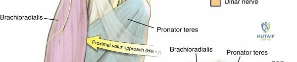
Preoperative setup requires meticulous attention to detail. Anesthesia is typically achieved via a regional popliteal or ankle block, combined with monitored anesthesia care (MAC) or general anesthesia, depending on patient preference and surgical duration. The patient is positioned supine on the operating table. A critical positioning nuance is the placement of a bump under the ipsilateral hip; this internally rotates the naturally externally rotated lower extremity to a neutral position, ensuring the plantar aspect of the foot rests flat and perpendicular to the operating table, providing the surgeon with a true AP perspective. A calf or thigh tourniquet is applied, and exsanguination is performed using an Esmarch bandage prior to inflation (typically set between 250 and 300 mmHg) to ensure a bloodless surgical field, which is vital for identifying the delicate terminal branches of the deep peroneal nerve and the intricate lateral capsular structures.
Step-by-Step Surgical Approach and Fixation Technique
Distal Soft Tissue Release (Modified McBride)
The distal soft tissue release is the foundational procedure upon which nearly all hallux valgus corrections rely. It systematically addresses the pathologically contracted lateral tethering structures, allowing for the mobilization of the sesamoid apparatus and the correction of the MTP joint subluxation.
The procedure begins with a dorsal longitudinal incision in the first web space, carefully deepened through the subcutaneous tissue. The surgeon must meticulously identify and retract the terminal branches of the deep peroneal nerve and the first dorsal metatarsal artery. Deep dissection exposes the conjoined tendon of the adductor hallucis as it inserts onto the base of the proximal phalanx and the fibular sesamoid. The adductor hallucis tendon is sharply transected at its insertion. Next, the deep transverse metatarsal ligament, which tethers the fibular sesamoid to the second metatarsal head, is identified and released.
Following this, the lateral sesamoid suspensory ligament is released, allowing the fibular sesamoid to mobilize medially. The surgeon must palpate the sesamoid to ensure it is completely free from lateral tethering. If a severe, rigid contracture persists, a controlled lateral capsulotomy of the first MTP joint is performed, typically through multiple puncture capsulotomies to preserve the lateral blood supply to the metatarsal head. A critical surgical pitfall is the routine excision of the fibular sesamoid; this practice has been largely abandoned in modern academic surgery as it significantly increases the risk of iatrogenic hallux varus and irreversibly alters the biomechanics of the FHB.
Distal Metatarsal Osteotomies (Chevron)
The distal Chevron osteotomy is the workhorse procedure for mild to moderate hallux valgus deformities. It is an intrinsically stable, V-shaped, horizontally directed displacement osteotomy of the metatarsal metaphysis.
A medial longitudinal incision is made directly over the midline of the first MTP joint, extending from the mid-diaphysis to the interphalangeal joint. An inverted L-shaped or longitudinal medial capsulotomy is performed, raising a full-thickness capsuloperiosteal flap to expose the medial eminence. The exostectomy is performed using a microsaw; the cut must be strictly parallel to the medial border of the foot. The surgeon must preserve the sagittal groove (crista) to avoid "staking the head," a technical error that destroys the medial buttress for the tibial sesamoid and inevitably leads to hallux varus.
The osteotomy is initiated by driving a 0.045-inch K-wire centrally into the metatarsal head, approximately 1 to 2 cm proximal to the articular surface, to mark the apex. A microsaw is used to create a 60-degree V-osteotomy. The dorsal arm exits proximal to the articular cartilage, and the plantar arm exits proximal to the sesamoid apparatus, ensuring the capsular vascular attachments to the capital fragment remain intact. The capital fragment is then translated laterally by 30% to 50% of the metatarsal width. The osteotomy is provisionally pinned and definitively secured with a single 2.5 mm or 3.0 mm headless compression screw directed from dorsal-proximal to plantar-distal. The medial capsule is then imbricated to correct residual pronation and secure the medial repair.
Diaphyseal Osteotomies (Scarf)
The Scarf osteotomy is a highly versatile, Z-step cut performed in the diaphysis of the first metatarsal. It allows for massive lateral translation, the ability to lengthen or shorten the metatarsal, and the capacity to correct the DMAA through medial rotation of the distal fragment.
An extended medial longitudinal incision is utilized to expose the entire medial shaft of the first metatarsal. The osteotomy design consists of three cuts. The primary longitudinal cut is made in the mid-diaphysis, strictly parallel to the plantar aspect of the foot to prevent unintended elevation or depression of the metatarsal head. The dorsal transverse cut is made distally, and the plantar transverse cut is made proximally. The plantar-distal fragment is then translated laterally to achieve the desired IMA correction. If a high DMAA is present, the distal fragment can be rotated medially on the proximal fragment to restore joint congruency.
Fixation is achieved using two cortical screws (typically 2.5 mm, 3.0 mm, or specialized Scarf screws) placed from dorsal to plantar, compressing the diaphyseal bone. Following rigid fixation, the overhanging medial cortical bone of the proximal fragment is resected flush with the translated distal fragment. A major surgical pitfall associated with the Scarf osteotomy is "troughing." This occurs when the hard cortical bone of the translated distal fragment collapses into the softer cancellous bone of the proximal metaphysis, leading to an unintended dorsiflexion elevation of the first ray and subsequent, severe transfer metatarsalgia.
First Tarsometatarsal Arthrodesis (Lapidus Procedure)
The Lapidus procedure is widely considered the gold standard for severe hallux valgus, particularly in the presence of clinical first ray hypermobility, TMT joint arthritis, or recurrent deformity following failed distal procedures. By fusing the base of the first metatarsal to the medial cuneiform, it provides powerful, permanent correction of the IMA and allows for multiplanar correction, including the critical coronal plane pronation.
A dorsal longitudinal incision is made over the first TMT joint, positioned lateral to the extensor hallucis longus (EHL) tendon to avoid neurovascular injury. The capsule of the first TMT joint is sharply incised. The articular cartilage of both the base of the first metatarsal and the medial cuneiform is meticulously resected. This can be achieved using a microsaw, sharp osteotomes, or curettes, ensuring that the subchondral bone plate is preserved but adequately fenestrated to promote robust osteogenesis and arthrodesis.
Correction is achieved by translating the first metatarsal base laterally and plantarflexing it to close the IMA and restore the medial longitudinal arch. Crucially, the pronation of the hallux is corrected by manually supinating the metatarsal before temporary fixation. Rigid internal fixation is mandatory to prevent nonunion. This is typically achieved using a combination of crossed solid or cannulated compression screws (3.5 mm or 4.0 mm), a dorsal or medial locking plate, or a modern plantar plating system designed to resist the tension forces on the plantar aspect of the foot. Local autograft harvested from the medial eminence resection is routinely packed into the arthrodesis site to augment healing.
Proximal Phalangeal Osteotomy (Akin Procedure)
The Akin osteotomy is an adjunctive procedure, almost never utilized in isolation for primary hallux valgus. It is a medial closing-wedge osteotomy of the proximal phalanx indicated for correcting hallux valgus interphalangeus or addressing residual clinical valgus after the metatarsal deformity has been fully corrected.
The medial incision used for the primary MTP joint procedure is extended distally over the proximal phalanx. The periosteum is elevated minimally to preserve blood supply. A medial closing wedge osteotomy is created approximately 5 to 10 mm distal to the MTP joint line. The critical technical aspect of the Akin procedure is that the lateral cortex of the proximal phalanx must remain intact to act as a tension band hinge.
Once the wedge of bone is removed, the osteotomy is closed, thereby rectifying the residual valgus angulation of the toe. The osteotomy is then secured. While historically pinned with K-wires or secured with intraosseous sutures, modern academic practice heavily favors rigid fixation with a single memory-metal staple, a small locking plate, or a 2.0 mm to 2.5 mm headless compression screw to allow for early mobilization without loss of correction.
Complications, Incidence Rates, and Salvage Management
Despite meticulous surgical technique, complications following hallux valgus reconstruction can and do occur. The academic surgeon must be intimately familiar with the etiology, prevention, and salvage management of these adverse events. Patient dissatisfaction often stems not from the primary procedure, but from the failure to rapidly recognize and appropriately manage a developing complication.
Iatrogenic hallux varus is perhaps the most devastating complication, characterized by medial deviation of the hallux and loss of MTP joint function. It is almost exclusively the result of surgical overcorrection. Etiologies include aggressive medial eminence resection that removes the sagittal groove ("staking the head"), over-tightening of the medial capsular imbrication, accidental or intentional complete fibular sesamoidectomy, or excessive lateral translation of the metatarsal head. Flexible varus deformities recognized early may be salvaged with soft tissue reconstruction, such as an EHL transfer routed under the deep transverse metatarsal ligament. However, rigid deformities or those complicated by secondary MTP joint arthrosis mandate a first MTP joint arthrodesis for definitive salvage.
Avascular necrosis (AVN) of the first metatarsal head is a catastrophic risk, particularly when a distal osteotomy (like a Chevron) is combined with an overly aggressive lateral soft tissue release. The blood supply to the metatarsal head is tenuous, relying heavily on capsular vessels entering the plantar-lateral metaphysis. Disruption of this extraosseous network, combined with the intraosseous disruption of the osteotomy, can lead to ischemic collapse. Early AVN may be managed conservatively with strict offloading and bone stimulators. However, severe collapse with fragmentation and secondary arthrosis inevitably necessitates a first MTP joint arthrodesis, often requiring structural bone grafting to restore metatarsal length.
Recurrence of the hallux valgus deformity is the most common long-term complication. It is frequently the result of an error in preoperative decision-making, such as utilizing a distal osteotomy for a severe deformity, failing to recognize and address a hypermobile first TMT joint, or undercorrecting the coronal plane pronation. Inadequate medial capsular imbrication or premature weight-bearing can also lead to early loss of fixation and recurrence. Revision surgery must address the underlying biomechanical failure; simply repeating a distal osteotomy is doomed to fail. Salvage typically requires a more proximal, powerful procedure, most commonly a Lapidus arthrodesis, to definitively stabilize the medial column.
| Complication | Estimated Incidence | Primary Etiology | Salvage Strategy |
|---|---|---|---|
| Iatrogenic Hallux Varus | 2% - 8% | "Staking the head"; Over-tightened medial capsule; Fibular sesamoidectomy. | Flexible: EHL transfer / Soft tissue release. Rigid/Arthritic: 1st MTP Arthrodesis. |
| Avascular Necrosis (AVN) | 1% - 3% | Extensive lateral release combined with distal osteotomy; Disrupted plantar-lateral vessels. | Early: Offloading. Late/Collapse: 1st MTP Arthrodesis (often with structural graft). |
| Recurrence of Deformity | 10% - 15% | Missed hypermobility; Undercorrection of IMA or pronation; Inadequate fixation. | Revision with a more proximal stabilizing procedure (e.g., Lapidus Arthrodesis). |
| Nonunion / Delayed Union | 3% - 5% (Lapidus) | Inadequate joint preparation; Non-rigid fixation; Patient non-compliance with NWB. | Revision arthrodesis with autogenous bone grafting and rigid plate fixation. |
| Transfer Metatarsalgia | 5% - 10% | Metatarsal shortening; Dorsiflexion malunion (troughing in Scarf); Unaddressed lesser toe pathology. | Plantarflexing opening wedge osteotomy of 1st MT; Weil osteotomies of lesser MTs. |
Phased Post-Operative Rehabilitation Protocols
The postoperative rehabilitation protocol is not a monolithic entity; it must be meticulously tailored to the specific surgical procedure performed, the stability of the fixation construct, and the patient's individual bone quality. A generalized approach risks either premature catastrophic failure of the osteotomy or prolonged, debilitating arthrofibrosis of the first MTP joint. The overarching goal is to protect the osseous healing while initiating early, controlled motion to optimize functional outcomes.
For patients undergoing distal metatarsal osteotomies (e.g., Chevron) combined with soft tissue balancing, the inherent stability of the V-cut allows for accelerated rehabilitation. Phase 1 (0-2 weeks) focuses on wound healing and edema control. Patients are typically allowed immediate heel-weight-bearing or flat-foot weight-bearing in a rigid-soled postoperative shoe. Crutches are utilized for balance. At the 2-week mark, sutures are removed. Crucially, the hallux is maintained in a toe spica taping or strapping in slight plantarflexion and neutral alignment for a full 6 weeks. This taping is not merely a dressing; it acts as an external ligament, protecting the delicate medial capsular repair from tension forces while it heals. Phase 2 (2-6 weeks) involves continued protected weight-bearing and the initiation of active and passive MTP joint range of motion exercises to prevent capsular adhesions. By Phase 3 (6-12 weeks), patients transition to wide, supportive athletic shoes and begin intrinsic foot strengthening and proprioceptive training.
Rehabilitation following diaphyseal and proximal osteotomies (e.g., Scarf, Ludloff) requires a more conservative approach due to the larger osteotomy surface area and the increased biomechanical leverage acting on the fixation. Phase 1 (0-2 weeks) typically involves strict non-weight-bearing or touch-down weight-bearing in a splint or controlled ankle motion (CAM) boot. Phase 2 (2-6 weeks) allows for progressive protected weight-bearing in the CAM boot, strictly contingent upon radiographic evidence of early callus formation. Range of motion exercises are initiated cautiously. Phase 3 (6-12 weeks) involves weaning from the CAM boot to a stiff-soled shoe, with aggressive physical therapy to restore MTP joint mobility and calf strength.
The Lapidus procedure demands the most stringent postoperative protocol, as a nonunion at the TMT joint is a highly morbid complication. Phase 1 (0-4 weeks) mandates strict, absolute non-weight-bearing in a short leg cast or locked CAM boot. The patient must utilize crutches, a knee scooter, or a wheelchair. Phase 2 (4-8 weeks) transitions the patient to progressive weight-bearing in a CAM boot, but only after clinical and radiographic evaluation confirms the absence of hardware failure and the presence of consolidating arthrodesis. Unlike MTP joint procedures, early MTP motion can be initiated as soon as the soft tissues allow, as the fusion is proximal. Phase 3 (8-12 weeks) involves transitioning to regular footwear with a rigid orthotic to support the midfoot. Return to high-impact sports or running is rarely permitted before 4 to 6 months postoperatively, ensuring complete cortical bridging of the fusion mass.
Summary of Landmark Literature and Clinical Guidelines
The evidence-based protocols, surgical algorithms, and biomechanical principles detailed throughout this chapter are not anecdotal; they are deeply rooted in decades of rigorous orthopedic literature. A comprehensive understanding of hallux valgus surgery requires familiarity with the landmark studies that have shaped modern clinical guidelines and defined the standard of care.
The anatomical and biomechanical foundation of hallux valgus was definitively outlined by Coughlin (1996) in his seminal instructional course lectures. His work elucidated the progressive nature of the deformity, the critical role of the sesamoid apparatus, and the biomechanical consequences of the failing windlass mechanism. This literature established the modern paradigm that hallux valgus is a complex soft-tissue and osseous derangement, moving the field away from simple, isolated exostectomies. Similarly, the early work of Johnson et al. (1979) on the Chevron osteotomy provided the clinical validation for distal metatarsal corrections in mild-to-moderate deformities, establishing the 60-degree V-cut as the gold standard for intrinsic stability.
The popularization and technical refinement of the Scarf osteotomy in the academic literature are largely credited to Barouk (2000). His extensive publications detailed the precise local anatomy, the geometric principles of the Z-step cut, and the critical techniques required to prevent complications such as troughing. Furthermore, the biomechanical superiority of various osteotomies was quantified by Acevedo et al. (
