Advanced Management of Fracture Nonunions: Biomechanics, Bone Grafting, and Surgical Reconstruction
Key Takeaway
Fracture nonunions represent a complex intersection of biological failure and mechanical instability. This comprehensive guide details the evidence-based management of nonunions, emphasizing the critical roles of orthobiologics, bone grafting, and advanced surgical reconstruction. From optimizing host biology to executing precise surgical techniques like exchange nailing and distraction osteogenesis, orthopedic surgeons will find actionable, textbook-level protocols to restore skeletal integrity and function in challenging clinical scenarios.
Introduction to Fracture Nonunion
The successful management of a fracture nonunion remains one of the most formidable challenges in orthopedic surgery. A nonunion is classically defined by the U.S. Food and Drug Administration (FDA) as a fracture that is at least nine months old and has not shown any radiographic signs of progression toward healing for three consecutive months. However, clinically, a nonunion is recognized when the surgeon determines that the fracture has no biologic potential to heal without further intervention.
The etiology of nonunion is multifactorial, representing a failure of the mechanical environment, the biological milieu, or a combination of both. Drawing upon decades of foundational research—from Urist’s discovery of Bone Morphogenetic Proteins (BMPs) to Ilizarov’s principles of distraction osteogenesis—this masterclass provides a comprehensive, evidence-based framework for the evaluation, biological optimization, and surgical reconstruction of fracture nonunions.
Clinical Pearl: Always approach a nonunion as infected until proven otherwise. Occult infection is present in up to 10-15% of presumed aseptic nonunions. Routine intraoperative tissue cultures (minimum of five distinct samples) are mandatory during any nonunion takedown.
Pathophysiology and Classification
Understanding the underlying pathophysiology is critical for dictating the surgical strategy. The Weber and Cech classification remains the gold standard, dividing nonunions based on their biological viability.
Hypertrophic Nonunions
Hypertrophic nonunions possess excellent biological healing potential but lack adequate mechanical stability. They are characterized by abundant callus formation that fails to bridge the fracture gap due to excessive strain (Perren’s Strain Theory).
* Elephant Foot: Abundant, hypertrophic callus.
* Horse Hoof: Mildly hypertrophic callus.
* Oligotrophic: Minimal callus, but biologically active (often confused with atrophic nonunions on plain radiographs; requires scintigraphy or clinical judgment to differentiate).
Atrophic Nonunions
Atrophic nonunions lack biological healing potential. Radiographically, the bone ends are osteopenic, sclerotic, or resorbed, with no evidence of callus formation. These require both biological augmentation (bone grafting, orthobiologics) and mechanical stabilization.
Infected Nonunions
Infected nonunions are characterized by the presence of pathogenic microorganisms, leading to bone necrosis, sequestrum formation, and persistent instability. Eradication of infection is the absolute prerequisite before definitive skeletal reconstruction.
Systemic Optimization and Risk Factor Modification
Surgical intervention is doomed to fail if host biology is compromised. The optimization of systemic factors is a non-negotiable phase of preoperative planning.
The Impact of Smoking and Nicotine
Nicotine is a potent vasoconstrictor that diminishes peripheral blood flow, while carbon monoxide competitively inhibits oxygen binding to hemoglobin, leading to cellular hypoxia. As demonstrated in the LEAP (Lower Extremity Assessment Project) study and extensive animal models, smoking significantly increases the risk of delayed union, nonunion, and postoperative infection.
* Protocol: Absolute smoking cessation is required for a minimum of 4 to 6 weeks prior to elective nonunion surgery. Serum cotinine levels should be drawn to confirm compliance.
Non-Steroidal Anti-Inflammatory Drugs (NSAIDs)
Prostaglandins (specifically PGE2) are essential for the early inflammatory phase of fracture healing and the differentiation of osteoblasts. Both non-selective NSAIDs and COX-2 specific inhibitors have been shown to impair endochondral ossification.
* Protocol: Discontinue all NSAIDs. Pain management should be transitioned to acetaminophen, neuromodulators, and judicious use of opioids.
Surgical Warning: Metabolic bone diseases, particularly Vitamin D deficiency, are rampant in nonunion populations. A comprehensive metabolic panel, including 25-OH Vitamin D, Calcium, Phosphorus, PTH, and Thyroid function tests, must be evaluated and corrected preoperatively.
Diagnostic Evaluation
Imaging Modalities
- Orthogonal Radiographs: The initial step. Assess for hardware failure, callus formation, and deformity (varus/valgus, apex anterior/posterior).
- Computed Tomography (CT): The gold standard for assessing bridging trabeculae. Thin-slice CT with multiplanar reconstruction is essential for evaluating articular involvement and metaphyseal defects.
- Nuclear Scintigraphy: Technetium-99m bone scans can differentiate between biologically active (hypertrophic/oligotrophic) and inactive (atrophic) nonunions. The addition of Gallium-67 or Indium-111 labeled white blood cell scans is highly specific for diagnosing occult osteomyelitis.
Principles of Bone Grafting and Orthobiologics
When biological augmentation is required (atrophic nonunions, segmental defects), the surgeon must utilize materials that provide osteogenesis (live cells), osteoinduction (growth factors), and osteoconduction (a structural scaffold).
Autologous Bone Graft
Autograft remains the "gold standard" as it possesses all three properties: osteogenic, osteoinductive, and osteoconductive.
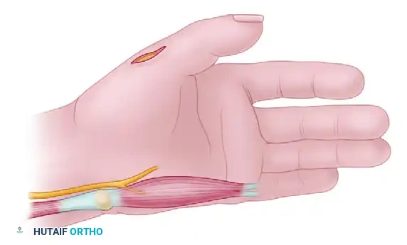
* Iliac Crest Bone Graft (ICBG): The traditional workhorse. Provides excellent cancellous bone rich in osteoprogenitor cells.
* Reamer-Irrigator-Aspirator (RIA): Utilized for harvesting large volumes of autograft from the femoral or tibial canal. It significantly reduces donor site morbidity compared to ICBG and provides a graft with equivalent or superior osteogenic potential.
Bone Morphogenetic Proteins (BMPs)
Discovered by Marshall Urist, BMPs are members of the TGF-beta superfamily that induce the differentiation of mesenchymal stem cells into osteoblasts.
* rhBMP-2 and rhBMP-7: Commercially available recombinant proteins used off-label in many nonunion scenarios. They are highly osteoinductive but lack structural integrity, necessitating combination with an osteoconductive carrier (e.g., allograft, demineralized bone matrix).
Bone Marrow Aspirate Concentrate (BMAC)
Percutaneous aspiration of bone marrow (typically from the anterior or posterior iliac crest) followed by centrifugation concentrates mesenchymal stem cells and growth factors. BMAC can be injected percutaneously into oligotrophic nonunions or mixed with structural allografts during open procedures.
Surgical Management Strategies: Step-by-Step
1. Management of Hypertrophic Nonunions
The primary issue is mechanical instability. The biological engine is running, but the clutch is slipping.
Surgical Technique: Exchange Nailing (Femur/Tibia)
* Indications: Diaphyseal hypertrophic nonunions previously treated with an intramedullary nail.
* Positioning: Supine on a fracture table or radiolucent flat table, depending on surgeon preference and associated deformities.
* Step 1: Hardware Removal. Remove the existing locking screws and the intramedullary nail.
* Step 2: Reaming. Over-ream the canal by 1.5 to 2.0 mm larger than the previous nail. This serves two purposes: it increases the working diameter for a stiffer, larger nail, and the reamings act as an autologous bone graft at the nonunion site.
* Step 3: Nail Insertion. Insert a larger diameter nail.
* Step 4: Locking. Dynamization or static locking depends on the fracture pattern. For axially stable patterns, dynamic locking allows controlled micromotion, stimulating secondary bone healing.
Pitfall: Do not open the nonunion site in a purely hypertrophic nonunion. Opening the site disrupts the established vascularity and the biologically active callus. Stability alone will lead to consolidation.
2. Management of Atrophic Nonunions
Atrophic nonunions require a complete biological overhaul combined with absolute mechanical stability.
Surgical Technique: Open Takedown, Decortication, and Plating
* Indications: Atrophic diaphyseal or metaphyseal nonunions.
* Step 1: Approach and Exposure. Utilize an extensile approach. Carefully elevate the periosteum, preserving soft tissue attachments as much as possible.
* Step 2: Takedown. Excisional debridement of the nonunion site is mandatory. Remove all fibrous tissue and sclerotic bone ends until punctate bleeding (the "paprika sign") is observed.
* Step 3: Judet Decortication. Perform osteoperiosteal decortication (shingling) of the host bone for 2-3 cm proximal and distal to the nonunion. This dramatically increases the vascular surface area for graft incorporation.
* Step 4: Mechanical Stabilization. Apply a heavy-duty construct, typically a broad dynamic compression plate (DCP) or locking compression plate (LCP). Achieve absolute stability (compression) if the fracture pattern allows.
* Step 5: Biological Augmentation. Pack the defect and the decorticated area with copious amounts of autograft (ICBG or RIA) or a composite of allograft and orthobiologics (e.g., BMP-2).
3. Management of Segmental Defects and Infected Nonunions
Massive bone loss (>4-6 cm) or active infection precludes standard internal fixation and grafting.
Surgical Technique: The Ilizarov Method and Distraction Osteogenesis
* Indications: Infected nonunions, massive segmental bone defects, nonunions with severe limb length discrepancy or complex deformities.
* Biomechanics: Tension-stress effect. Gradual distraction of a living tissue creates steady tension that stimulates new bone formation.
* Step 1: Radical Debridement. Excise all infected and necrotic bone. This often creates a massive segmental defect.
* Step 2: Frame Application. Apply a circular external fixator (Ilizarov or Taylor Spatial Frame) using tensioned fine wires and half-pins. Ensure orthogonal fixation for maximum stability.
* Step 3: Corticotomy. Perform a low-energy percutaneous corticotomy in the healthy metaphysis (proximal or distal to the defect). Preserve the periosteal and endosteal blood supply.
* Step 4: Latency Phase. Wait 7 to 10 days postoperatively before initiating distraction. This allows the initial inflammatory phase of fracture healing to commence.
* Step 5: Distraction Phase. Distract the corticotomy site at a rate of 1.0 mm per day (typically divided into four 0.25 mm increments). This generates the regenerate bone.
* Step 6: Docking and Consolidation. Once the transported segment docks at the target site, the frame is locked. The consolidation phase typically takes twice as long as the distraction phase.
Clinical Pearl: At the docking site, fibrous union is common. Plan for a secondary procedure involving docking site preparation, decortication, and autologous bone grafting to ensure definitive bony union.
Biophysical Stimulation
Adjuvant non-invasive therapies can be utilized to upregulate the biological response in delayed unions or recalcitrant nonunions.
Pulsed Electromagnetic Fields (PEMF)
PEMF induces a time-varying magnetic field that generates an electrical current within the bone. This current stimulates osteoblast proliferation, increases the synthesis of extracellular matrix proteins, and upregulates BMP expression. It is FDA-approved for the treatment of nonunions and is typically worn for 3 to 10 hours daily.
Direct Current (DC) Stimulation
Invasive or semi-invasive systems that implant a cathode directly into the nonunion site. The cathode lowers the local oxygen tension and increases the local pH, creating an osteogenic environment. While highly effective, it requires surgical implantation and removal.
Extracorporeal Shockwave Therapy (ESWT)
High-energy acoustic waves induce microtrauma at the nonunion site, stimulating neovascularization (angiogenesis) and the release of osteogenic growth factors. It is particularly useful for hypertrophic and oligotrophic nonunions.
Postoperative Protocols and Rehabilitation
The postoperative management of a nonunion is as critical as the surgical execution. Protocols must be highly individualized based on the fixation construct and the biological quality of the bone.
- Weight-Bearing Status:
- Intramedullary Nailing: Often allows for immediate weight-bearing as tolerated, which promotes dynamization and secondary bone healing.
- Plate Fixation (Atrophic Nonunions): Strict non-weight-bearing or touch-down weight-bearing for 6 to 8 weeks until radiographic evidence of graft incorporation and bridging callus is observed.
- Circular External Fixators: Immediate weight-bearing is encouraged. Axial loading through the frame stimulates regenerate bone consolidation via the tension-stress effect.
- Radiographic Monitoring: Serial orthogonal radiographs should be obtained at 4, 8, 12, and 24 weeks postoperatively. Assess for hardware integrity, graft incorporation, and progressive obliteration of the fracture lines.
- Physical Therapy: Early mobilization of adjacent joints is mandatory to prevent arthrofibrosis. Muscle strengthening (isometric initially, progressing to isotonic) optimizes the mechanical environment surrounding the healing bone.
Conclusion
The management of fracture nonunions requires a masterful understanding of orthopedic biomechanics, host biology, and advanced surgical techniques. By meticulously classifying the nonunion, optimizing the patient systemically, selecting the appropriate orthobiologic adjuncts, and executing precise, mechanically sound surgical reconstructions, the orthopedic surgeon can successfully navigate these complex clinical scenarios and restore function to the compromised limb.
You Might Also Like