Surgical Hip Approaches: Master the Tensor Fasciae Latae Anatomy
Key Takeaway
Your ultimate guide to Surgical Hip Approaches: Master the Tensor Fasciae Latae Anatomy starts here. The tensor fasciae latae is a superficial muscle of the thigh, enveloped by the fascia lata, originating from the anterior iliac crest and inserting into the iliotibial tract. In hip surgery, its relationship with the gluteus medius defines the intermuscular plane utilized in the anterolateral approach, allowing access to the hip joint when retracted.
Introduction and Epidemiology
The surgical management of hip pathology represents a cornerstone of modern orthopedic practice, encompassing a broad spectrum of interventions from arthroplasty to fracture fixation and tumor resection. As the demand for hip procedures continues to rise, driven by an aging population and advancements in implant technology, the judicious selection and meticulous execution of surgical approaches remain paramount for optimizing patient outcomes and minimizing morbidity. This guide serves as an exhaustive reference for orthopedic surgeons, residents, and medical students, focusing on the critical applied anatomy relevant to anterior, lateral, and anterolateral hip approaches, with particular emphasis on the tensor fasciae latae (TFL).
The evolution of hip surgery has seen a continuous refinement of access strategies, moving towards less invasive techniques that aim to preserve muscle integrity and reduce soft tissue disruption. Understanding the nuanced anatomical relationships, particularly the internervous planes, is fundamental to achieving these goals. While posterior approaches historically dominated, anterior and lateral approaches have gained significant traction, each offering distinct advantages and challenges. The ability to navigate these approaches safely and effectively hinges upon a profound mastery of regional anatomy, especially the intricate interplay of the fascia lata, the TFL, and the gluteal musculature.
Surgical Anatomy and Biomechanics
A thorough understanding of the topographical and functional anatomy of the hip region is non-negotiable for any surgeon undertaking hip procedures. The fascia lata, a robust envelope of deep fascia, forms a critical structural component of the thigh, compartmentalizing muscles and providing attachment points. Its strategic relationships with key muscles — the sartorius, tensor fasciae latae, and gluteus maximus — dictate the planes of surgical dissection for anterior, lateral, and anterolateral hip approaches.
Fascia Lata and Associated Musculature
The fascia lata encases the entire thigh musculature. Proximally, it attaches to the iliac crest, sacrum, coccyx, inguinal ligament, pubic arch, and ischial tuberosity. Distally, it contributes to the patellar retinacula and invests the knee. Its formidable strength is particularly evident laterally, where it forms the iliotibial (IT) tract.
The seed content highlights the fascia lata's specialized role in enclosing specific muscles. It covers the sartorius muscle anteriorly. More profoundly, it splits into a superficial and deep layer to envelop the tensor fasciae latae and gluteus maximus muscles. This bilaminar arrangement around the TFL and gluteus maximus is crucial for understanding the initial layers encountered during approaches involving these muscles. The gluteus medius, in contrast, arises from the outer wing of the ilium and is covered by the fascia lata, rather than being enclosed by it in the same manner as the TFL or gluteus maximus.

Tensor Fasciae Latae Anatomy and Relationships
The tensor fasciae latae is a spindle-shaped muscle situated in the anterolateral aspect of the thigh, critical to both hip kinematics and surgical access.
* Origin: It originates from the anterior part of the outer lip of the iliac crest, the anterior superior iliac spine (ASIS), and the outer surface of the ilium posterior to the ASIS.
* Insertion: Distally, it inserts into the anterior aspect of the iliotibial tract approximately one-third of the way down the thigh. The IT tract, in turn, inserts into the lateral condyle of the tibia (Gerdy's tubercle).
* Innervation: The TFL is innervated by the superior gluteal nerve (L4, L5, S1), a branch of the sacral plexus, which also innervates the gluteus medius and minimus. This shared innervation is a key consideration for internervous planes.
* Blood Supply: It receives blood supply from the ascending branch of the lateral circumflex femoral artery.
* Function: The TFL acts as a hip abductor, flexor, and internal rotator. It also provides tension to the IT tract, stabilizing the knee in extension.
The relationship between the TFL and the gluteus medius is particularly central to the anterolateral approach. As described in the seed content, the TFL originates from the anterior portion of the outer lip of the iliac crest, while the gluteus medius arises from the outer wall of the ilium, between the anterior and posterior gluteal lines. Their origins are nearly continuous, but the TFL lies slightly more superficial (lateral) and anterior to the gluteus medius. As these muscles course distally towards their insertions, the TFL maintains an even more superficial position relative to the gluteus medius. The TFL inserts into the IT tract, whereas the gluteus medius inserts into the anterior and lateral facet of the greater trochanter. This anatomical divergence creates the potential for an intermuscular or intramuscular plane of dissection.

Gluteus Medius and Minimus
These are the primary hip abductors, crucial for maintaining pelvic stability during gait.
* Gluteus Medius: Originates from the outer surface of the ilium between the anterior and posterior gluteal lines. Inserts into the lateral aspect of the greater trochanter. Innervated by the superior gluteal nerve.
* Gluteus Minimus: Lies deep to the gluteus medius. Originates from the outer surface of the ilium between the anterior and inferior gluteal lines. Inserts into the anterior aspect of the greater trochanter. Also innervated by the superior gluteal nerve.
Damage to the superior gluteal nerve or significant disruption of the gluteus medius/minimus complex can lead to abductor insufficiency, resulting in a Trendelenburg gait.

Neurovascular Structures of Concern
Several critical neurovascular structures are in proximity to the common hip approaches and demand meticulous identification and protection.
* Lateral Femoral Cutaneous Nerve (LFCN): Arises from the lumbar plexus (L2, L3) and typically crosses the iliac crest near the ASIS, often passing under the inguinal ligament. It provides sensation to the anterolateral thigh. Highly variable in its course, it is particularly vulnerable during the anterior approach, leading to meralgia paresthetica if injured.
* Superior Gluteal Nerve and Artery: These structures emerge from the pelvis through the greater sciatic foramen, superior to the piriformis muscle. They course between the gluteus medius and minimus, supplying both muscles. They are at risk during the lateral and anterolateral approaches, especially with excessive posterior retraction of the gluteus medius or deep splitting into the gluteal muscles.
* Femoral Nerve, Artery, and Vein: Located medially to the TFL and sartorius, within the femoral triangle. While generally protected by these muscles, deep or medial dissection during the anterior approach requires caution to avoid injury.
* Sciatic Nerve: Located posteriorly, exiting the pelvis inferior to the piriformis. Although not directly exposed in anterior, lateral, or anterolateral approaches, aggressive posterior retraction or limb positioning can put it at stretch.

Internervous Planes for Hip Approaches
The principle of internervous plane dissection is fundamental to minimizing muscle damage and preserving function.
* Anterior Approach (Hueter): This approach exploits the internervous plane between the sartorius (innervated by the femoral nerve) and the tensor fasciae latae (innervated by the superior gluteal nerve). The deep fascia is incised longitudinally, revealing this interval. This allows direct access to the hip capsule without detaching major muscles, thereby minimizing abductor muscle injury.
* Anterolateral Approach (Watson-Jones): This approach utilizes the interval between the tensor fasciae latae and the gluteus medius. Both muscles are innervated by the superior gluteal nerve. Therefore, this is an intermuscular split within the same nerve territory rather than a true internervous plane. Dissection proceeds through the superficial fascia, incising the fascia lata lateral to the TFL. The TFL is retracted anteriorly, and the gluteus medius is retracted posteriorly, or an intramuscular split is made within the anterior fibers of the gluteus medius.
* Lateral Approach (Hardinge): This approach involves detaching the anterior one-third to one-half of the gluteus medius and minimus muscles from their insertion on the greater trochanter, reflecting them superiorly and posteriorly. While this provides excellent exposure, it violates the abductor mechanism directly, risking abductor weakness, gluteal lurch, and potential for non-union of the reattached abductors. The superior gluteal nerve entry point into the gluteus medius must be meticulously protected during this reflection.

Indications and Contraindications
The choice of surgical approach to the hip is dictated by numerous factors, including the specific pathology, patient characteristics, surgeon experience, and desired biomechanical outcomes.
Indications for Surgical Intervention
| Operative Indications | Non-Operative Indications |
|---|---|
| End-stage osteoarthritis of the hip (primary/revision THA) | Early-stage osteoarthritis (mild pain, minimal functional loss) |
| Inflammatory arthropathies (rheumatoid arthritis) | Trochanteric bursitis or tendinopathy (initial management) |
| Avascular necrosis of the femoral head | Minor labral tears (asymptomatic or manageable with PT) |
| Acute femoral neck fractures (THA, hemiarthroplasty, ORIF) | Femoroacetabular impingement (FAI) without significant cartilage damage (PT) |
| Acetabular fractures (ORIF) | Low-grade adductor or hamstring strains |
| Periprosthetic fractures of the hip | Early osteochondral lesions (conservative management, activity modification) |
| Developmental dysplasia of the hip (THA, osteotomy) | Mild piriformis syndrome (physical therapy, injections) |
| Septic arthritis of the hip (irrigation and debridement) | |
| Resection of benign or malignant tumors | |
| Synovial conditions (synovectomy, biopsy) |
Contraindications for Surgical Intervention
Absolute Contraindications:
* Active infection in the hip joint or surrounding tissues.
* Severe medical comorbidities that preclude safe anesthesia or surgery (e.g., uncontrolled cardiac failure, severe coagulopathy).
* Neuropathic arthropathy with active joint destruction.
* Skeletal immaturity for procedures requiring definitive arthroplasty.
Relative Contraindications:
* Morbid obesity (increased complication rates, difficult exposure).
* Severe soft tissue compromise or poor skin integrity in the operative field.
* Previous surgery with extensive scarring that may obscure anatomy or increase dissection difficulty.
* Active psychiatric conditions that would impede adherence to postoperative protocols.
* Uncontrolled diabetes mellitus (increased risk of infection).
* Known neurovascular abnormalities or anatomical variants that significantly complicate standard dissection.
Factors Influencing Approach Selection
The selection among anterior, anterolateral, or lateral approaches is a critical preoperative decision influenced by:
* Pathology: Certain fractures (e.g., anterior column acetabular fractures) lend themselves better to anterior approaches. Revision arthroplasty may utilize existing incisions or require extended approaches.
* Patient Habitus: Obese patients may present challenges with superficial dissection and retraction for some approaches.
* Surgeon Preference and Experience: Proficiency with a particular approach often correlates with better outcomes.
* Prior Surgery: Existing scars and altered anatomy necessitate careful planning.
* Associated Deformities: Fixed flexion or adduction contractures may influence exposure.
* Equipment Availability: Specialized tables (e.g., traction table for direct anterior THA) may be required.
Pre Operative Planning and Patient Positioning
Meticulous preoperative planning is fundamental to ensuring a safe and successful surgical outcome, regardless of the chosen approach.
Preoperative Assessment
- Clinical Evaluation: A comprehensive history, including previous hip pathology, surgeries, and comorbidities, is essential. A thorough physical examination assesses range of motion, muscle strength, gait, neurovascular status, and skin integrity. Identification of nerve palsies or specific tendinopathies is crucial.
- Imaging: Standard anteroposterior pelvis and lateral hip radiographs are mandatory. For arthroplasty, these are used for templating implant size and position. Computed tomography (CT) scans are invaluable for complex acetabular or proximal femoral fractures, tumor resections, and revision cases to delineate bone loss and component malposition. Magnetic resonance imaging (MRI) is useful for soft tissue assessment, avascular necrosis, or occult fractures.
- Templating: For hip arthroplasty, digital templating allows for preoperative estimation of component sizes, leg length, and offset, guiding intraoperative decisions and improving accuracy. This also helps anticipate potential challenges related to bone stock or deformity.
- Medical Optimization: All comorbidities must be optimally managed. This includes glycemic control in diabetics, cardiac clearance for high-risk patients, and appropriate anticoagulation management.
Patient Positioning and Preparation
Proper patient positioning is critical for optimal exposure, prevention of iatrogenic injury, and efficient workflow.
-
Anterior Approach (Hueter):
- Positioning: Supine on a standard operating table or a specialized traction table (e.g., Hana table for direct anterior THA). The affected hip is brought to the edge of the table. A bump may be placed under the ipsilateral hip to allow for easier extension and external rotation, though this is often unnecessary with a traction table.
- Padding: All pressure points (heels, sacrum, contralateral limb) are meticulously padded.
- Draping: Draping should allow for full range of motion of the hip, particularly extension, adduction, and external rotation for femoral head dislocation. A clear view of the ASIS and greater trochanter is essential for incision planning.
- Precaution: Careful attention to the lateral femoral cutaneous nerve (LFCN) is paramount, minimizing direct pressure or excessive traction on the skin in its vicinity.
-
Anterolateral Approach (Watson-Jones) and Lateral Approach (Hardinge):
- Positioning: Lateral decubitus position is standard, with the affected hip superior. The contralateral limb is typically flexed at the hip and knee and secured in a well-padded beanbag or supports to ensure stability.
- Padding: Axillary roll placed, pressure points (malleoli, knee, peroneal nerve at fibular head, ulnar nerve at elbow, dependent ear) are thoroughly padded.
- Draping: Draping should permit adequate access to the entire greater trochanter, iliac crest, and proximal femur, allowing for manipulation of the limb into flexion, extension, abduction, adduction, and rotation.
- Precaution: Ensure stability of the patient in the beanbag to prevent rolling. Confirm all lines and tubes are secure and not under tension.
Prophylaxis
- Antibiotic Prophylaxis: Administered preoperatively (e.g., Cefazolin 1g IV within 60 minutes of incision) according to institutional protocols to reduce surgical site infection risk.
- Thromboprophylaxis: Deep vein thrombosis (DVT) and pulmonary embolism (PE) prophylaxis are initiated as per guidelines, considering mechanical (TED stockings, SCDs) and/or pharmacological agents.
- Heterotopic Ossification (HO) Prophylaxis: For high-risk patients or procedures (e.g., revision surgery, ankylosing spondylitis, significant soft tissue trauma), NSAIDs (e.g., Indomethacin) or radiation therapy may be considered.
Detailed Surgical Approach and Technique
A precise understanding of surgical anatomy, particularly the TFL's role, is critical for each approach.
Anterior Approach Hueter
The direct anterior approach (DAA) utilizes an internervous plane, theoretically minimizing muscular damage and facilitating potentially faster recovery.
- Incision: A longitudinal incision is made, typically 8-15 cm in length, centered over the ASIS and extending distally along the line connecting the ASIS to the lateral border of the patella. The distal portion of the incision is often shifted slightly medially to better expose the rectus femoris. The incision is placed medial to the TFL.
- Superficial Dissection: The subcutaneous tissue is incised. Meticulous dissection is required to identify and protect the lateral femoral cutaneous nerve (LFCN), which typically emerges from under the inguinal ligament near the ASIS. Its course is highly variable, and it may be encountered superficially or deeper. If identified, it should be carefully mobilized and retracted.
- Deep Fascia Incision: The fascia covering the sartorius and TFL is incised longitudinally. The internervous plane is found between the sartorius (medially, innervated by femoral nerve) and the TFL (laterally, innervated by superior gluteal nerve).
- Muscle Interval Dissection: The sartorius is retracted medially, and the TFL is retracted laterally. This exposes the underlying rectus femoris and its tendinous origin (direct head from ASIS, indirect head from superior acetabular rim). The ascending branch of the lateral circumflex femoral artery and vein, which lie deep to the rectus femoris, must be identified and ligated to prevent excessive bleeding.
- Rectus Femoris Management: The rectus femoris can either be retracted medially or detached from its origin. For THA, partial detachment of the direct head (from ASIS) and full detachment of the indirect head (from superior acetabular rim) provides excellent exposure. If detached, the rectus femoris must be repaired at closure.
- Capsular Exposure: Beneath the rectus femoris, the iliopsoas muscle and the hip joint capsule are visible. The iliopsoas may be released from its insertion on the lesser trochanter if it is tight, though this is not always necessary.
- Capsulotomy: A T-shaped or H-shaped capsulotomy is performed to expose the femoral head and neck. Care is taken to identify and protect the medial femoral circumflex artery, which lies on the posterior aspect of the femoral neck.
- Femoral Head Dislocation: The hip is dislocated by extending, adducting, and externally rotating the limb.
- Preparation and Implantation: Femoral head osteotomy, acetabular reaming, cup implantation, femoral canal preparation, stem implantation, and trial reduction are performed.
- Closure: The capsule is often repaired, though capsular repair after DAA remains controversial. The rectus femoris, if detached, is reattached. The deep fascia is closed, followed by subcutaneous layers and skin.

Anterolateral Approach Watson-Jones
This approach offers direct access to the hip joint by splitting the abductor muscles. While often referred to as an "internervous" approach, it involves a split between muscles (TFL and gluteus medius) supplied by the same nerve (superior gluteal nerve).
- Incision: A curvilinear or straight longitudinal incision is made, beginning 2-3 cm posterior to the ASIS, extending distally along the anterior border of the greater trochanter for 8-15 cm. It follows the natural curvature of the proximal TFL.
- Superficial Dissection: The subcutaneous fat is incised. The fascia lata is exposed.
- Deep Fascia Incision: The fascia lata is incised longitudinally along the interval between the anterior border of the gluteus medius and the posterior border of the tensor fasciae latae.
- Muscle Interval Dissection: The TFL is retracted anteriorly, and the gluteus medius is retracted posteriorly. The interval can be challenging to define, especially in muscular patients. The anterior fibers of the gluteus medius are often split longitudinally in line with its fibers, rather than simply retracting. This split exposes the gluteus minimus.
- Gluteus Minimus Management: The gluteus minimus, lying deep to the medius, is then either split or partially released from the greater trochanter to expose the hip capsule. Care must be taken to avoid damaging the superior gluteal neurovascular bundle, which lies further posteriorly and superiorly, supplying both gluteus medius and minimus.
- Capsular Exposure: The hip capsule is exposed beneath the gluteus minimus.
- Capsulotomy: A T-shaped capsulotomy or excision of a portion of the capsule allows access to the femoral head and neck.
- Femoral Head Dislocation: The hip is dislocated by flexing, adducting, and internally rotating the limb.
- Preparation and Implantation: Similar steps to the anterior approach for arthroplasty components.
- Closure: The capsule is repaired if possible. The gluteus minimus and gluteus medius are meticulously repaired, ensuring anatomical reapproximation. The fascia lata is then closed, followed by subcutaneous layers and skin.
Lateral Approach Hardinge (Direct Lateral Approach)
This approach provides excellent exposure to the acetabulum and proximal femur but involves detachment of a portion of the hip abductors.
- Incision: A straight or curvilinear incision is made, centered over the greater trochanter, extending 8-15 cm proximally and distally. The incision starts roughly 5 cm proximal to the tip of the greater trochanter and extends distally along the axis of the femur.
- Superficial Dissection: The subcutaneous tissue is incised, exposing the fascia lata and the iliotibial tract.
- Deep Fascia Incision: The fascia lata and the IT tract are incised longitudinally over the greater trochanter. This exposes the underlying vastus lateralis, which originates from the lateral femoral shaft distal to the greater trochanter.
- Abductor Detachment: The key step of the Hardinge approach involves detaching the anterior one-third to one-half of the gluteus medius and gluteus minimus from their insertion on the greater trochanter. An osteotomy of the greater trochanter can also be performed, but this is less common with this specific approach. The detached portion of the abductor complex is then reflected superiorly and posteriorly. This maneuver provides direct access to the hip capsule.
- Neurovascular Protection: Careful attention is paid to the superior gluteal neurovascular bundle, which enters the gluteus medius posteriorly. Excessive posterior reflection of the abductors must be avoided to prevent nerve injury.
- Capsular Exposure: The hip capsule is exposed.
- Capsulotomy: An H-shaped or T-shaped capsulotomy is performed.
- Femoral Head Dislocation: The hip is dislocated by internally rotating and adducting the limb.
- Preparation and Implantation: Similar steps for arthroplasty components.
- Closure: The detached abductor muscles are meticulously reattached to the greater trochanter using strong non-absorbable sutures through drill holes or transosseous sutures. Secure repair is paramount to prevent abductor dysfunction and trochanteric bursitis. The fascia lata and IT band are then repaired, followed by subcutaneous layers and skin.
Complications and Management
Surgical approaches to the hip, while often successful, are associated with a distinct set of potential complications. A thorough understanding of these risks and their management strategies is essential for all orthopedic surgeons.
Common Complications and Management Strategies
| Complication | Incidence (Approximate) | Salvage Strategies / Management |
|---|---|---|
| Lateral Femoral Cutaneous Nerve (LFCN) Injury (anterior approach) | 10-20% paresthesia, 1-5% persistent dysesthesia | Conservative management (reassurance, observation) for mild cases. Neuropathic pain medications (gabapentin, pregabalin). Local corticosteroid injections. For severe, persistent cases: surgical neurolysis, neurectomy, or nerve grafting (rare). |
| Superior Gluteal Nerve Injury (anterolateral/lateral approaches) | < 1-5% (higher with aggressive retraction/detachment) | Abductor muscle weakness (Trendelenburg gait). Physical therapy to strengthen remaining abductors and improve gait mechanics. Assistive devices (cane). For severe, persistent deficits: abductor transfer procedures (e.g., gluteus maximus transfer) or tendon reconstruction. |
| Heterotopic Ossification (HO) | 5-10% (clinically significant); higher in high-risk groups | Prophylaxis in high-risk patients: NSAIDs (Indomethacin 25mg TID for 3-6 weeks post-op) or single-dose radiation therapy (700-800 cGy within 24-72 hours post-op). Symptomatic HO: surgical excision once mature, often with repeat prophylaxis. |
| Periprosthetic Joint Infection (PJI) | 0.5-2% (primary); higher in revision | Early infection (<3 weeks): Irrigation and debridement with retention of components, pathogen-specific IV antibiotics. Late/chronic infection: Two-stage revision arthroplasty (explantation, antibiotic spacer, prolonged IV antibiotics, reimplantation). Single-stage revision (selected cases). Girdlestone resection arthroplasty (salvage). |
| Hip Dislocation | 1-5% (approach dependent) | Closed reduction under sedation/anesthesia. Assessment for component malposition or soft tissue impingement. Recurrent dislocation: revision surgery to address component malposition, offset, or soft tissue balance. Dual-mobility implants. Constrained liners. |
| Periprosthetic Fracture | 0.1-5% (intraoperative/postoperative) | Intraoperative: often managed with cerclage wires or cables, or revision to a longer stem. Postoperative: depends on fracture pattern (Vancouver classification). ORIF with plates/screws, or revision arthroplasty (long-stem revision, femoral component exchange). |
| Abductor Dysfunction / Trochanteric Pain Syndrome (lateral approaches) | 5-10% (up to 30% for persistent pain) | Physical therapy, NSAIDs, local corticosteroid injections. For persistent cases: surgical revision of abductor repair, or debridement of adventitious bursa. Endoscopic abductor repair. Tendon transfer. |
| Vascular Injury | < 0.1% | Immediate recognition and vascular surgical consultation for repair. Hematoma evacuation. |
| Deep Vein Thrombosis (DVT) / Pulmonary Embolism (PE) | 0.5-2% (symptomatic with prophylaxis) | Prophylaxis (mechanical, chemical). Diagnosis with duplex ultrasound (DVT) or CT pulmonary angiography (PE). Anticoagulation (LMWH, oral anticoagulants). IVC filter for recurrent PE or contraindication to anticoagulation. |
| Nerve Palsy (Sciatic, Femoral) | < 1% | Immediate recognition and removal of potential compressive factors (e.g., tight retraction, cement). Observation, neurophysiology studies (EMG/NCS). Neuropathic pain management. Surgical exploration and neurolysis (rarely). |
Approach-Specific Complication Considerations
- Anterior Approach: While promoting faster initial recovery, it carries a higher risk of LFCN injury and a learning curve for appropriate retraction without causing neurovascular damage. Exposure can be limited in muscular or obese patients.
- Anterolateral Approach: Offers a good balance of exposure and muscle preservation compared to the direct lateral. However, it still involves some dissection within the abductor muscles, potentially leading to mild abductor weakness if the split is extended or the superior gluteal nerve is compromised.
- Lateral Approach (Hardinge): Provides robust exposure but directly violates the abductor mechanism. The risk of abductor weakness, gluteal lurch, and non-union of the abductor repair is higher. Trochanteric bursitis is also a more common sequela due to irritation from the abductor repair.
Post Operative Rehabilitation Protocols
Postoperative rehabilitation is an integral component of the surgical journey, crucial for restoring function, optimizing implant longevity, and preventing complications. Protocols are tailored to the specific surgical approach, patient's bone quality, implant fixation (cemented vs. uncemented), and any intraoperative findings.
General Principles
- Pain Management: Effective multimodal analgesia is paramount to facilitate early mobilization and patient participation in therapy.
- Early Mobilization: Encouraging out-of-bed activity on postoperative day 0 or 1 minimizes risks of DVT, pneumonia, and deconditioning.
- Wound Care: Strict adherence to wound care protocols reduces infection risk.
- Education: Patients receive clear instructions regarding precautions, activity levels, and symptoms requiring immediate medical attention.
Phase 1 Acute Postoperative (Weeks 0-6)
- Weight Bearing:
- Cemented Components: Full weight-bearing (FWB) as tolerated is typically allowed immediately.
- Uncemented Components: Generally, FWB is allowed, but some surgeons may opt for protected weight-bearing (toe-touch or partial) for 2-6 weeks depending on bone quality, press-fit stability, and patient risk factors (e.g., osteopenia, revision surgery).
- Abductor Repair (Lateral Approach): Often requires protected weight-bearing (e.g., toe-touch to 50% PWB) for 4-6 weeks to allow abductor healing, followed by gradual progression.
- Range of Motion (ROM):
- Anterior Approach: Typically fewer formal restrictions on hip precautions, emphasizing early, gentle ROM exercises within comfort. Avoidance of extreme hyperextension and external rotation during the initial phase.
- Anterolateral Approach: Similar to anterior, but with caution regarding extreme adduction and internal rotation.
- Lateral Approach: Precautions to protect the abductor repair, often limiting active abduction against gravity initially. Passive ROM is initiated cautiously.
- Strengthening:
- Isometrics of quadriceps, hamstrings, and gluteals are initiated.
- Ankle pumps and calf raises to promote circulation.
- Gentle hip flexion, extension, abduction, and adduction within pain-free limits.
- Gait Training: Progressive ambulation with assistive devices (walker, crutches), focusing on proper gait mechanics.
Phase 2 Subacute Rehabilitation (Weeks 6-12)
- Weight Bearing: Progress to full weight-bearing, gradually weaning from assistive devices as strength and balance improve.
- Range of Motion: Continue to work on improving pain-free ROM.
- Strengthening:
- Progressive resistive exercises for all hip muscle groups, emphasizing abductors (gluteus medius/minimus), extensors (gluteus maximus), and core stabilizers.
- Closed-chain exercises (mini-squats, lunges, step-ups) are introduced.
- Balance and proprioceptive training.
- Functional Activities: Introduce functional tasks relevant to daily living, such as stair climbing, getting in/out of chairs.
Phase 3 Advanced Rehabilitation and Return to Activity (Weeks 12+)
- Strengthening: Continue advanced strengthening exercises, incorporating sport-specific or work-specific movements.
- Endurance and Agility: Introduce low-impact aerobic activities (swimming, cycling) and gradually progress to higher-impact activities if appropriate and cleared by the surgeon.
- Return to Sport/Work: Guided return to recreational activities or demanding occupations based on individual progress, muscle strength, balance, and pain levels. High-impact sports are generally discouraged after total hip arthroplasty due to increased wear and risk of component loosening.
Approach-Specific Postoperative Considerations
- Anterior Approach: Patients often demonstrate faster initial recovery of gait and less noticeable gait deviations. However, some may experience persistent numbness or dysesthesia from LFCN irritation.
- Lateral Approach: Due to the abductor detachment, patients may experience a slower recovery of abductor strength and a higher incidence of Trendelenburg gait or persistent trochanteric pain. Rehabilitation focuses heavily on abductor strengthening and careful monitoring of abductor repair integrity.
Summary of Key Literature and Guidelines
The literature comparing different surgical approaches to the hip is vast and continually evolving. While surgeon experience and preference often play a significant role, evidence-based data guides modern practice.
Studies comparing direct anterior (DAA), anterolateral (AL), and posterior (PA) approaches for total hip arthroplasty (THA) have yielded mixed results regarding superior outcomes.
* Early Recovery: Several meta-analyses and systematic reviews suggest that DAA may be associated with faster early functional recovery, shorter hospital stays, and lower narcotic use in the immediate postoperative period compared to PA and AL approaches. This is often attributed to the "muscle-sparing" nature of the internervous dissection.
* Dislocation Rates: Traditionally, PA has been associated with higher dislocation rates, particularly in older literature. However, with modern soft tissue repair techniques, dislocation rates for PA are now comparable to DAA and AL approaches in experienced hands. Some studies suggest DAA may have a slightly lower dislocation rate, but this often requires specific patient selection and a learning curve for the surgeon.
* Complications: DAA has a recognized learning curve and is associated with a higher incidence of lateral femoral cutaneous nerve injury and potentially greater intraoperative blood loss during the early part of a surgeon's experience. AL approaches carry a risk of superior gluteal nerve injury and abductor weakness, though typically less severe than with traditional lateral approaches involving complete abductor detachment.
* Abductor Function: While DAA is often touted for preserving abductor function, objective measures of abductor strength and gait kinematics often show minimal long-term differences among approaches. However, lateral approaches involving abductor detachment generally result in a higher incidence of abductor weakness and Trendelenburg gait compared to DAA and PA.
* Leg Length Discrepancy (LLD): All approaches carry a risk of LLD. However, proponents of the DAA on a traction table argue for more accurate intraoperative assessment of leg length and offset. Recent advancements in navigation and robotic-assisted surgery for all approaches also aim to mitigate LLD.
* Revision Surgery: The choice of approach for revision THA is often dictated by the original incision, type of revision (acetabular vs. femoral), and extent of bone loss or soft tissue scarring. Extended trochanteric osteotomy (ETO) can be utilized with various approaches for complex femoral revisions.
Professional societies such as the American Academy of Orthopaedic Surgeons (AAOS) provide guidelines on hip arthroplasty, often emphasizing shared decision-making with patients, consideration of surgeon experience, and recognition of the varying risks and benefits associated with each approach. The consensus is that no single approach is definitively superior in all contexts, and the surgeon's familiarity and expertise with a chosen approach are critical determinants of success.
In conclusion, the mastery of surgical hip approaches, particularly the nuances of the tensor fasciae latae anatomy and its relationship to the gluteal muscles and neurovascular structures, is paramount for orthopedic surgeons. Each approach – anterior, anterolateral, and lateral – offers distinct advantages and challenges. A deep anatomical understanding, meticulous preoperative planning, precise surgical execution, and structured postoperative rehabilitation are all essential to minimize complications and achieve optimal functional outcomes for patients undergoing hip surgery. The ongoing evolution of surgical techniques and evidence-based medicine continues to refine our understanding and practice, underscoring the importance of lifelong learning in this specialized field.
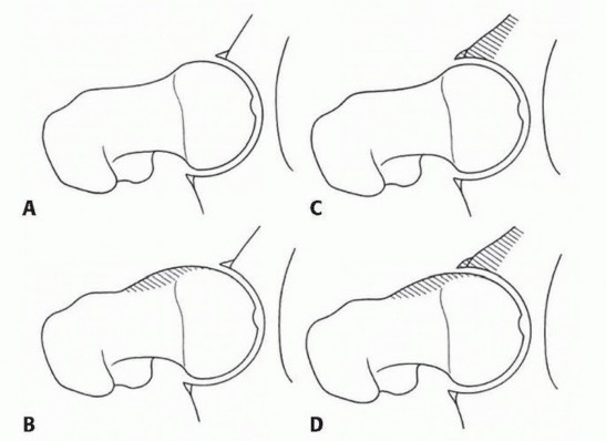
Clinical & Radiographic Imaging


You Might Also Like