Orthopedic Board Review MCQs: Foot & Ankle, Trauma & Sports Medicine | Part 136

Key Takeaway
This page provides Part 136 of an interactive, high-yield MCQ bank for orthopedic residents and surgeons. Designed for OITE and AAOS/ABOS board exam preparation, it features 100 verified questions mirroring official formats, covering critical topics such as Ankle, Foot, and Knee to enhance exam readiness and knowledge.
About This Board Review Set
This is Part 136 of the comprehensive OITE and AAOS Orthopedic Surgery Board Review series authored by Dr. Mohammed Hutaif, Consultant Orthopedic & Spine Surgeon.
This set has been strictly audited and contains 100 100% verified, high-yield multiple-choice questions (MCQs) modelled on the exact format of the Orthopaedic In-Training Examination (OITE) and the American Academy of Orthopaedic Surgeons (AAOS) board examinations.
How to Use the Interactive Quiz
Two distinct learning modes are available:
- Study Mode — After selecting an answer, you immediately see whether you are correct or incorrect, together with a full clinical explanation and literature references.
- Exam Mode — All feedback is hidden until you click Submit & See Results. A live timer tracks elapsed time. A percentage score and detailed breakdown are displayed upon submission.
Pro Tip: Use keyboard shortcuts A–E to select options, F to flag a question for review, and Enter to jump to the next unanswered question.
Topics Covered in Part 136
This module focuses heavily on: Ankle, Dislocation, Foot, Fracture, Knee, Nerve, Tendon.
Sample Questions from This Set
Sample Question 1: Which statement is true with respect to acetabular fracture surgery as the time between injury and surgery increases?...
Sample Question 2: 03 Which of the following findings is one of the diagnostic criteria for diffuse idiopathic skeletal hyperostosis?...
Sample Question 3: A tendon repair is thought to be weakest during which phase of tendon healing?...
Sample Question 4: -What is the diagnosis?...
Sample Question 5: A 24-year-old man reports the development of a foot drop following a knee dislocation 1 year ago. The common peroneal nerve was found to be in continuity at the time of surgical reconstruction of the posterolateral corner of the knee joint....
Why Active MCQ Practice Works
Evidence consistently demonstrates that active recall through spaced MCQ practice yields substantially greater long-term retention than passive reading alone (Roediger & Karpicke, 2006). All questions in this specific module have been algorithmically verified for clinical integrity and complete explanations.
Comprehensive 100-Question Exam
00:00
Start Quiz
Question 1
Which statement is true with respect to acetabular fracture surgery as the time between injury and surgery increases?

Explanation
Question 2
03 Which of the following findings is one of the diagnostic criteria for diffuse idiopathic skeletal hyperostosis?
Explanation
Diffuse idiopathic skeletal hyperostosis is a common disease, most prevalent in those over 50 years of age. The usual presentation is a middle-aged or older patient with chronic mild pain in the middle to lower back, spinal stiffness, and typical radiographic changes in the thoracic spine. Diffuse idiopathic skeletal hyperostosis is predominantly a radiographic diagnosis with 3 major diagnostic criteria. 1. Flowing ossification along the anterolateral aspect of at least four contiguous vertebrae. 2. Preservation of disk height in the involved vertebral segment; the relative absence of significant degenterative changes, such as marginal sclerosis in vertebral bodies or vacuum phenomenon. 3.
Absence of facet-joint ankylosis; absence of sacroiliac erosion, sclerosis, or intra-articular osseous fusion. Treatment is typically non-operative, with anti-inflammatories, activity modification and PT.
back to this question next question
Question 3
A tendon repair is thought to be weakest during which phase of tendon healing?
Explanation
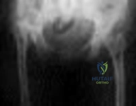
Question 4
-What is the diagnosis?
Explanation
Salter-Harris type II fracture of the proximal humerus
Question 5
A 24-year-old man reports the development of a foot drop following a knee dislocation 1 year ago. The common peroneal nerve was found to be in continuity at the time of surgical reconstruction of the posterolateral corner of the knee joint. He would like to eliminate the need for an ankle-foot orthosis. What is the best option to achieve elimination of the orthosis?
Explanation
REFERENCES: Pinzur MS, Kett N, Trilla M: Combined anteroposterior tibial tendon transfer in post-traumatic peroneal palsy. Foot Ankle 1988;8:27l-275.
Lipscomb P, Sanchez J: Anterior transplantation of the posterior tibial tendon for persistant palsy of the common peroneal nerve. J Bone Joint Surg Am 1961;43:60-66.
Question 6
Following a vertebroplasty of L2, cement is noted to protrude directly anterior to the L2 vertebral body. The cement is closest to which of the following structures?
Explanation
and duodenum are anterior to the aorta. The aorta lies in the midline just in front of the
vertebral body.
REFERENCES: Clement CD: Anatomy: A Regional Atlas of Human Anatomy, ed 3.
Baltimore, MD, Munich, Germany, Urban and Schwarzberg, 1987, Figure 331.
Netter FH: Atlas of Human Anatomy. Summit, NJ, Ciba-Geigy, 1989, plate 328.
Question 7
A 38-year-old man sustains a terrible triad injury consisting of an elbow dislocation, comminuted and displaced radial head fracture, and a type I coronoid fracture. Intraoperative findings after radial head replacement and lateral collateral ligament complex repair reveal persistent instability consisting of medial opening on valgus stress and posteromedial subluxation of the ulnohumeral and radiocapitellar joints. What is the best next step?
Explanation
normally correct the medial instability. Articulated versus static external fixation can be considered if
restoration of the ligamentous constraint of the medial side of the elbow cannot be accomplished surgically.
Question 8
Which of the following is a true statement regarding thoracic disk herniations? Review Topic
Explanation
common in the lower third near the thoracolumbar region. Posterior laminectomy and disk excision has the highest rate of neurologic deterioration and is not recommended. Multiple studies have shown that herniated thoracic disks can be found at one or more levels in 40% of asymptomatic individuals.
Question 9
Figure below depicts the radiograph obtained from a 52-year-old woman who has leg-length inequality and chronic, activity-related buttock discomfort. This problem has been lifelong, but it is getting worse and increasingly causing back pain. What is the best current technique for total hip arthroplasty?
Explanation
A high hip center is not recommended for Crowe type IV hips because of the lack of acetabular bone and altered hip biomechanics. An anatomic center is a better option but necessitates a technique to address the tight soft-tissue envelope. A trochanteric osteotomy with progressive femoral shortening has been described but can be prone to trochanter nonunion. Iliofemoral lengthening prior to surgery has been described but may not be tolerated by all patients. A shortening subtrochanteric osteotomy avoids trochanter nonunion and allows adjustment of femoral anteversion. Fixation of the osteotomy can include a stem with distal rotational control, plate fixation, a step versus oblique cut, or strut grafts.
Question 10
When compared with patients having a body mass index (BMI) lower than 35, patients with a BMI above 40 who undergo primary total hip arthroplasty (THA) and total knee arthroplasty (TKA) are likely to have
Explanation
The obesity epidemic is increasing, and the number of patients with a BMI higher than 35 undergoing THA and TKA also is growing. Controversy exists over the optimal BMI cutoff and the ability to perform joint replacements safely in patients who are morbidly obese. Several clinical series and national database analyses have shown that morbidly obese patients undergoing THA or TKA are at increased risk for wound complications as well as 30-day and 90-day readmissions. These patients’ incisions are typically larger because of the size of the soft-tissue envelope. Although the clinical scores following successful THA or TKA often are lower than the scores of controls, the overall changes in clinical function and satisfaction are equivalent in nonobese and obese patients.
Question 11
Osteonecrosis of the large joints may develop in patients with which of the following conditions?
Explanation
REFERENCES: Tektonidou MG, Malagari K, Vlachoyiannopoulos PG, et al: Asymptomatic avascular necrosis in patients with primary antiphospholipid syndrome in the absence of corticosteroid use: A prospective study by magnetic resonance imaging. Arthritis Rheum 2003;48:732-736.
Liu YF, Chen WM, Lin YF, et al: Type II collagen gene variants and inherited osteonecrosis of the femoral head. N Engl J Med 2005;352:2294-2301.
Question 12
If a laminectomy for spinal stenosis is performed, which of the following is an indication for concomitant arthrodesis at that level?
Explanation
REFERENCES: Herkowitz HN, Kurz LT: Degenerative lumbar spondylolisthesis with spinal stenosis: A prospective study comparing decompression with decompression and intertransverse process arthrodesis. J Bone Joint Surg Am 1991;73:802-807.
Garfin SR, Rauschning W: Spinal stenosis. Instr Course Lect 2001;50:145-152.
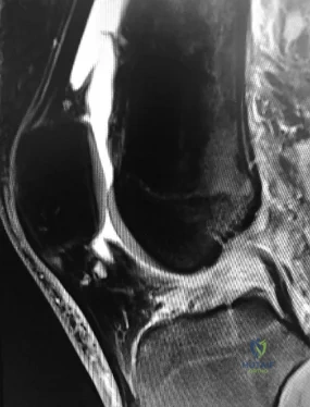
Question 13
Which of the following physical examination findings is most likely present in the condition producing the MRI findings shown in Figure 92?
Explanation
REFERENCES: Vellet AP, Marks PH, Fowler PJ, et al: Occult posttraumatic osteochondral lesions of the knee: Prevalence, classification, and short-term sequelae evaluated with MR imaging. Radiology 1991;178:271-276.
Cone R: Imaging sports-related injuries of the knee, in DeLee J, Drez D, Miller M (eds): DeLee & Drez’s Orthopaedic Sports Medicine: Principles and Practice, ed 2. Philadelphia, PA, WB Saunders, 2003, vol 2, pp 1595-1652.
Question 14
After performing an open distal clavicle excision and resecting 15 mm of distal clavicle, which potential concern for shoulder function could result?
Explanation
AC joint arthritis often is marked by pain along the anterior and superior aspects of the shoulder. It can occasionally radiate into the trapezius and the anterolateral neck region. A patient may have tenderness to palpation directly at the AC joint or pain with the cross-body adduction stress test and the O'Brien active compression test. During the cross-body adduction test, this patient has pain when the examiner lifts his arm in 90 degrees of forward flexion and maximally adducts it across his body. Although the cross-body adduction test is the most sensitive provocative test for AC joint osteoarthritis at 77%, the O’Brien active compression test has been shown to be most specific at 95%.
Physical therapy, rest, activity modification, and other nonsurgical treatments might not reverse osteoarthritis changes at the AC joint, but these interventions can often help improve pain, range of motion, and function. A corticosteroid injection into the AC joint may be an option if nonsurgical treatments do not work, although Wasserman and associates demonstrated that only 44% of AC joint injections accurately entered the joint.
Persistent pain is the most common complication following distal clavicle excision. Although the exact amount of distal clavicle that should be resected is a topic of debate, resection of 10 mm or more of the distal clavicle may lead to instability of the AC joint, especially if the AC capsule is sectioned.
Question 15
One of the serious potential complications of repair of distal biceps tendon ruptures is limited pronation and supination as a result of synostosis. What surgical approach and technique presents the highest risk for development of this complication?
Explanation
REFERENCE: Norris TR: Orthopaedic Knowledge Update: Shoulder and Elbow. Rosemont, IL, American Academy of Orthopaedic Surgeons, 1997, p 342.
Question 16
Figure 37 shows the clinical photograph of a 1-day-old infant who weighed 10.25 lb at birth. Examination reveals an absent right Moro reflex and limited active motion of the right shoulder, elbow, and wrist, but flexion of the fingers. Passive range of motion of the shoulder and elbow is normal. What is the most likely diagnosis?
Explanation
REFERENCES: Hoffer MM, Phipps GJ: Closed reduction and tendon transfer for treatment of dislocation of the glenohumeral joint secondary to brachial plexus birth palsy. J Bone Joint Surg Am 1998;80:997-1001.
Pearl ML, Edgerton BW: Glenoid deformity secondary to brachial plexus birth palsy. J Bone Joint Surg Am 1998;80:659-667.
Waters PM, Smith GR, Jaramillo D: Glenohumeral deformity secondary to brachial plexus birth palsy. J Bone Joint Surg Am 1998;80:668-677.
Question 17
An obese 4-year-old boy has infantile Blount’s disease. Radiographs reveal a metaphyseal-diaphyseal angle of 18 degrees and a depression of the medial proximal tibial physis. Management should consist of
Explanation
REFERENCES: Raney EM, Topoleski TA, Yaghoubian R, Guidera KJ, Marshall JG: Orthotic treatment of infantile tibia vara. J Pediatr Orthop 1998;18:670-674.
Loder RT, Johnston CE: Infantile tibia vara. J Pediatr Orthop 1987;7:639-646.
Question 18
Figures 5a and 5b show the radiographs of a 21 -year-old wrestler who reports that his leg was rolled over while wrestling. The patient has decreased sensation and function in the distribution of the peroneal nerve, and he has absent pulses. What is the most appropriate initial management at this time?
Explanation
intervention of the artery, but documentation with studies would be appropriate. Delayed reconstruction of injured structures is appropriate.
REFERENCES: Fanelli GC, Orcutt DR, Edson CJ: The multiple- ligament injured knee: Evaluation, treatment, and results. Arthroscopy 2005;21:471 -486.
McDonough EB Jr, Wojtys EM: Multiligamentous injuries of the knee and associated vascular injuries.
Am J Sports Med 2009;37:156-159.
Wascher DC: High-velocity knee dislocation with vascular injury: Treatment principles. Clin Sports Med 2000;19:457-477.
Question 19
Figure 81 is the radiograph of a healthy 72-year-old man who has a 3-month history of medial knee pain. He denies any specific trauma. Until 3 months ago when the pain began, he had been an avid runner for many years. Initial treatment should be oral anti-inflammatory medication
Explanation
Question 20
- A 40-year-old woman has had pain in the metatarsophal joint of the second toe for the past 6 months despite nonsurgical treatment. A dorsalplantar stress test reproduces the pain, and there is 10 mm of dorsal subluxation of the toe. Radiographs show a normal second metatarsophalangeal joint. Surgical treatment should consist of synovectomy and
Explanation
Question 21
-Images from an MRI scan of this patient’s left hip are shown in Figure 30c through 30e. What is the most likely cause of his acute pain?
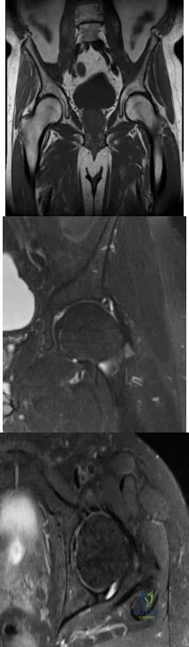
Explanation
This patient has cam-type femoroacetabular impingement. Decreased internal rotation and a positive impingement test (forced flexion, adduction, and internal rotation) are classic findings. The lack of pain with resisted hip flexion makes hip flexor strain unlikely, and the lack of tenderness at the greater trochanter renders trochanteric bursitis unlikely. Although athletic pubalgia can be a source of longstanding groin pain, he lacks the pain with a resisted sit-up and tenderness along the pubic ramus that is frequently noted in patients with pubalgia. His radiographs reveal a focal femoral neck prominence consistent with cam impingement, although pistol grip deformities and flattening of the lateral femoral head are often present as well. His MRI scan shows a labral tear, which is common in cam impingement.Surgical treatment for cam impingement can be effective for symptomatic patients. Even among highlevel athletes, open surgical dislocation of the hip has been shown to have good results. Most patients with cam impingement can be treated with arthroscopic osteoplasty and achieve results comparable to those realized with open surgical dislocation. The literature describes success in terms of athletes returning to sports (even professional athletes) to be approximately 90% after arthroscopic treatment.Byrd and Jones described 5 patients who developed transient neurapraxias that resolved uneventfully.The patients in his series who had concomitant microfracture had a 92% return to sports within the follow-up period. Cam impingement has long been thought to be associated with a history of a slipped capital femoral epiphysis. The capitis in these patients is displaced posteriorly, resulting in a prominent anterior femoral neck and decreased hip internal rotation. Pincer impingement is associated with a deep acetabulum, such as protrusion acetabula and acetabular retroversion. A patient who underwent a periacetabular osteotomy can develop a more retroverted acetabulum as well.
Question 22
They used three outcome tools, SF-36, WOMAC, and Modified Boston Children's Hospital Grading System to evaluate the the two groups at a minimum of 2 years from injury. The foot injury group, including all types of foot fractures, had a poor outcome when using any of these measures. Turchin concludes that “Foot injuries cause significant disability to multiply injured patients. More attention should be given to these injuries, and more
Explanation
Excessive bleeding into joints and muscles is a common manifestation of hemophilia. The iliacus muscle is a frequent site of hemorrhage in patients with severe or moderate hemophilia. Intramuscular hematoma of the iliacus muscle is likely to occur following play or sporting events that include forceful contraction of the hip flexor muscles. As the hematoma expands, it may
compress the adjacent femoral nerve, potentially resulting in complete femoral nerve palsy. Femoral nerve compression typically includes paresthesias in the distribution of the terminal saphenous nerve branch.
Gilbert et al. review the complex relationship between recurrent bleeding, synovitis, and the development of arthritis in the patient with hemophilia. They discuss both conservative and surgical treatment modalities in these patients and recommend arthroscopic synovectomy for the knee and ankle joints. They conclude that the greatest risk to these procedures is a decreased range of motion.
Kuo et al. reports on a fourteen-year-old healthy boy with an 11-day history of pain and weakness in the right lower limb following a fall. They report pain in the right lower extremity, numbness of the anterior aspect of the right thigh and medial border of the right leg and foot, inability to ambulate and
weakened quadriceps muscle strength. MRI revealed an iliacus hematoma with a complete femoral nerve palsy. He underwent CT-guided percutaneous drainage for decompression with complete resolution of the palsy.
Illustration A is a diagram of dermatomal distribution. Illustration B shows the lumbar plexus demonstrating the intimate relationship of the femoral nerve to the iliacus muscle.
Incorrect Answers:
A 45-year-old male trauma patient presents with multiple extremity injuries including the foot injury shown in Figure A. The foot fracture is treated surgically, and heals without any initial complications. At a minimum of 12 months, this patient will be expected to have which of the following scores compared to a
Patients with pauciarticular juvenile rheumatoid arthritis (JRA), specifically the subgroup with elevated antinuclear antibody (ANA) titers, are associated with the highest incidence (~75%) of anterior uveitis. As a result, referral for an ophthalmology consultation is recommended.
Pauciarticular JRA is the most common subgroup of JRA and typically presents between the ages of 2 to 4 years with mild swelling of one to four joints. The diagnosis is typically one of exclusion as laboratory studies, including erythrocyte sedimentation rate and rheumatoid factor, are usually within normal limits. In JRA, iridocyclitis, a type of anterior uveitis typically occurs following the onset of synovitis but may precede the joint symptoms. This iridocyclitis is frequently indolent but requires immediate ophthalmologic consultation for a slit-lamp examination because if left untreated, anterior uveitis may progress to loss of vision.
Foeldavri et al. review JRA anterior uveitis. They report an overall incidence of
10%, but this is dependent on the JRA subtype. They noted that a large proportion of children with JRA develop uveitis in the first year of disease and
90% after 4 years. They state that early age of JRA onset, oligoarticular subtype, and ANA reactivity are the main risk factors for the development of uveitis. They conclude that JRA-associated uveitis is important to recognize and treat early to prevent any visual damage.
Hawkins et al. review bilateral chronic anterior uveitis in JRA. They report that female gender, oligoarthritis, and presence of antinuclear antibodies are risk factors.
They report on treatment options, including the use of biologics. They conclude that stepwise immunomodulatory therapy is indicated, with new biologic drugs being used in cases of refractory uveitis.
Incorrect Answers:
Anterior 4: Pompe disease is a glycogen storage disease which may lead to ptosis (drooping of the upper eyelid), not anterior uveitis
A 9-year-old male with hemophilia A presents with severe groin pain, parasthesias over the medial aspect of the distal tibia, and difficulty ambulating several hours after a soccer game. He is believed to have an intramuscular hematoma surrounding the iliacus muscle. Which nerve is MOST likely to be compressed?
Which of the following conditions places the patient at highest risk for anterior uveitis and necessitates referral to an ophthalmologist?
Salmonella is a classic cause of osteomyelitis in patients with sickle cell disease.
Sickle cell disease is a genetic disorder of hemoglobin synthesis. The disease occurs in two phenotypes: sickle cell anemia (most severe) and sickle cell trait (most common). The two most common causes of osteomyelitis in children with sickle cell disease are
Staphylococcus aureus and Salmonella. Although S. aureus is the most common cause of osteomyelitis in the general population, the literature varies on which is the most common in patients with sickle cell disease. The increased risk in these patients may be associated with gastrointestinal microinfarcts, poor circulation of blood in bone, and splenic infarcts that predispose patients to infection by encapsulated bacteria (i.e., Salmonella).
Piehl et al. analyzed records of seven hundred seventeen patients with sickle cell disease treated over a thirteen-year period. They identified and retrospectively reviewed sixteen cases of osteomyelitis in fifteen patients. The authors found Salmonella to be the causative organism in thirteen cases with Proteus mirabilis, Escherichia coli, and Staphylococcus aureus all affecting one patient each. The authors report the annual incidence of osteomyelitis in their series as 0.36%.
Givner et al. reviewed sixty-eight cases of osteomyelitis in children with sickle cell disease and positive cultures over a ten year period. Of the sixty-eight, 50 (75%) yielded Salmonella and Staphylococci was isolated 7 (10%). In
addition, the authors report non-speciated gram-positive cocci were isolated in
11 (16%), non-speciated gram-negative rods in 5 (7%), and non-specified bacteria in 2 (3%). The authors conclude Salmonella is the most common pathogen causing osteomyelitis in patients with major sickle hemoglobinopathies.
Epps et al. reviewed fifteen patients with sickle cell disease and osteomyelitis. Staphylococcus aureus was isolated in eight cases (53%), Salmonella in six (40%), and Proteus mirabilis in one (7%). The authors conclude S. aureus, not Salmonella, may be the most common cause of osteomyelitis associated in patients with sickle-cell disease.
Figure A demonstrates an osteolytic lesion of the distal tibia and Figure F demonstrates sickle-shaped erythrocytes.
Incorrect Answers
Low toughness is a disadvantage of ceramic bearings in total hip arthroplasty.
Ceramic is a non-metal that demonstrates excellent wear characteristics when used with polyethylene in total hip arthroplasty. Although it has a high Young's modulus, it has a low fracture toughness. Subsequently, ceramic is poorly resistant to crack formation. In contrast, UHMWPE has a high fracture toughness because of the presence of very long hydrocarbon chains.
Santavirta et al. review alternative bearing materials to improve wear in total hip arthroplasty. Alumina ceramics are noted to be biostable and bioinert. The best wear properties are noted with ceramic-on-ceramic bearings. For current ceramic constructs, fracture risk is less than 1 per 1000.
Lang et al. review the use of ceramics in total hip replacement. The authors note that ceramic has high compressive strength and high wettability. Low fracture toughness and linear elastic behavior increase the risk of breakage of ceramic components under stress. Processing improvements, enhanced head- neck interfaces and liner modifications have lead to a decrease in the rate of ceramic fracture.
Illustration A shows a compromised ceramic head as a manifestation of the low fracture toughness of the material.
Incorrect Answers:
An 8-year-old African American female presents with lower extremity pain and subjective fever. On exam there is tenderness about the distal tibia. Further workup reveals elevated inflammatory markers and a lytic lesion (Figure A). An aspirate is obtained and cultures grow Salmonella. Additional investigation is most likely to reveal which of the following findings (Figure B-F)?
An ideal fluid film lubrication regime minimizes friction. A larger head size results in a greater development of full-film lubrication due to the increased relative sliding velocity of the larger bearing surfaces. Increased surface roughness inhibits the formation of the film lubrication. The most important factor influencing the predicted lubrication film thickness
has been found to be the radial clearance between the ball and the socket.
Jin et al report that slight clearance, not complete congruence, is optimal for formation of the optimal fluid film lubrication. They note that full fluid film lubrication may be achieved in these hard/hard bearings provided that the surface finish of the bearing surface and the radial clearance are chosen correctly and maintained.
Dumbleton reviewed the literature of metal-on-metal THA and concluded that the current literature does not show any clinical benefit of metal-on-metal compared to metal on poly. Metal-on-metal has been shown to have higher metal ion level in blood, and measurement of these levels is recommended to help identify those at risk of adverse effects from metal on metal prostheses.
Low toughness is a disadvantage of which of the following bearing surfaces used in total hip arthroplasty?
This attending did not fully disclose that the resident would be performing the cementing portions of the case unsupervised. This represents an ethically unsound scenario as the patient was misled regarding involvement of the resident in their surgery.
The informed consent process is grounded in the ethical principle of autonomy. Informed consent represents a shared decision making process where a
patient understands all the risks and benefits of a surgery fully and makes an informed decision. However, the patient's choice of surgeon is felt to be critical
to the informed consent process and any variation from that surgeon performing the surgery should be discussed explicitly. A surgeon who performs surgery or part of surgery on the patient without prior consent may be held liable for battery.
Kocher presents three cases demonstrating the spectrum of "ghost surgery". They state the substitution of an authorized surgeon with an unauthorized surgeon or allowing surgical trainees to operate without appropriate guidance constitutes "ghost surgery".
Deviation from what is explicitly discussed has been justified in an emergency scenario or if the treatment is aimed at an overall condition.
Bhattacharyya et al reviewed malpractice claims for factors that positively correlated with successful defense. They found that those who performed informed consent in the office had lower risk of malpractice payment. They conclude surgeons can decrease their risk of malpractice claims by performing informed consent in the office and documenting the discussion.
Incorrect Answers:
Which of the following features of metal-on-metal total hip arthroplasty does not allow for improved fluid film lubrication between the components?
The patient sustained a fragility fracture with lab work consistent with primary hyperparathyroidism.
Hyperparathyroidism is commonly caused by increased activity of the parathyroid glands resulting in high levels of PTH. Increased circulating levels of PTH leads to calcium being "sucked" out of bone and into the serum. This
alteration in calcium hemostasis leads to low-density bone and a predisposition to fragility type fractures. When present, lab values are much different from standard age-related osteoporosis. Furthermore, referral to medical and surgical endocrinology specialists for directed treatments may improve overall bone quality and prevent further fragility fractures.
Fraser summarizes primary and secondary hyperparathyroidism. He describes the normal physiologic response to low calcium of an increase in PTH. Increased PTH has three downstream effects of increased tubular resorption of calcium by the kidneys, increased osteoclast activity to harvest calcium from bone, and increased active vitamin D levels leading to increased bowel absorption of calcium.
Singhal et al. reviewed hyperparathyroidism and what the orthopedic surgeon should know. They state when a patient presents with a pathologic fracture and elevated serum calcium levels, an appropriate lab workup for hyperparathyroidism should be done. They stated when surgery is needed for hyperparathyroidism and fracture, surgery can safely be performed simultaneously as demonstrated by 3 case examples.
Figure A exhibits a left femoral neck fracture, which is a fragility fracture associated with poor bone density. Illustration A is a figure from Fraser's article exhibiting the
feedback loop from the hypothalamus, pituitary, adrenal/glandular axis.
Incorrect answers:
Prior to undergoing a total knee arthroplasty at an academic medical center a patient is told during informed consent by the attending surgeon that resident involvement in the case will be limited to retracting. During the case the attending is present up to trialing of the selected components. The surgeon leaves prior to cementing to start trialing components in another case while the chief resident remains alone in the room for the completion of the case. Which of the following is true regarding the ethics of this practice?
Patients in factorial randomized control trials (RCT) are assigned to groups that receive a specific combination of interventions and non-interventions.
In factorial RCTs, patients are randomized to groups receiving treatment A and B, treatment A or control, treatment B or control, or no treatment. This study design is useful because two interventions can be assessed with the same
study population and any interaction between the treatments can be determined (for example, does treatment A work differentially when combined with treatment B). Other randomized control trial designs include parallel, cluster, and crossover. Parallel studies are performed by having two or more groups that exclusively have one intervention without group overlap.
Crossover studies have each group receive each intervention in a random sequence. Cluster design studies have pre-existing groups of participants
(such as schools, or cities) that are randomly selected to receive or not receive an intervention.
Karlsson and the International Society of Arthroscopy, Knee Surgery and Orthopaedic Sports Medicine published an exhaustive guide to research for evidence-based medicine in a step-wise fashion. They cover levels of evidence, design for randomized control trials and the CONSORT checklist. They also describe proper study design of cohort, case- control, case series, systematic review, meta-analysis studies. The second half of the guide discusses appropriate outcome measures, statistical analyses, and data interpretation, reporting complications, and concludes with steps to writing a scientific article.
Incorrect Answers:
A 66-year-old woman falls from standing and sustains the injury shown in Figure A. Her most recent T score was -1.9, 3 months prior to presentation. Preoperative lab work reveals elevated serum calcium, elevated alkaline phosphatase, decreased serum phosphorus, and elevated parathyroid hormone (PTH). Which of the following correctly describes the underlying etiology of her osteopenia?
The most recent update of the CDC guidelines for the prevention of SSI issues a category IA strong recommendation stating that "in clean and clean- contaminated procedures, do not administer additional antimicrobial prophylaxis doses after the surgical incision is closed in the operating room, even in the presence of a drain."
The previous 2002 CDC guidelines for the prevention of SSI focused on three performance parameters: (1) initiation of parenteral antibiotics within 1 hour of the surgical incision, (2) selection of an appropriate antibiotic, and (3) discontinuation of antibiotics within 24 hours. The most recent updated 2017
CDC guidelines for the prevention of SSI has several notable changes with an emphasis that additional doses of antibiotics after initial prophylaxis are no longer recommended.
Berrios-Torres et al. review the 2017 updates to the CDC guidelines for prophylaxis against SSI. Strong recommendations include that in clean and clean-contaminated cases, additional antimicrobial prophylaxis should not be administered after the surgical incision is closed in the operating room, even in the presence of a drain. Furthermore, the authors discuss that there is no evidence that re-dosing intraoperative antibiotics or continuation of antibiotics until surgical drains have been removed provides any additional protection against surgical site infection.
O'Hara et al. highlights the key updates in the most recent CDC guidelines for prevention of SSI. The authors present specific suggestions for translating these recommendations into evidence-based policies and practices. They conclude that the implementation of new and existing guidelines in SSI prevention requires thoughtful and careful collaboration with several inter- professional and interdisciplinary teams.
Incorrect Answers:
Which of the following study designs describes a randomized controlled trial in which two interventions are applied separately or in combination to study groups?
The patient has an allergy to cephalosporins and a history of an MRSA infection. Of the choices listed, vancomycin is the best preoperative antibiotic for this patient.
The choice of preoperative antibiotics is of great interest given the large
medical and economic cost of periprosthetic infections. Standard preoperative prophylaxis in patients undergoing total joint arthroplasty consists of cefazolin or cefuroxime. In patients with beta-lactam allergies, the treatment options include clindamycin or vancomycin. Vancomycin is often the antibiotic of choice given it's higher efficacy with regard to MRSA prevention. In those patients who are considered at risk for MRSA infection and a beta-lactam allergy, vancomycin can be supplemented with an aminoglycoside (gentamicin) or aztreonam.
Bratzler et al. review antimicrobial prophylaxis for surgery and state for orthopedic joint replacement procedures cefazolin or cefuroxime is the recommended antibiotic. For patients with a confirmed beta-lactam allergy, they recommend vancomycin or clindamycin. They also state antibiotics should be stopped within 24hrs after surgery.
Dellinger et al. review antibiotics for surgical prophylaxis. They state the standard antibiotics for orthopedic procedures are cefazolin or cefuroxime. They state if there is also a concern for MRSA infection vancomycin can be added in addition to the above antibiotics.
Incorrect Answers:
Which of the following is STRONGLY recommended by the most recent (2017) Centers for Disease Control and Prevention (CDC) Guidelines with regard to antimicrobial prophylaxis for the prevention of surgical site infection (SSI)?
Clindamycin is a bacterial protein synthesis inhibitor by inhibiting ribosomal translocation at the 50S subunit.
Clindamycin is primarily bacteriostatic but may be bactericidal at higher concentrations.
Side effects of clindamycin may include a hypersensitivity reaction and pseudomembranous colitis. Resistance to clindamycin is conferred by a plasmid that alters the 50s ribosome binding site for clindamycin. The D- zone test is used to determine whether an organism has inducible resistance
to clindamycin.
Marcotte and Trzeciak published a review on community-acquired methicillin- resistant Staphylococcus aureus (CA-MRSA). They reported that CA-MRSA
does not have predictable susceptibility to clindamycin. They conclude that clindamycin also presents a risk for the development of Clostridium difficile colitis and inducible clindamycin resistance for which a D-zone test should be performed when culture results reveal erythromycin resistance.
Steward et al. performed a lab study to determine the efficacy of testing for induced clindamycin resistance in erythromycin-resistant Staphylococcus aureus. They reported that resistance to erythromycin and clindamycin can occur through methylation of their ribosomal target site (50s), which is mediated by erm genes. They conclude that disk diffusion is the preferred method for testing S. aureus isolates for inducible clindamycin resistance.
Incorrect Answers:
A 68-year-old man is scheduled to undergo total hip arthroplasty. He states he had an anaphylactic reaction after taking cefazolin for an MRSA hand infection 10 years ago. Which of the following best describes the preoperative antibiotic that should be administered for this patient?
Advanced glycation end-products (AGEs) cause excessive cross-linking of collagen in aging articular cartilage. As a result, the stiffness is increased.
AGEs are produced by spontaneous nonenzymatic glycation of proteins when sugars (glucose, fructose, ribose) react with lysine or arginine residues. The most abundant matrix protein in cartilage is Type II collagen. AGEs cause changes to the aging cartilage matrix and the aging chondrocyte. The increased cross-linking of Type II collagen results in an increase in cartilage stiffness (i.e. increase in the modulus of elasticity) and an increase in brittleness (i.e. less strain needed to go from the yield point to the fracture point on the stress-strain curve). As a result of the change in the aging cartilage’s biomechanical properties, it's susceptible to fatigue failure. Additionally, AGEs decrease the anabolic response of chondrocytes from autocrine signaling via TGF-beta, IGF-1, BMP-7, and OP-1. These two initial mechanisms contribute to aging cartilage to eventually lead to the development of osteoarthritis.
Li et al. reviewed age-related changes in cartilage and seek to define the different
mechanisms between aging cartilage and osteoarthritis. They state that with AGEs, there is excessive collagen cross-linking increases cartilage stiffness, while shortening/degradation of aggrecan leads to loss of sugar side chains and water-binding ability. Additionally, increased levels of AGEs are associated with a decline in anabolic activity. They state that these changes to cartilage make it more vulnerable to damage and therefore the onset of osteoarthritis. This is contrast to the initial steps in the mechanism of osteoarthritis which is characterized by cell proliferation, formation of chondrocyte clusters, increased synthesis of irregular cartilage matrix, and eventually a pro-catabolic and pro-inflammatory state that results in an imbalance in cartilage homeostasis and cartilage matrix breakdown.
Anderson et al. reviewed the relationship between osteoarthritis and aging.
They state that knee cartilage thins with aging, especially on the femoral and patellar sides, suggesting a gradual loss of cartilage matrix. AGEs formation leads to modification of type II collagen by cross-linking of collagen molecules, increasing stiffness and brittleness and increasing susceptibility to fatigue failure. Furthermore, describe the senescent phenotype of the chondrocyte
and its similarities with osteoarthritic chondrocyte phenotype.
Incorrect Answers:
Which of the following antibiotics works by binding to the 50S ribosomal subunit?
The patient has clinical signs and symptoms of gout. Figure D would correspond to this diagnosis as it shows negatively birefringent needle-shaped monosodium urate crystals.
Gout is an idiopathic disorder of nucleic acid metabolism that leads to hyperuricemia and deposition of monosodium urate crystals, most commonly in the joints of the lower limb (knee, ankle, and classically the 1st metatarsophalangeal joint). Diagnosis can be confirmed with joint arthrocentesis revealing negatively birefringent needle-shaped crystals. Treatment of acute gout flares is generally comprised of NSAIDs and colchicine, and chronic gout is treated with allopurinol to prevent the build-up
of uric acid.
Shmerling et al. prospectively analyzed the synovial fluid test results of 100 consecutive patients undergoing diagnostic arthrocentesis. They noted that synovial fluid white blood cell count (WBC) and the percentage of polymorphonuclear cells performed well as discriminators between inflammatory and noninflammatory diseases. Given the diagnostic value of synovial WBCs, the authors concluded that ordering of chemistry studies of synovial fluid should be discouraged because they are likely to provide misleading or redundant information.
Chiodo et al. review the use of intra-articular aspiration and injections for both diagnosis and treatment of disorders of the lower extremity such as infectious arthritis, gout, pigmented villonodular synovitis (PVNS), rheumatoid arthritis, and hemophilia. The authors discuss the importance of knowledge of regional anatomy, procedural indications, and appropriate techniques for successful aspiration/injection. The authors review safe and effective aspiration and injection techniques for the lower extremity, including the hip, knee, foot, and ankle.
Figure A reveals hemosiderin stained multinucleated giant cells consistent with PVNS. Figure B is a gram stain revealing gram-positive cocci in clusters consistent with Staphylococcus aureus. Figure C reveals rhomboid-shaped, positively birefrigerant crystal consistent with calcium pyrophosphate/pseudogout. Figure D reveals negatively birefringent needle- shaped crystals of monosodium urate/gout. Figure E reveals a collection of histiocytes and inflammatory cells around prominent intimal hyperplasia.
Incorrect Answers
An increase in advanced glycation end-products (AGEs) is characteristic of which of the following clinical conditions and results in which pathologic process?
Regardless of the number of level I studies included in a systematic review, having one study with <80% follow-up decreases the level of evidence for this review from level I to level II.
After classifying the type of study (e.g. therapeutic study, prognostic study, diagnostic study, economic analysis, or decision analysis) the “level of evidence” is then determined. The level of evidence (on a scale of I through V) for medical research is determined. It is important to consider the characteristics of a study’s design. This would include the percent follow-up, utilization of a control group, presence of blinding, heterogeneity of results, and process of randomization. Specific to meta-analyses and systematic reviews, it is important to know that the lowest quality study used in the review determines the level of evidence. In evidence-based medicine, higher levels of evidence have a larger impact on clinical recommendations.
Bhandari et al. analyzed the interobserver agreement among reviewers in categorizing the type of study, level of evidence, and subclassification for different clinical studies. The authors had 6 different surgeons with different levels of training in epidemiology analyzed 51 separate papers published in JBJS. The results demonstrated that the interobserver absolute agreement for the type of study and the level evidence was 82% and 67%, respectively. The epidemiology-trained reviewers had nearly perfect agreement in categorizing the type of study, level of evidence, and subclassification.
Wright et al. published an editorial introducing the different types of study designs and defined the different levels of evidence. Illustration A is a figure from this editorial.
Incorrect Answers:
A 55-year-old male, alcoholic, presents to the ER with acute right knee swelling and pain x 3 days. He admits to prior episodes of this pain that resolve after a few days. Serum labs reveal an ESR of 40 mm/hr and CRP of 5 mg/dl. He undergoes right knee aspiration and based on the results, he is discharged home on colchicine with the presumed diagnosis of gout. Which of the following images of the aspiration results are consistent with this diagnosis?
conclude that the patient populations and outcomes measure are homogenous and you do not have any concerns with randomization. You notice one of
the studies included had 70% follow-up, yet the remaining studies had
>80% follow-up. Knowing this, you appropriately assign what level of evidence to the systematic review?
The correct sequence of events should be the surgeon reads the surgical information on the consent to the patient, then the surgeon marks the surgical site with the patient’s assistance, then allows the anesthesia team to perform their procedure, and then performs a final Time-Out with the surgical team immediately prior to the surgical incision.
Orthopedic surgical patients are at risk of surgical errors due the number of procedures that can be performed on the bilateral extremities. The responsibility to identify the correct surgical procedure at the correct location has expanded beyond only the surgeon. The entire surgical team is
responsible for confirming the patient, surgical site, and surgical procedure. All members on the surgical team should be valued and emboldened to “speak up’ and actively participate. To help improve communication and reduce complications, surgical safety checklists have become common. In a statement
published by the AAOS is 2015, they support the use of standardized surgical systems, including the use checklists, as it is critically important to keep patients safe. In 1998, the AAOS introduced the “Sign Your Site” safety program to reduce wrong-site surgeries through improved site identification. Permanent ink should be used to mark the site(s) with the patient's assistance prior to surgery, and the site(s) should be confirmed by the surgical team during the Time-Out immediately before the start of the surgical procedure.
Singer et al. performed a study to evaluate the association between surgical teamwork and surgery safety checklist performance. Their results emphasized the importance of surgeon buy-in and clinical leadership to initiating and maintaining surgical safety checklists. In addition to surgeon buy-in and clinical leadership, factors that help maintain high-quality and consistent surgical teamwork were communication, coordination, respect, and assertiveness.
Incorrect Answers:
You are reviewing a systematic review on the 90-day complication rate and outcome for same day total joint arthroplasty for publication. After you analyze the methodology of the 6 randomized controlled trials included in the review, you
preoperative paperwork outside the room. The patient is taken to surgery and receives an interscalene block on the left shoulder after sedation. At the final Time- Out, the surgeon realizes a discrepancy with the laterality when the consent is read aloud. The surgeon aborts the case and wakes the patient. What is the correct sequence of events that should have happened to prevent this error? A: The surgeon begins
the surgery B: The surgical team performs a Time-Out C: The surgeon marks the surgical site D: The surgeon reads the surgical information on the consent to the patient E: The anesthesia team administers a local extremity block
Enchondral ossification occurs with relative stability constructs, which is represented by the bridge plate in figure C.
Fracture healing is a complex process that occurs in several key steps. The type of healing that occurs is dependent on the stability and strain of the fracture environment. In constructs with very little strain, also referred to as absolute stability, there is primary bone healing through Haversian remodeling. This produces very little callus and does not rely on a cartilage precursor. Relative stability constructs with higher strains produce a cartilage precursor, which subsequently ossifies in later stages of healing, also referred to as enchondral ossification.
Perren reviewed the biological mechanisms of fracture healing. The author discussed the importance of skeletal stiffness for limb function in addition to the healing process that utilizes soft tissue compensatory mechanisms to aid
in fracture healing. The author concluded that the goal of fracture healing is to obtain a functional limb to allow for daily mobility and activity.
Gerstenfeld et al. investigated the effect of non-selective and COX-2 selective NSAIDs effects on bone healing in a rat model. They reported a significantly higher nonunion rate in valdecoxib treated rats compared to the ketorolac group. They also noted that withdrawal of either drug at six days resulted in prostaglandin E2 levels returning to normal levels after 14 days. The authors concluded that COX-2 specific NSAIDs inhibited bone healing greater than nonspecific NSAIDs with the magnitude of the effect dependent on the duration of treatment, but the effects on prostaglandin E2 levels appear reversible with discontinuation of the drug.
Figure A is the AP radiograph of the left distal tibia with three lag screws through a spiral fracture. Figure B is the lateral radiograph of the right elbow with an olecranon plate.
Figure C is the AP radiograph of the right distal femur with a lateral bridge plate. Figure D is an AP radiograph of the left ankle with a lag screw and neutralization plate on the distal fibula. Figure E is the lateral radiograph of the forearm with a compression plate on the radius.
Incorrect Answers:
A 31-year-old man is scheduled to undergo a right shoulder arthroscopic labral repair. The surgeon is running behind and hurries to the preoperative holding area. The surgeon greets the patient and verbally confirms the operative site with the patient. The surgeon leaves the patient’s room and completes the appropriate
The yield point is the transition point between elastic and plastic deformation. The yield strength is defined as the amount of stress necessary to produce a specific amount of permanent deformation.
Stress is the amount of force applied to a material and strain is the deformation resulting
from that stress. This is graphically depicted as a stress- strain curve, where the X-axis represents strain and the Y-axis represents stress. The elastic modulus of a material is the linear region of the graph (rise over run/stress on strain). Remember, an elastic material is one that resists a change in shape (less strain or deformation under increasing stress). Non- linear regions include the toe region for some materials (tendons/ligaments) and the plastic zone, which occurs after the yield point.
Mantripragada et al. provide a review of recent advances in designing orthopaedic implants. Of note, they discuss modifications to metallic implants to reduce unwanted effects, such as nickel-free stainless steel. They also go over newer alloys with desirable mechanical and biological properties, such as tantalum, niobium, zirconium, and magnesium.
Kennedy et al. provide a classic in-vitro tension study of the human knee ligaments. They used an Instron Tension Analyzer to test the ultimate failure of the medial collateral, lateral collateral, anterior cruciate, and posterior cruciate ligaments at different loading rates. They found that the posterior cruciate ligament was the strongest (the other ligaments were all of
comparable strength) and that microscopic failure occurred before macroscopic failure. Illustration A represents a stress-strain curve.
Incorrect Answers:
is a phenomenon especially associated with a ductile material; the diameter of the material is diminished prior to fracture.
material can absorb before fracture and is the area under the stress-strain curve. Answer 5: The toe region is seen in materials such as ligaments and tendons and represents the straightening of the crimped ligament fibers.
Which of the following fixation constructs would achieve fracture healing through enchondral ossification?
The preosteoclast (precursor to the osteoclast) is the only cell of myeloid origin. The remainder of the cells involved in bone formation, remodeling, and metabolism are of mesenchymal origin.
Osteoclast signaling, function, and biology have grown increasingly well understood over the past few decades. Osteoclast activity is regulated by
osteoblasts, thereby coupling bone formation and resorption. Osteoclast differentiation from myeloid precursor cells is stimulated by key molecules including RANK, PU-1, and CSF-1. An understanding of these molecular pathways is essential to developing effective directed anti-resorptive therapies.
Zaidi et al. present a comprehensive review of proliferation, differentiation, and hormonal regulation of cells of the bone. The authors specifically discuss the unique origin of the osteoclast from the myeloid lineage and conversely the mesenchymal origin of the osteoblast. Furthermore, they highlight the
most recent understanding of the molecular mechanisms involved in osteoclast formation
and signaling, including M-CSF and RANKL.
Caterson et al. discusses mesenchymal differentiation in the context of musculoskeletal regeneration. The authors review the growth factors and bioactive signaling molecules involved in directed differentiation itno the various mesodermal lineages including bone, cartilage, muscle, tendon, marrow, and adipose. They emphasize the importance of understanding these pathways to regenerative medicine.
Illustration A is a diagram illustrating the difference between mesenchymal and myeloid lineages.
Incorrect answers:
The point on a stress-strain curve that separates the plastic and elastic regions is defined as which of the following:
Due to the risk of inducible clindamycin resistance in erythromycin-resistant MRSA, a D-test should be performed.
Isolates of MRSA that are resistant to erythromycin have been shown to become resistant to clindamycin through a process called inducible resistance, which is conferred by a plasmid that alters the 50S ribosome binding site for both clindamycin and erythromycin. Thus, when culture results reveal erythromycin-resistant MRSA, a D-zone test should be performed to check for inducible clindamycin resistance. The D-zone test is performed by
placing an erythromycin disk in proximity to a clindamycin disk on an agar plate inoculated with methicillin-resistant S aureus (MRSA). A zone of inhibition in the shape of the letter "D" is seen with an inducible strain and is considered a positive test. If the D- zone test is positive, then clindamycin should not be used because the strain of MRSA can become resistant to the treatment.
Marcotte et al. published a review on community-acquired methicillin-resistant Staphylococcus aureus (CA-MRSA). They reported that clindamycin has activity against Streptococcus species, but it is not as predictable against CA- MRSA. Clindamycin also presents a risk for the development of Clostridium difficile colitis and inducible clindamycin resistance. for which a D-zone test should be performed when culture results reveal erythromycin resistance.
Steward et al. performed a study to determine the efficacy of testing for induced clindamycin resistance in erythromycin-resistant Staphylococcus aureus. They reported that resistance to erythromycin and clindamycin can occur through methylation of their ribosomal target site (25), which is typically mediated by erm genes. They found that disk diffusion is the preferred method for testing S. aureus isolates for inducible clindamycin resistance.
Illustration A is an image of a positive D-zone test, which indicates inducible clindamycin resistance.
Incorrect Answers:
Which of the following cells involved in bone metabolism derives from a myeloid origin?
Enoxaparin primarily exerts its effects by inhibiting Factor Xa, which is labeled C in Figure A.
Enoxaparin is a low molecular weight heparin (LMWH) that primarily exerts its effects by inhibiting Factor Xa. It achieves this by binding to antithrombin to form a complex that irreversibly inactivates clotting factor Xa. Enoxaparin has the advantage of not requiring laboratory monitoring and can be reversed with protamine sulfate. However, it is important to note that protamine sulfate is less effective in reversing enoxaparin compared to unfractionated heparin (UFH).
Hyers published a review on the past, present, and future management of venous thromboembolism. He found that, for the most part, LMWH and other newer anticoagulants have been shown to be superior to UFH in terms of the venographic endpoint. He also reports that several meta-analyses have demonstrated that LMWH offers superior benefit to UFH for VTE prevention in hip and knee surgery patients.
Tørholm et al. performed a study to determine outcomes of thromboprophylaxis using LMWH compared to placebo in elective hip surgery. They found that 9 (16%) patients in the treatment group and 19 (35%) in the placebo group developed deep venous thrombosis. The risk of thrombosis in the placebo group was increased with prolonged surgery and occurred more frequently during the first 4 postoperative days. They concluded that LMWH offers safe and easily administered thromboprophylaxis in total hip replacement.
Figure A is an image of the coagulation cascade. Illustration A is an image of the
coagulation cascade with the sites of action of the various anticoagulants labeled.
Incorrect Answers:
A 42-year-old IV drug user presents to the emergency department with a large abscess on his forearm. A bedside I&D is performed and he is started on broad-spectrum IV antibiotics. Initial results from his cultures demonstrate methicillin-resistant Staphlycoccus aureus (MRSA) that is also resistant to erythromycin. The team would like to transition him to oral clindamycin. Prior to transitioning him to clindamycin, which additional laboratory test should be performed?
Teriparatide promotes bone formation in patients at high risk of fractures due to severe osteoporosis that is refractory to multiple treatments, including bisphosphonates and cement augmentation. Teriparatide is a human recombinant N-terminal parathyroid hormone.
Teriparatide administered in daily injections results in bony formation, whereas continuous infusion results in bony resorption. In rat models, teriparatide caused an increase in the incidence of osteosarcoma, and thus should only be prescribed for patients for whom the potential benefits outweigh the potential risk. It can be administered in isolation or as an adjunct treatment during bisphosphonate therapy. However, in patients on long-term bisphosphonate therapy, discontinuation of bisphosphonates are advised to reduce potential complications of atypical femur fractures and jaw osteonecrosis.
Watts et al. published a review article on postmenopausal osteoporosis. They reported that bisphosphonates can accumulate in bone, thus after a period of treatment, lower- risk patients should be offered a drug holiday. Denosumab, on the other hand, is not sustained when treatment is discontinued, so no drug holiday is warranted. They concluded that, although there are safety
concerns regarding atypical femoral fracture and osteonecrosis of the jaw with long term use, the benefit of hip fracture risk reduction far outweighs the risk of these relatively uncommon side effects.
Song et al. performed a meta-analysis to investigate the effect of teriparatide monotherapy and the additive effect of teriparatide on antiresorptive agents in postmenopausal women with osteoporosis. They reported that teriparatide monotherapy significantly improved bone mineral density (BMD) in the lumbar spine, total hip, and femoral neck compared with placebo; the additive effect
of teriparatide over hormone replacement therapy (HRT) and denosumab agents was evident in all 3 skeletal sites; however, teriparatide plus alendronate did not demonstrate additive effect in total hip and femoral neck. They concluded that, for patients with osteoporosis who were at high risk for fracture, BMD increased more in patients receiving teriparatide than in those receiving alendronate.
Saag et al. compared the use of teriparatide or alendronate in the management of glucocorticoid-induced osteoporosis. They reported that BMD had increased more in the teriparatide group than in the alendronate group in the lumbar spine and total hip at 6 and 12 months, respectively. They also reported significantly fewer new vertebral fractures in the teriparatide group compared to the alendronate group. They concluded that in severely osteoporotic patients at high risk for fracture, BMD increased more in patients receiving teriparatide than in those receiving alendronate.
Figure A depicts multiple vertebral insufficiency fractures in the setting of a prior cement augmentation procedure.
Incorrect Answers:
Where in the coagulation cascade shown in Figure A does enoxaparin primarily exert its effects?
This patient is presenting with signs of a septic nonunion after open reduction and internal fixation (ORIF) of a radial shaft fracture. Of the choices listed, C- reactive protein (CRP) is the best predictor of infection in the setting of nonunion.
Nonunions after fracture fixation may occur from infection. The most sensitive and readily-available laboratory marker to detect infection is the CRP. CRP is an acute phase reactant that significantly rises within 6 hours after tissue damage or onset of clinical infection. CRP then peaks 2-3 days later and returns to normal levels 5-21 days after the inciting event if it is treated (e.g. antibiotics for cellulitis). In septic nonunions, the chance of fracture healing is low if the infection is not properly treated, and chronic infection can lead to substantially elevated CRP values.
Wang et al. evaluated the effectiveness of laboratory tests in the diagnosis of
infected nonunion. They reported that the sensitivity and specificity of CRP for detection of infected nonunions are both higher than those of IL-6. They concluded that the diagnostic utility of CRP was superior to IL-6, which is contrary to similar studies comparing these markers in prosthetic joint infection patients.
Stucken et al. performed a study to investigate the utility of a standardized protocol to rule out infection in high-risk patients and to evaluate the efficacy of each component of the protocol. They reported that the ESR and the CRP levels were both independently accurate predictors of infection. They
concluded that their protocol can help surgeons to risk-stratify patients prior to the surgical treatment of a nonunion, allowing them to counsel patients more appropriately.
Figure A depicts a nonunion of a radial shaft fracture after ORIF. Incorrect Answers:
An 85-year-old woman presents with severe back pain and the CT shown in Figure A. Her history is notable for prior vertebral compression fractures for which she underwent a cement augmentation procedure. She has been on bisphosphonates for the last 5 years, without improvement of her osteoporosis. She has no history of malignancy. What is the mechanism of action of the medication that should be prescribed for her refractory osteoporosis?
A receiver operating characteristic (ROC) curve is used to determine responsiveness.
Responsiveness is a measure of the diagnostic ability of different tests. It can be determined by calculating the C-statistic, which represents the area under a
Receiver Operating Characteristic (ROC) curve. On a ROC curve, the false positive rate (1 - specificity) is plotted on the x-axis, while the true positive rate (sensitivity) is plotted on the y-axis. The higher the area under the curve, the more responsive the outcome measure. A value of 0.5 indicates a random chance and a therefore useless test, while values above 0.75 usually are considered to be adequately responsive.
Kocher et al. published a review on clinical epidemiology and biostatistics for orthopaedic surgeons. They reported that the relationship between the sensitivity and specificity of a diagnostic test can be portrayed with use of a receiver operating characteristic (ROC) curve. A ROC graph shows the relationship between the true- positive rate (sensitivity) on the y-axis and the false-positive rate (1 − specificity) on the x-axis plotted at each possible cutoff. Overall diagnostic performance can be evaluated on the basis of the area under the ROC curve.
Hanley et al. published a review on the meaning and use of the area under a receiver operating characteristic (ROC) curve. They reported that it represents the probability that a randomly chosen diseased subject is (correctly) rated or ranked with greater suspicion than a randomly chosen non-diseased subject.
Illustration A is an example of a ROC curve. Illustration B is an example of a funnel plot. Illustration C is an example of a Kaplan-Meier curve. Illustration D is a table outlining the interpretation of the Cronbach alpha coefficient. Illustration E is an example of a forest plot.
Incorrect Answers:
A 32-year-old man underwent open reduction and internal fixation for an open radial shaft fracture 6 months ago. He is now experiencing fevers and chills at night and pain and swelling over the surgical site. A current radiograph is depicted in Figure A. What is the most accurate laboratory test for assessing his most likely diagnosis?
The Patient-Reported Outcomes Measurement Information System (PROMIS) has been shown to have reduced floor and ceiling effects compared to other assessment tools.
The PROMIS system was developed to produce a highly reliable, precise, and versatile assessment of outcomes. When administered in a computerized adaptive mode, each question that is answered is followed with a customized follow-up question based on the previous response, which allows for reduced testing items and time. Further, the results of the assessment are reported in T-scores with 50 being the population norm and with a standard deviation of
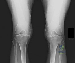
Question 23
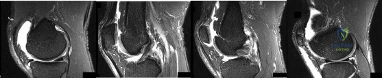
Figures 1 through 4 are selected sagittal MR images of an otherwise healthy 20-year-old collegiate football running back who was tackled during a game and has immediate onset of right knee pain. Video analysis of the injury shows that his flexed knee impacted the field. He is not able to return to play. On examination in the training room the following morning, he has a moderate effusion, no patellar instability, minimal joint line tenderness, and is stable to varus and valgus stress at 30° of knee flexion. A dial test is also negative. He has increased laxity in the anterior to posterior direction. What is the most appropriate next step in treatment?
Explanation
This athlete sustained an isolated PCL injury. The mechanism of injury is typical for a PCL injury. When a PCL injury is identified, one must rule out other ligamentous injuries to the knee. The patient has a stable examination to varus and valgus and a negative dial test, so the lateral collateral, medial collateral, and posterolateral corner (respectively) are intact. It is common to have increased anterior to posterior translation in isolated PCL injuries, even with an intact ACL, as the tibia will rest posterior to the medial femoral condyle. Treatment of isolated PCL injuries is typically nonoperative, with an initial focus on quadriceps strengthening. Hamstring strengthening and rehabilitation is added at a later time, as this places increased stress on the healing PCL. The images reveal an isolated PCL injury with intact menisci and ACL, ruling out ACL reconstruction using autograft tissue and PCL reconstruction using autograft tissue.
Question 24
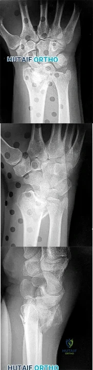
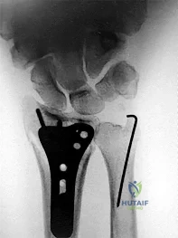
Figures 1 through 3 are the radiographs of a 65-year-old man who sustained a fracture from a fall. The patient elects open reduction and internal fixation of the distal radius. After plating the distal radius, the distal radioulnar joint (DRUJ) is examined and found to be unstable in both pronation and supination. What is the best next step?
Explanation
styloid fracture were found to increase risk for DRUJ instability. An ulnar styloid base fracture involves the insertion of the radioulnar ligaments and can cause DRUJ instability if displaced. If persistent instability is present after fixation of the ulnar styloid, DRUJ pinning is a reasonable option. Early ROM
The initial radiographs show a comminuted displaced distal radius fracture, along with a displaced fracture of the base of the ulnar styloid. The displacement is best seen on the oblique view. After reduction and fixation of the radius, DRUJ stability should be assessed. The majority of scenarios that involve this injury pattern will not be unstable because of the oblique band of the interosseous ligament. When DRUJ instability is present after fixation of the radius, reduction and fixation of the ulnar styloid fracture is the best option to provide stability of the distal radioulnar joint (DRUJ) (Figure below). A study by Lawton and associates revealed that all distal radius fractures complicated by DRUJ instability were accompanied by an ulnar styloid fracture. A fracture at the ulnar styloid’s base and substantial displacement of an ulnar
with splinting would not allow reduction or healing of the ulnar styloid and would result in persistent instability. Short-arm casting also would not allow stability of the DRUJ and would be a less reliable method with which to achieve healing of the ulnar styloid.
Question 25
Up to what time frame are the risks minimized in anterior revision disk replacement surgery?
Explanation
REFERENCE: Tortolani JP, McAfee PC, Saiedy S: Failures of lumbar disc replacement. Sem Spine Surg 2006;18:78-86.
Question 26
What tendon is closest to an appropriately placed anterolateral portal for ankle arthroscopy?
Explanation
REFERENCE: Ogut T, Akgun I, Kesmezacar H, et al: Navigation for ankle arthroscopy: Anatomical study of the anterolateral portal with reference to the superficial peroneal nerve. Surg Radiol Anat 2004;26:268-274.
Question 27
An active, right-handed 71-year-old woman fell on her left shoulder and sustained the injury shown in the radiographs in 52a and 52b and the CT scan in 52c. Management should consist of
Explanation
Question 28
When comparing arthroscopic and open rotator cuff repairs, which of the following tears shows a decreased recurrent tear rate in the open versus the arthroscopic group? Review Topic
Explanation
Question 29
A 19-year-old man was in a motorcycle accident. He sustained a grade IIIB open tibia fracture with a wide zone of injury to the surrounding soft tissue and a closed-head injury. The patient was treated emergently with irrigation, debridement, and external fixation. What is the most accurate statement regarding long-term functional and financial outcomes?
Explanation
Lower Extremity Assessment Project data suggest that long-term functional outcomes and patient satisfaction at 7 years are equivalent between those
who undergo limb-salvage and primary amputations. Return to work is essentially the same between the 2 groups. The projected lifetime healthcare cost for patients treated with amputation is nearly 3 times higher than costs for those who are treated with limb-salvage procedures.
RECOMMENDED READINGS
Busse JW, Jacobs CL, Swiontkowski MF, Bosse MJ, Bhandari M; Evidence-Based Orthopaedic Trauma Working Group. Complex limb salvage or early amputation for severe lower-limb injury: a meta-analysis of observational studies. J Orthop Trauma. 2007 Jan;21(1):70-6. PubMed PMID: 17211275. View Abstract at PubMed
MacKenzie EJ, Jones AS, Bosse MJ, Castillo RC, Pollak AN, Webb LX, Swiontkowski MF, Kellam JF, Smith DG, Sanders RW, Jones AL, Starr AJ, McAndrew MP, Patterson BM, Burgess AR. Health-care costs associated with amputation or reconstruction of a limb-threatening injury. J Bone Joint Surg Am. 2007 Aug;89(8):1685-92. PubMed PMID: 17671005. View Abstract at PubMed
CLINICAL SITUATION FOR QUESTIONS 85 THROUGH 88
Figures 85a and 85b are the plain radiographs of a 38-year-old man who fell off the roof of a 2-story house and sustained an isolated injury to his right knee. Examination reveals a swollen leg with a knee effusion. The skin is intact, but there are some abrasions and an obvious deformity. His neurovascular examination reveals active dorsiflexion and plantar flexion with some pain and symmetric palpable pulses
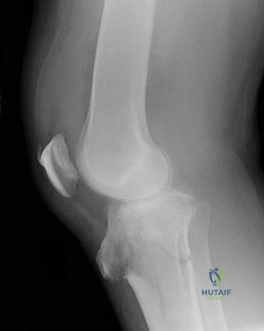
85A
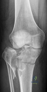
B
Question 30
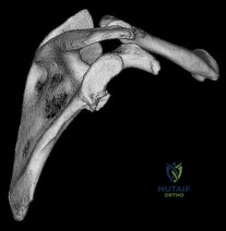
A 23-year-old man reports a 6-year history of recurrent instability in the right dominant shoulder. He has not undergone surgery and has essentially stopped all of his sporting activities. On examination, he has instability and apprehension in the midrange of motion (abduction of 45 to 60 degrees with external rotation) and a palpable clunk representing a transient dislocation over the anterior glenoid rim. A three-dimensional CT scan is shown in Figure 31. What is the most appropriate surgical intervention to provide him with reliable stability postoperatively? Review Topic
Explanation
Question 31
Tension band wire fixation is best indicated for which of the following types of olecranon fractures?
Explanation
Question 32
Which of the following is considered the most appropriate shoe modification following transmetatarsal amputation?
Explanation
REFERENCES: Philbin TM, Leyes M, Sferra JJ, Donley BG: Orthotic and prosthetic devices in partial foot amputations. Foot Ankle Clin 2001;6:215-228.
Marks RM: Mid-foot/mid-tarsus amputations. Foot Ankle Clin 1999;4:1-16.
Question 33
Figure 33 shows the oblique radiograph of an 11-year-old boy who has a mild left flatfoot deformity. Examination reveals that subtalar motion is limited and painful. Despite casting for 6 weeks, the patient reports foot pain that limits participation in sport activities. A CT scan shows no subtalar joint abnormalities. Management should now include
Explanation
has been described for treatment of the peroneal spastic flatfoot without demonstrable
tarsal coalition.
REFERENCES: Gonzalez P, Kumar SJ: Calcaneonavicular coalition treated by resection and interposition of the extensor digitorum brevis muscle. J Bone Joint Surg Am 1990;72:71-77.
Vincent KA: Tarsal coalition and painful flatfoot. J Am Acad Orthop Surg 1998;6:274-281.
Luhmann SJ, Rich MM, Schoenecker PL: Painful idiopathic rigid flatfoot in children and adolescents. Foot Ankle Int 2000;21:59-66.
Question 34
A 35-year-old skiier presents with pain in the left buttock and proximal posterior thigh after a fall. His clinical appearance is shown in Figure A. He is enrolled in 8 weeks of physical therapy after 2 weeks of rest, icing and NSAIDS. He returns for follow-up 6 months after his injury and has persistent ischial tuberosity pain with running. Examination confirms focal ischial tuberosity tenderness. MRI images are seen in Figures B and C. Which surgical option is most appropriate? Review Topic
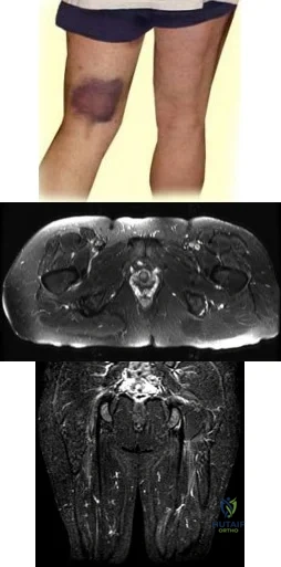
Explanation
Untreated partial hamstring ruptures may present with residual pain, weakness and hamstring dysfunction. The mechanism is eccentric lengthening (sprinting or cutting) A proposed treatment algorithm is: (1) Nonoperative management for single tendon avulsion with <2cm retraction. The ruptured tendon scars to intact tendons. (2) Repair for acute 3-tendon rupture (semitendinosus, semimembranosus, biceps femoris) with retraction >= 2cm. (3) Surgery for young (<50y) patients with 2 tendon avulsion and retraction >= 2cm.
Bowman et al. examined the outcomes of operative management of partial hamstring tears in 17 patients. They found no postoperative difficulties with ADLs, and no recurrent surgery was required. All patients returned to their preoperative level of activity. They concluded that surgery can lead to good function with low complications and is reserved for patients who have failed nonoperative management.
Hofmann et al. retrospectively reviewed 19 patients with nonoperatively managed complete hamstring avulsions. They found diminished SF-12 scores, diminished hamstring strength at 45° and 90° of flexion (62% and 66%, respectively) compared with the normal side. They concluded that nonsurgical management leads to both subjective functional and objective strength deficits.
Figure A shows pronounced bruising from hematoma tracking following the injury. Figures B and C are coronal and axial images showing partial avulsion of the right hamstring insertion. The images correspond with Illustration B, with arrows pointing to the "sickle sign" . Illustration A shows the origins of the hamstring tendons. Illustration C shows the origins of the hamstring group (bf, biceps femoris; st, semitendinosus; sm, semimembranosus; qf, quadratus femoris; am, adductor magnus)
Incorrect Answers:
Question 35
A 72-year-old woman has a painful right hip, and left hip issues are discovered on the radiographs shown in Figures 1 and 2. An arthroplasty was done 24 years previously. Her left hip is pain-free, but she reports occasional clicking and grinding on the left side. She wishes to avoid major revision surgery. Considering this, what is the best next step to address the left hip?
Explanation
Question 36
Figures below show the radiographs obtained from a 19-year-old woman with a 3-year history of progressive hip pain in the left groin with activity, which is unresponsive to activity modification and physical therapy. Examination reveals normal range of motion, with pain on anterior impingement testing. What treatment is associated with the best long-term results?
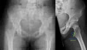
Explanation
This patient has symptomatic femoroacetabular impingement as well as clinical and radiographic signs of acetabular retroversion, including a cross-over sign, ischial spine sign, and posterior wall sign bilaterally. Good midterm to long-term outcomes have been reported with reverse (anteverting) Bernese periacetabular osteotomy (PAO). In patients with less retroversion, open or arthroscopic rim trimming with labral refixation have shown good short-term results, but longer-term results have yet to be fully delineated. Isolated hip arthroscopy and labral repair would not be indicated without addressing the retroversion deformity. Femoral varus rotational osteotomy plays no role in the treatment of this pathology. Open surgical dislocation with rim trimming could be considered in patients with less deformity, but some studies have shown inferior long-term results compared with reverse PAO.
Question 37
Figure 3 shows the radiographs of a 32-year-old man who fell 12 feet onto his outstretched arm and sustained a fracture-dislocation of the elbow. Initial management consisted of closed reduction of the dislocation. Surgical treatment should now include repair or reduction and fixation of the
Explanation
REFERENCES: Norris TR (ed): Orthopaedic Knowledge Update: Shoulder and Elbow. Rosemont, IL, American Academy of Orthopaedic Surgeons, 1997, pp 345-354.
Kasser JR (ed): Orthopaedic Knowledge Update 5. Rosemont, IL, American Academy of Orthopaedic Surgeons, 1996, pp 283-294.
O’Driscoll SW, Jupiter JB, King GJ, Hotchkiss RN, Morrey BF: The unstable elbow. Instr Course Lect 2001;50:89-102.
Question 38
Figures 1 through 4 are the CT scans and intraoperative image of a 17-year-old boy who sustained a gunshot wound to his knee. What is the most appropriate definitive surgical management for his articular cartilage defect?
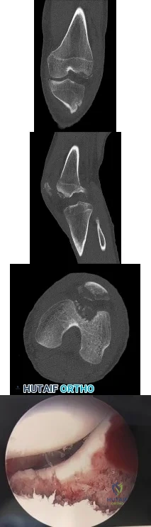
Explanation
Question 39
A 79-year-old woman reports a history of left leg pain with walking. Her pain is exacerbated with walking and stair climbing, and her symptoms are improved by standing after she stops walking. Lumbar flexion does not provide any significant improvement of the symptoms and sitting does not significantly change symptoms. Her leg pain is worse at night and she obtains relief by hanging her leg over the side of the bed. The neurologic examination is essentially normal. Examination of the lower extremities demonstrates mild early trophic changes, and her pulses distally are palpable but are diminished bilaterally. Radiographs are shown in Figures 15a and 15b. What is the next most appropriate step in management?
Explanation
REFERENCES: Aufderheide TP: Peripheral arteriovascular disease, in Rosen P, Barkin R (eds): Emergency Medicine: Concepts and Clinical Practice, ed 4. St Louis, MO, Mosby, 1998,
pp 1826-1844.
Mirkovic S, Garfin SR: Spinal stenosis: History and physical examination. Instr Course Lect 1994;43:435-440.
Question 40
Figure 16 shows the radiograph of a 23-year-old man who has severe right shoulder pain after his motorcyle hit a bridge guardrail. He is neurologically intact. Nonsurgical management will most likely result in
Explanation
REFERENCES: Egol KA, Connor PM, Karunakar MA, Sims SH, Bosse MJ, Kellam JF: The floating shoulder: Clinical and functional results. J Bone Joint Surg Am 2001;83:1188-1194.
Williams GR Jr, Naranja J, Klimkiewicz J, et al: The floating shoulder: A biomechanical basis for classification and management. J Bone Joint Surg Am 2001;83:1182-1187.
Edwards SG, Whittle AP, Wood GW: Nonoperative treatment of ipsilateral fractures of the scapular and clavicle. J Bone Joint Surg Am 2000;82:774-779.
Question 41
What clinical finding is associated with the least favorable prognosis in an adolescent patient who has been diagnosed with a high-grade osteosarcoma of the distal femur?
Explanation
REFERENCE: Mirra JM: Bone Tumors: Clinical, Radiologic, and Pathologic Correlation. Philadelphia, PA, Lea and Febiger, 1989, pp 344-350.
Question 42
A 45-year-old woman with a long-standing history of diabetes mellitus has a large draining plantar ulcer of the right foot. Examination reveals some local cellulitis and erythema surrounding the ulcer. A clinical photograph is shown in Figure 7. Based on these findings, what is the most appropriate antibiotic?
Explanation
REFERENCES: Pinzur MS, Slovenkai MD, Trepman E: Guidelines for diabetic foot care. Foot Ankle Int 1999;20:695-702.
Eckman MH, Greenfield S, Mackey WC, et al: Foot infections in diabetic patients: Decision and cost-effectiveness analyses. JAMA 1995;273:712-720.
Question 43
Percutaneous placement of a lateral proximal tibial locking plate that extends down to the distal third of the leg is associated with postoperative decreased sensation of which of the following distributions?

Explanation
Question 44
A 72-year-old woman who was doing well after undergoing total shoulder arthroplasty for arthritis 4 months ago is suddenly unable to elevate her arm. Examination reveals 70 degrees of external rotation compared with 45 degrees on the uninvolved side, and she is unable to lift her hand off her lower back. Radiographs are shown in Figures 43a through 43c. Treatment should consist of
Explanation
REFERENCES: Moeckel BH, Altchek DW, Warren RF, Wickiewicz TL, Dines DM: Instability of the shoulder after arthroplasty. J Bone Joint Surg Am 1993;75:492-497.
Gerber C, Hersche O, Farron A: Isolated rupture of the subscapularis tendon. J Bone Joint Surg Am 1996;78:1015-1023.
Question 45
A 10-year-old girl with a monoarticular pattern of juvenile rheumatoid arthritis (JRA) has had a 3-cm limb-length discrepancy since age 8 years when inflammation in the right knee came under good medical control. Because her right leg is longer, the patient states that she would like her legs to be close to equal in length in the future. A growth-remaining chart is shown in Figure 14. Management should consist of
Explanation
REFERENCES: Simon S, Whiffen J, Shapiro F: Leg-length discrepancies in monoarticular and pauciarticular juvenile rheumatoid arthritis. J Bone Joint Surg Am 1981;63:209-215.
Ansell BM, Bywaters EGL: Growth in Still’s disease. Ann Rheum Dis 1956;15:295-319.
Question 46
A football player injures his knee when he is tackled and falls awkwardly. He does not note any discreet “pop,” but pain prevents him from returning to the game. An effusion is noted the following day and an MRI scan is ordered. Selected images are shown in Figures 1 through 3. Based on these images, physical examination findings likely include
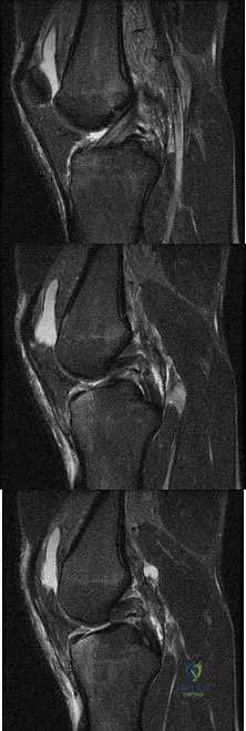
Explanation
Question 47
-Assuming that the lesion can be covered appropriately and there is no drainage from the lesion, when should the patient be allowed to safely return to wrestling?
Explanation
This patient has cellulitis, which is typically caused by group A Streptococcus or Staphylococcus. The patient’s lack of improvement with first-line antibiotics is concerning for methicillin-resistant Staphylococcus aureus (MRSA) infection. MRSA cellulitis is becoming more prevalent in young athletes,and a high index of suspicion is required to provide appropriate intervention during this
aggressive disease process. The diagnosis is typically made clinically without the use of cultures. Oral trimethoprimsulfamethoxazole (a sulfonamide-class drug) double strength twice daily for 10 to 14 days or doxycycline (a tetracycline-class drug) 100 mg twice daily for 10 to 14 days are recommended for first-line treatment of suspected MRSA cellulitis. There is no indication to proceed with irrigation and debridement; however, if the patient develops a soft-tissue abscess or the underlying joint becomes involved, this would be an appropriate intervention. Switching the athlete to an IV cephalosporin (cefazolin) is not likely to be effective against the presumed resistant bacteria.
Ciprofloxacin (a fluoroquinolone-class drug) is effective against many bacteria, but not MRSA. The current recommendation for wrestlers with cellulitis is that return to competition be allowed after 72 hours of antibiotic treatment if there has been no extension of the cellulitis for 48 hours, the lesion can be covered, and there is no drainage from the lesion. The other responses are not current recommendations for return to competition.
Question 48
Figures 21a and 21b show the radiograph and CT scan of a 14-year-old patient with thigh pain. The next most appropriate step in management should consist of
Explanation
REFERENCES: Frassica FJ, Waltrip RL, Sponseller PD, Ma LD, McCarthy EF Jr: Clinicopathologic features and treatment of osteoid osteoma and osteoblastoma in children and adolescents. Orthop Clin North Am 1996;27:559-574.
Kneisl JS, Simon MA: Medical management compared with operative treatment for osteoid osteoma. J Bone Joint Surg Am 1992;74:179-185.
Rosenthal DI, Hornicek FJ, Wolfe MW, Jennings LC, Gebhardt MC, Mankin HJ: Percutaneous radiofrequency coagulation of osteoid osteoma compared with operative treatment. J Bone Joint Surg Am 1998;80:815-821.
Question 49
A 21-year-old man who was injured in a snowboarding accident 18 months ago now reports wrist pain. An MRI scan is shown in Figure 37. Based on the image findings, what is the most likely diagnosis?
Explanation
REFERENCE: Perlik PC, Guilford WB: Magnetic resonance imaging to assess vascularity of scaphoid nonunions. J Hand Surg Am 1991;16:479-484.
Question 50
Figures 34a and 34b show the axial and sagittal MRI scans of a 36-year-old man who reports the insidious onset of pain in the right shoulder. What is the most appropriate description of the acromial morphology? Review Topic
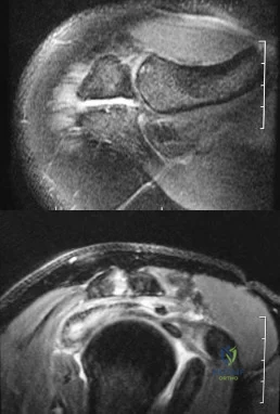
Explanation
Question 51
An otherwise healthy year-old woman has pain 2 years after total hip arthroplasty. The clinical photograph in below demonstrates her skin envelope, and associated radiograph. Her C-reactive protein level is normal, and her erythrocyte sedimentation rate is mildly elevated. The white blood cell count is normal. Hip aspiration attempted under fluoroscopy generates no fluid. What is the best definitive treatment?
Explanation
This patient clearly has a chronically infected total hip arthroplasty, indicated by the open, draining sinus, as well as trochanteric bone resorption on radiographs, and two years of pain. Recently, specific guidelines have been published to better help the clinician define infection. Repeating the hip aspiration is unnecessary, because infection is already evident. Initiating a wound care consult would not address the underlying infection. The determination whether to retain the components or perform a two-stage exchange is based more on the acuity of infection. In this particular case, the patient is chronically infected. Irrigation and debridement with a liner exchange and retention of the components are reserved for the acute setting.
Question 52
A 31-year-old male sustains an ipsilateral displaced transverse acetabular fracture, pubic rami fractures, and a sacroiliac joint dislocation. What structure should be reduced and stabilized first?

Explanation
The referenced article by Matta reviewed 259 patients with acetabular fractures treated within 21 days of injury and found that the hip joint can be preserved and post-traumatic osteoarthrosis can be avoided if an anatomical reduction is achieved.
Question 53
A 20-year-old male lacrosse player sustains an anterior dislocation of the shoulder. He is extremely concerned about recurrent dislocations. Which of the following treatments has been shown to reduce the risk of recurrent dislocation?
Explanation
REFERENCES: Itoi E, Hatakeyama Y, Kido T, et al: A new method of immobilization after traumatic anterior dislocation of the shoulder: A preliminary study. J Shoulder Elbow Surg 2003;12:413-415.
Itoi E, Sashi R, Minagawa H, et al: Position of immobilization after dislocation of the glenohumeral joint: A study with use of magnetic resonance imaging. J Bone Joint Surg Am 2001;83:661-667.
Question 54
During an anterior approach to the bicipital 53 tuberosity, you encounter a nerve overlying the brachioradialis fascia (Figure 58). It provides innervation to the
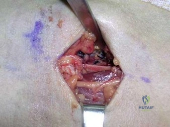
Explanation
The structure shown is the lateral antebrachial cutaneous nerve (LABC). It is the terminal sensory branch of the musculocutaneous nerve and runs superficial to the brachioradialis. It supplies sensation to the anterolateral surface of the forearm. The flexor pollicis longus is innervated by the anterior interosseous nerve. The extensor indicis proprius is innervated by the radial nerve. The LABC does not innervate the skin of the anteromedial forearm. Careful
identification and protection of this nerve is critical to prevent the most common nerve injury during distal biceps repair.
RECOMMENDED READINGS
Agur AM. Grant's Atlas of Human Anatomy. 10th ed. Philadelphia, PA: Lippincott Williams & Wilkins; 1999:460.
Hoppenfeld S, deBoer P. Surgical Exposures in Orthopedics. 2nd ed. Philadelphia, PA: Lippincott Williams & Wilkins; 1994:118-125.
RESPONSES FOR QUESTIONS 59 THROUGH 61
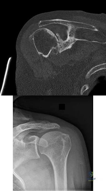
Please select the image that represents the most appropriate response to the question or statement below.
Question 55
A 60-year-old man with diabetes mellitus is referred for evaluation of nonhealing ulcers of his left foot. Nonsurgical management has failed to provide relief, and a below-the-knee amputation is being considered. Which of the following studies best predicts successful amputation wound healing?
Explanation
REFERENCES: Wyss CR, Harrington RM, Burgess EM, et al: Transcutaneous oxygen tension as a predictor of success after amputation. J Bone Joint Surg Am 1988;70:203-207.
Dwars BJ,van den Broek TA, Rauwerda JA, et al: Criteria for reliable selection of the lowest level of amputation in peripheral vascular disease. J Vasc Surg 1992;15:536-542.
Question 56
Figure 48 shows an MRI scan of the knee. The arrow is pointing to what structure?
Explanation
REFERENCES: Gray H: Anatomy of the Human Body. Philadelphia, PA, Lea and Febiger, 1918, 2000.
Netter FH: Atlas of Human Anatomy. Summit, NJ, Ciba-Geigy, 1989, pp 464-465.
Question 57
50%
Explanation
Enneking et al. conducted both radiographic and histologic studies of sixteen massive retrieved human allografts four to sixty-five months after implantation. Analysis of the articular cartilage revealed no evidence that any chondrocytes had survived, even when the graft had been cryoprotected before it was preserved by freezing.
A 64-year-old woman with a longstanding history of rheumatoid arthritis complains of finger dysfunction for the past 6 months. Figure A displays her hand during active extension of all fingers. Figure B displays her hand maintaining her fingers extended following passive extension. What is the next most appropriate treatment for the ring finger?
Spiral oblique retinacular ligament reconstruction
Sagittal band reconstruction
Lateral band reconstruction
Central slip reconstruction
Triangular ligament and transverse retinacular ligament reconstruction
Sagittal band disruption is often associated with rheumatoid arthritis. When this patient attempts to actively extend the affected digit, the extensor tendon
subluxates ulnarly as a result of the sagittal band rupture, and is left with an extensor lag. If one passively extends the finger fully, the patient is able to maintain this position, as the tendon is intact.
Sagittal band reconstruction can be performed with Watson's technique of creating a distally based tendon graft harvested from the central third of the extensor tendon, passed deep to the intermetacarpal ligament and sutured back to itself. Illustration A depicts an intraoperative view of the tendon before reconstruction and Illustration B displays tendon following sagittal band reconstruction. Illustration C displays all of the anatomic locations
of the options listed above.
A splenectomy is performed in a 7-year-old boy following a motor vehicle accident. All of the following are recommended for long-term management EXCEPT:
Pneumococcal vaccination
Haemophilus influenza type B vaccination
Meningococcal group C vaccination
Lifelong prophylactic antibiotics
Hepatitis A vaccination
All of the responses are correct except the need for Hepatitis A vaccine. Hepatitis A is a virus with tropism for hepatocytes which causes infection from fecal-oral contaminated food/water, and shows no increased rate of either infectivity or morbidity in patients with hyposplenism.
Basic recommendations for splenectomized patients include:
All splenectomized patients and those with functional hyposplenism should receive pneumococcal immunization.
Patients not previously immunized should receive haemophilus influenza type B vaccine.
Patients not previously immunized should receive meningococcal group C conjugate vaccine.
Influenza immunization should be given.
Lifelong prophylactic antibiotics are still recommended (oral phenoxymethylpenicillin or erythromycin). This is seemingly despite lack of good data demonstrating a role for lifelong chemoprophylaxis and the acknowledgement that long-term compliance may be problematic.
Davies et al review the current level of evidence supporting these guidelines
for infection prevention in patients with hyposplenism. New to these guidelines are issues regarding occupational exposure and the use of the meningococcal group C and the seven-valent pneumococcal vaccine in non-immunized hyposplenic patients.
Gandhi et al evaluated their nonoperative management of blunt splenic injury in pediatric trauma care. They found compared to historical controls, children with blunt splenic injuries who were hemodynamically stable could be safely monitored with a protocol which required 4 days of inpatient care, 3 weeks of quiet home activities, and 3 months of light activity. This protocol seems to allow for safe return to unrestricted activity.
Incorrect Answer:
Ligaments attach to bone by both direct insertion and indirect insertion. Which of the following most accurately describes the order of the four transition zones of direct insertion?
Ligament > fibrocartilage > mineralized fibrocartilage > bone
Ligament > mineralized fibrocartilage > fibrocartilage > bone
Ligament > mineralized fibrocartilage > periosteum > bone
Ligament > Sharpey's fiber > periosteum > bone
Ligament > periosteum > fibrocartilage > bone
There are two types of tendon/ligament insertion into bone: direct and indirect insertion. The more common, indirect insertion, occurs when the superficial ligament fibers insert into the periosteum. Direct insertion of tendon/ligaments
into bone occurs through a transition of 4 distinct phases: 1) ligament, 2) fibrocartilage, 3) mineralized fibrocartilage, and 4) bone.
While flexing the elbow to perform a biceps curl, what type of muscle contraction is occuring?
Isometric
Isokinetic
Plyometric
Eccentric
Concentric
Concentric muscle contractions occur when a muscle shortens during contraction, as in the upward motion when performing a biceps curl. An eccentric contraction occurs when a muscle lengthens with contraction, as in the "negative" or lowering motion of a biceps curl. An example of an isometric (muscle contracts while maintaining constant length) contraction would be pushing against an immovable object. An example of an isokinetic (muscle has constant speed of contraction) occurs with specialized equipment like Cybex machines. Plyometric contractions occur when a muscle rapidly lengthens just prior to contraction - like during repetitive box jumping.
Woo and Buckwalter describe the mechanisms, barriers, and molecular processes involved in ligament and tendon injury and repair.
A 34-year-old laborer has her left foot crushed in a piece of farming equipment as shown in Figure A. All of the following are reasons for a poor outcome following a crush injury to the foot EXCEPT:
Workers compensation injury
Development of reflex sympathetic dystrophy (complex regional pain syndrome)
Delayed soft-tissue coverage in mangled extremities
Immediate skeletal stabilization
Ongoing litigation
This patient has a mangled extremity. Rigid skeletal stabilization is recommended to enhance soft-tissue healing.
Level 4 evidence from Myerson et al found that delayed soft-tissue coverage in mangled extremities correlated with poor outcome. Poor results also occurred
if treatment was not immediately initiated (immediate debridement shown in Illustration A), if patients subsequently had neuritis or reflex sympathetic dystrophy, or if patients were involved in ongoing workers' compensation and litigation. Neuroischemia following substantial soft-tissue injury likely plays a role in the development of chronic pain after crush injuries to the foot, either through direct trauma to the peripheral nerves or by intraneural or extraneural fibrosis. This trauma to the nerve may cause chronic neuritis, which then triggers a sympathetically mediated reflex sympathetic dystrophy (complex regional pain syndrome).
A 65-year-old man undergoes total knee replacement and is found to have deep vein thrombosis two days later. What molecule is
thought to be involved in this process when it is released during surgical dissection?
Prothrombin
RANKL
IL-1b
Thromboplastin
Factor XI Corrent answer: 4
Thromboplastin is also known more commonly as Tissue Factor (TF), which is involved in the Extrinsic Pathology of the coagulation cascade.
During surgical dissection, insults occur to the endothelial walls of blood vessels. There are three ways in which the body reacts to form a clot so that the patient does not bleed excessively. One is via vessel contraction, another is by collagen release, and a third is by tissue thromboplastin release. Thromboplastin release is part of the extrinsic coagulation pathway (see
Illustration A). Thromboplastin release activates Factor VII which activates Factor X which converts prothrombin to thrombin. Thrombin is the catalyst for converting fibrinogen to fibrin which induces clot formation. While this is useful for decreasing bleeding, it is the same mechanism by which a deep venous thrombosis (DVT) develops.
Which of the following materials has a Young's modulus of elasticity that is most similar to cortical bone
Titanium
Zirconia
Stainless steel
Ceramic (Al2O3)
Alloy (Co-Cr-Mo)
Of the materials listed Titanium has an elastic moduli closest to cortical bone. Titanium is extra-ordinarily light, strong, highly ductile, and corrosion resistant. Titanium is however very notch sensitive and has poor wear resistance.
Young Modulus of Elasticity is defined as the stiffness (ability to maintain
shape under external loading) of a material. On the stress vs. strain curve it is defined as the slope of the line in the elastic zone (see Illustration A). Young’s modulus is constant and different for each material. The relevant moduli (unit GPa) are approximated below:
Question 58
006%-3.4 %. The typical skin flora includes staph and strep as well as P. acnes, which has a propensity for the shoulder. Because it is an anaerobic organism, cultures may only become positive after 7-21 days.
Explanation

A 47-year-old, healthy, active patient presents with a sub-acute, full-thickness supraspinatus tear. His physical examination reveals significant weakness and pain with abduction. There was no glenohumeral instability. Radiographs demonstrate a type 1 acromion. An MRI scan shows a crescent shaped tear with 2-cm of tendinous retraction and no tendinous fatty changes. A subacromial corticosteroid injection 6 weeks ago provided him with 24 hours of pain relief but no improvement in strength. What would be the most appropriate treatment option?
Repeat subacromial corticosteriod injection

Biological augmentation of rotator cuff with porcine small intestine xenograft Rotator cuff repair

Rotator cuff repair plus acromioplasty

Rotator cuff repair, remplissage procedure, bicep tenodesis and distal clavicle excision
This patient has an isolated supraspinatus rotator cuff tear with symptomatic weakness. The most appropriate treatment would be isolated rotator cuff repair.
The primary purpose of rotator cuff repair is to restore muscle function. Secondary outcomes include reduction of pain and prevention of irreversible cuff changes, specifically muscular atrophy. Non-operative treatment ( exercise, therapy and pain medications) are recommended for partial thickness tears. The indication of surgical repair includes, isolated supraspinatus weakness +/- pain
that correlates with MRI imaging of a respective full thickness tear. Routine acrominoplasty is not recommended in conjunction with rotator cuff repair, especially with no previous symptoms of impingement.
Pedowitz et al. developed clinical practice guidelines for the treatment of rotator cuff pathology. The strongest supporting evidence in current literature was given a grade of 'moderate' with four treatment recommendations. These were,
Exercise and non-steroidal anti-inflammatory drugs can be used to manage partial thickness tears,

Routine acromioplasty is not required the time of cuff repair,

Non-cross-linked, porcine small intestine submucosal xenograft patches should not be used to manage cuff tears, and

Surgeons can advise patients that workers' compensation status correlates with a less favorable outcome after rotator cuff surgery.
Illustration A shows the different shapes of rotator cuff tears. Incorrect Answers:
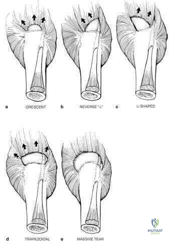
A 12-year-old baseball pitcher describes progressive worsening of medial elbow pain on
his throwing side. Examination reveals normal elbow range of motion. He is tender over the medial elbow to palpation. A dynamic ultrasound of his elbow shows no evidence of medial widening with valgus stress. His radiograph is shown in Figure A and an MRI is shown in Figure B. What is the most likely cause of his symptoms?
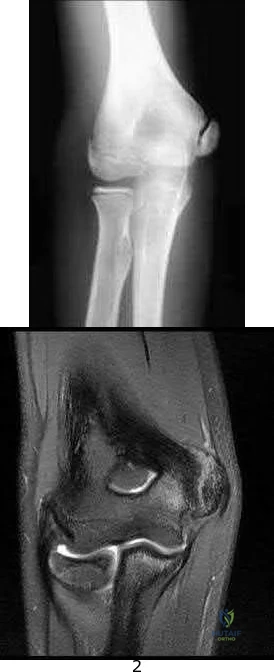
Displaced medial epicondyle avulsion fracture Medial apophysitis

Medial ulnar collateral ligament tear

Valgus extension overload with olecranon osteophytes Ulnar neuritis
The clinical presentation is consistent with Little League Elbow caused by medial apophysitis. Little League elbow is a general term explaining medial elbow pain in adolescent pitchers. The underlying pathology can include medial epicondyle stress fractures, avulsion fractures of the medial epicondyle, ulnar collateral ligament (UCL) injuries, or medial epicondyle apophysitis. In order to identify the underlying cause it is important to first rule out injury to the MCL by looking for medial widening on stress radiographs or dynamic ultrasound, or valgus instability on physical exam. Radiographs are useful to look for avulsion fractures or subtle physeal widening commonly seen with apophysitis.
Wei et al. obtained radiographs and magnetic resonance imaging on nine adolescent pitchers with a clinical diagnosis of Little League Elbow. They found radiographic findings in 4/9 and MRI findings in 6/9 patients. They emphasized that the MRI did not change management in any patients. Cain et al. review the different elbow conditions seen in throwing athletes. They emphasize the need to understand the underlying pathophysiology in order to treat and make appropriate changes to the biomechanics of the pitching technique.
Figure A shows an AP radiograph with slight widening of the apophysis, but no evidence of avulsion fracture. Figure B is an MRI which shows signal consistent with edema of the medial epicondyle apophysis.
Incorrect Answers:
The other responses are all typical throwing elbow conditions, but are much less common than apophysitis in the adolescent thrower.
What is the primary function of the structure labeled with an asterisk in Figure A?
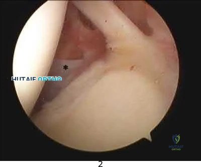
Prevents inferior translation of the humerus with the arm by the side Provides internal rotation of the humerus

Prevents anterior translation of the humerus with the arm in 45 degrees of abduction Prevents anterior translation of the humerus with the arm in 90 degrees of abduction Provides supination of the forearm and elbow flexion
The labeled structure is the middle glenohumeral ligament (MGHL) of the shoulder. The primary function of the MGHL is to prevent anterior translation of the humeral head with the arm in 45-60 degrees of abduction.
This structure originates from the glenoid labrum and inserts medial to the lesser tuberosity running obliquely across the subscapularis. The size of the structure may be variable and there are recognized normal anatomic variants ( including a cord like MGHL in the Buford complex). It is important to be able to recognize the MGHL and differentiate this from the subscapularis, IGHL, SGHL, and other intraarticular structures in the shoulder to be able to perform effective and precise arthroscopic procedures.
Burkhart et al. describe the function of the glenohumeral ligaments in anterior shoulder instability, noting that the MGHL provides a restraint to anterior translation with the arm in 45-60 degrees of abduction.
Wang et al. discuss microdamage to the inferior glenohumeral ligament from a basic science perspective, indicating that over time it may stretch and compromise it's function in restraining humeral translation.
Figure A is an arthroscopic image of the intraarticular structures of the shoulder with an asterisk on the MGHL.
Incorrect Answers (these are labeled on Illustration A, with the exception of the subscapularis which is difficult to visualize):

In which of the following clinical circumstances would it be appropriate to eccentrically ream the anterior glenoid?

year-old male undergoing a shoulder arthroplasty due to rotator cuff arthropathy 65-year-old female with a glenoid retroversion of 13-degrees undergoing shoulder arthroplasty

year-old female with humeral anteversion of 13-degrees undergoing shoulder arthroplasty

year-old female with glenoid retroversion of 25-degrees undergoing shoulder arthroplasty

year-old male with significant glenoid bone stock deficiency and severe osteoarthritis
The surgeon should consider eccentrically reaming the anterior glenoid when performing a total shoulder arthroplasty on a patient with a retroverted glenoid due to posterior deficiency associated with osteoarthritic changes which is most consistent with answer choice #2.
Normal version of the glenoid is 0-3 degrees of retroversion, but when doing a total shoulder the goal should be to place the glenoid component in neutral to slight anteversion. Reaming the anterior glenoid to neutral is a technique to be considered by the operative surgeon when presented with a patient undergoing total shoulder arthroplasty with a retroverted glenoid, as failure to perform this step increases the chance for glenoid loosening. If reaming down the anterior glenoid will take away too much bone stock (down to the coracoid process), one may consider bone grafting the posterior glenoid. To perform a total shoulder arthroplasty patients will need a functioning rotator cuff and appropriate glenoid bone stock.
Clavert et al. performed cadaveric analysis to simulate glenoid retroversion of greater than 15 degrees and found that retroversion to this degree cannot be safely corrected with eccentric anterior reaming when using a glenoid component with peripheral pegs due to penetration into the glenoid vault.
Nowak et al. used 3D-CT models of patients with advanced shoulder osteoarthritis with varying degrees of glenoid retroversion and simulated glenoid resurfacing. They found that smaller size glenoid components may allow for greater version correction when using in-line pegged components, as they would be less likely to result in peg penetration.
Illustration A shows >25 degrees of glenoid retroversion seen by axial radiograph of the shoulder in a patient with advanced osteoarthritis. In this case, anterior glenoid reaming is not the correct answer and a posterior glenoid allograft reconstruction would be appropriate.
Incorrect Answers:
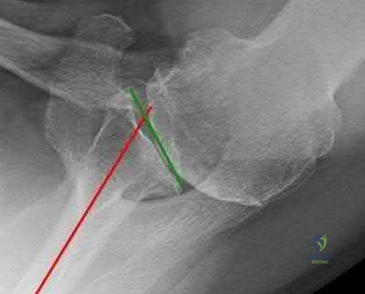
A 44-year-old left-hand dominant carpenter experienced immediate left elbow pain after trying to stop a heavy object from falling two days ago. Figure A shows a clinical image of the patient upon presentation. Physical exam shows full strength with wrist flexion, wrist extension, and pronation, but notable weakness with supination of the forearm. Sensory exam shows no deficits in the forearm or hand. There is a negative milking maneuver test and a positive hook test. Radiographs are shown in Figure B. What is the next most appropriate step in management?
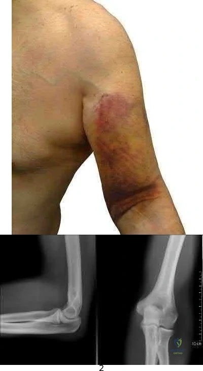
Sling use as needed for comfort and progressive physical therapy Allograft reconstruction of the distal biceps tendon

Ulnar collateral ligament reconstruction Distal biceps tendon avulsion repair Brachioradialis and ECRB avulsion repair
Distal biceps tendon avulsion repair is the most appropriate next step in management.
Distal biceps tendon ruptures occur most commonly in middle-aged men and usually involve the dominant extremity. The mechanism of injury is usually a single traumatic event with eccentric force on the flexed elbow.
Sutton et al. authored a Level 5 review of distal biceps tendon ruptures. They discuss that nonsurgical management of distal biceps tears is appropriate in the low-demand or medically ill patient. Surgical repair improves elbow flexion strength by 30% and supination strength by 40% compared to nonoperative management.
O'Driscoll et al. conducted a Level 2 study examining the accuracy of the hook test for distal biceps rupture diagnosis. They found that the hook test was abnormal in 33 of 33 (100%) patients with complete biceps avulsions, and intact in 12 of 12 (100%) with partial detachments.
Figure A is a clinical image demonstrating ecchymosis in the distal arm and antecubital fossa. Figure B shows normal elbow radiographs. Illustration A shows a normal hook test with an intact distal biceps insertion.
Incorrect Answers:
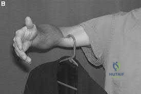
Early reverse total shoulder designs (before the development of the Grammont-style prosthesis) had a high failure rate due to early loosening of the glenoid component. What biomechanical feature accounted for this problem?

Glenoid component did not have a neck Humeral component too horizontal Center of rotation too lateral

Center of rotation too anterior Center of rotation too inferior
Early reverse ball-and-socket designs failed because their center of rotation remained lateral to the scapula, which limited motion and produced excessive torque on the glenoid component, leading to early loosening. The first modern reverse prosthesis was designed by Paul Grammont. According to Boileau et al., Grammont's design "introduced 2 major innovations (1) a large glenoid hemisphere with no neck and (2) a small humeral cup almost horizontally oriented with a nonanatomic inclination of 155 degrees, covering less than half of the glenosphere. This design medializes the center of rotation compared to earlier versions which minimizes torque on the glenoid component. Furthermore, the humerus is lowered relative to the acromion, restoring and even increasing deltoid tension. The Grammont reverse prosthesis imposes a new biomechanical environment for the deltoid muscle to act, thus allowing it to compensate for the deficient rotator cuff muscles." According to Gerber, "moving the center of rotation more medial and distal as well as implanting a large glenoid hemisphere that articulates with a humeral cup in 155 degrees of valgus are the biomechanical keys to sometimes spectacular short- to midterm results".
Which of the following preoperative factors is a contraindication to total shoulder arthroplasty?

Passive external rotation less than 10 degrees Eccentric posterior glenoid erosion

A 2-cm full-thickness supraspinatus tendon tear Inflammatory arthritis

A preganglionic brachial plexus injury
A preganglionic brachial plexus palsy, otherwise known as a root avulsion injury, presents with a flail arm and has a poor prognosis for recovery of motor function. Patients with brachial plexus palsies are not candidates for total shoulder arthroplasty due to the substantial motor and sensory deficits associated with these injuries.
In contrast, patients with a preoperative loss of passive external rotation, posterior glenoid erosion, a reparable full-thickness rotator cuff tear isolated to the supraspinatus tendon, and inflammatory arthritis are not contraindicated for a total shoulder arthroplasty.
Iannotti et al. performed a Level I prospective study in 118 patients who underwent either a total shoulder arthroplasty or a shoulder hemiarthroplasty for primary osteoarthritis. The presence of a reparable full-thickness rotator cuff tear did not adversely affect outcomes in either group but rather provided better active external rotation in the cohort receiving total shoulder arthroplasties. The authors concluded that a reparable tear of supraspinatus is not a contraindication to the use of a glenoid component.
Norris et al. compared outcomes of total shoulder arthroplasty and hemiarthroplasty performed for primary osteoarthritis in 160 patients. There were no differences in postoperative pain, function, ASES scores, or range of motion between groups for patients with reparable rotator cuff tears. The authors concluded that minor thinning and small tears of the rotator cuff can be adequately addressed at the time of surgery without adversely affecting outcomes.
Illustration A is a cervical T2 axial MRI which shows a cervical root avulsion, a form of preganglionic brachial plexus injury. Notice the perineural hyperintensity.
Incorrect Answers:
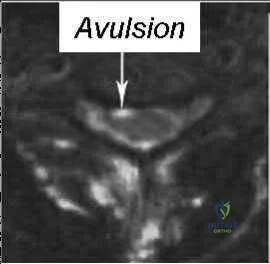
A 42-year-old male sustains a flail chest injury and subsequently undergoes operative stabilization of his chest wall. At first follow-up, the inferior angle of his ipsilateral scapula translates medially with any attempt at overhead activity. Injury to which of the following structures would cause this abnormality?

Spinal accessory nerve C8 and T1 nerve roots

Upper and lower subscapular nerves Thoracodorsal nerve

Long thoracic nerve
The clinical vignette describes medial scapular winging, which is seen after injury to the long thoracic nerve.
Medial scapular winging due to a long thoracic nerve palsy can be seen after repetitive stretching in athletes, with direct compression injury, or even iatrogenically during surgical procedures to the lateral thorax. Injury to the long thoracic nerve will eliminate the function of the serratus anterior,
which acts to protract the scapula laterally and upward and stabilize the vertebral border of scapula. This results in upper extremity weakness in forward elevation or abduction as the scapula is not stabilized against the thorax.
Meininger et al. report that lesions of the long thoracic nerve and spinal accessory nerves are the most common cause of scapular winging, although numerous underlying etiologies have been described. They report patients describe diffuse neck pain, shoulder girdle discomfort, upper back pain, and weakness with abduction and overhead activities. They also report that most cases are treated nonsurgically.
Wiater et al. review injuries to the spinal accessory nerve which causes dysfunction of the trapezius and subsequent lateral scapular winging. They note that the superficial course of the spinal accessory nerve in the posterior cervical triangle makes it susceptible to injury, and iatrogenic injury to the nerve after a surgical procedure is one of the most common causes of trapezius palsy. Most injuries are treated nonoperatively, but the Eden-Lange procedure, in which the insertions of the levator scapulae, rhomboideus minor, and rhomboideus major muscles are transferred, relieves pain, corrects deformity, and improves function in patients with irreparable injury to the spinal accessory nerve.
Illustration A shows a clinical photo of medial scapular winging, while illustration B shows a clinical photo of lateral scapular winging. Illustration C shows the long thoracic nerve during a rib fixation procedure, with the nerve sitting directly on top of the serratus anterior. The trapezius is overlying the scapula at the bottom of the photo, and the patient's head is to the right of the photo. Incorrect Answers:
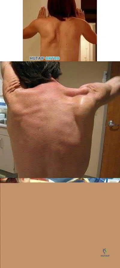
A patient sustains a distal biceps brachii tendon rupture. If treated non-operatively, the greatest loss of strength would be seen with which activity?

Forearm supination Forearm pronation

Elbow flexion

Shoulder forward flexion Shoulder internal rotation
While both elbow flexion and forearm supination strength are affected, there is a greater percentage loss of supination strength. Patients may complain of weakness and fatigue with rotational activities such as using a screwdriver. The primary elbow flexor is actually the brachialis, and therefore less weakness in flexion is reported.
Patterson reviewed distal biceps ruptures and found nonsurgical treatments had 21 55% loss of supination strength and 8 36% loss of flexion strength.
Klonz reviewed anatomic and non-anatomic repairs and found better results with anatomic repairs with 91% return of supination strength and 96% return of flexion strength. Supination strength after nonanatomic repair did not improve in 4 of 8 patients (42%-56% of the uninjured arm).
A 27-year-old right hand dominant construction worker falls off a scaffold onto his outstretched arm. Figure A exhibits the radiograph taken at a local emergency room.
Following treatment, he is placed in a sling and follows up at your office two weeks later. He complains of a feeling that his arm is going to 'pop out'. Which specific physical examination finding is likely to be present?
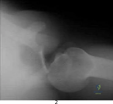
Hornblower's Test Jobe's Test

Apprehension Sign with shoulder abducted and externally rotated Speed's Test

Kim's Test
The patient suffered a posterior shoulder dislocation, likely injuring the posterior capsule and/or labrum. Out of all the answer choices, Kim's test assesses posterior structures. Thus, Kim's test is the physical examination finding most likely to be present.
Posterior dislocations occur less frequently than anterior dislocations, and are often missed. Following closed reduction, persistent instability can occur, usually associated with posterior capsular or labral pathology. Posteriorly directed provocative maneuvers, such as the Kim test can be positive.
Robinson et al. performed an epidemiologic analysis on 120 posterior dislocations. Recurrent instability occurred at a rate of 17.7%. Risk factors for recurrent instability included age less than 40-years-old, dislocation during seizure, and a large reverse Hill-sachs (>1.5 cm3). Kim et al. describe the Kim lesion, a separation between the posteroinferior labrum and the articular cartilage without complete detachment of the labrum, which cause persistent posterior instability.
Figure A depicts a posterior dislocation on xray. Illustration A depicts the Kim test, which is performed by having the patient seated, arm at 90° abduction, followed by flexing the shoulder to 45° forward flexion while simultaneously applying axial load on the elbow and posterior-inferior force on the upper humerus. The test is positive when there is pain. Video 1 depicts the proper way to perform a Kim Test.
Incorrect answers:
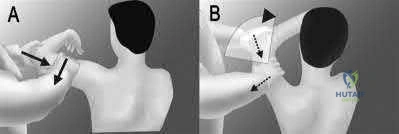
A 27-year-old male bodybuilder presents to the office with vague, deep shoulder pain and weakness with his bench press. His examination is somewhat difficult due to his large size, but no significant abnormal findings are noted. Radiographs are normal, and an MRI arthrogram is shown in Figure A. Which of the following is the most likely etiology of his complaints?
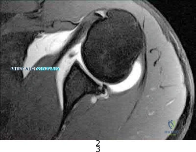
Pectoralis major rupture Supraspinatus partial thickness tear SLAP lesion

Tendonitis of the long head of the biceps Posterior labral tear
The clinical presentation and MRI are consistent with a Posterior labral tear.
Posterior labral tears are commonly seen in individuals that have repeated posteriorly-directed stress across their glenohumeral joint (football linemen, bodybuilders). These patients will often present with ill-described pain deep in their shoulder joint, along with decreases in shoulder strength. Focused shoulder examinations, such as the Jahnke Jerk Test or Push-pull test, can elicit pain from posterior labral tears; however, the sensitivity and specificity of these tests remain under question.
Mair et al. reviewed the outcome of posterior labral injuries in nine athletes who underwent arthroscopic repair with a bioabsorbable tack after failure of conservative management; all were
able to return to contact sports. They note that posteriorly applied forces can result in a shear-type vector that can cause posterior labral tears without capsular injury.
Bradley et al. reviewed 91 athletes with unidirectional recurrent posterior shoulder instability that were treated with an arthroscopic posterior capsulolabral reconstruction. They found that significant improvements in stability, pain, and function at a mean of 27 months postoperatively. Eightynine percent of the patients were able to return to their sport.
Figure A shows an axial MRI arthrogram of the shoulder with a posterior labral tear and an associated paralabral cyst. Illustration A is another axial shoulder MRI arthrogram cut showing a posterior labral tear (red arrow) and an associated paralabral cyst (yellow arrows).
Incorrect Answers:
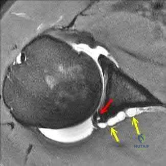
A patient sustains a full thickness tear of their teres minor. Which of the following test/signs would most likely be positive in this patient?

Jobe's test Belly press test

Internal rotation lag sign Hornblower's sign Hawkin's sign
Hornblower's test is completed by asking the patient to hold their shoulder in 90 degrees of abduction and 90 degrees of external rotation. The test is positive if the arm falls into internal rotation or they are unable to actively externally rotate against resistance. This suggests teres minor pathology.
There are various tests/signs used by clinicians to detect rotator cuff pathology. The teres minor is innervated by the axillary nerve and functions to externally rotate the humerus. The hornblower's test/sign has various descriptions, but all act to determine external rotation weakness. In addition to being sensitive and specific for teres minor pathology, it can also be positive with posterior supraspinatus tears.
Walch et al. review 54 patients that underwent repair of combined supraspinatus and infraspinatus rotator-cuff tears. They found that the hornblower's sign was highly sensitive and specific for irreparable degeneration of the teres minor, while the dropping-sign was highly sensitive and specific for irreparable degeneration of the infraspinatus.
Hertel et al. prospectively review 100 patients with painful shoulders and impingement syndrome. They compared various lag signs (ERLS-external rotation lag sign, IRLS-internal rotation lag sign, drop sign) to the Jobe and lift-off signs. The ERLS was less sensitive but more specific than the
Jobe sign for the supraspinatus/infraspinatus. The drop sign was the least sensitive but was as specific as the ERLS. The IRLS was as specific but more sensitive than the lift-off sign for subscapularis tears.
Illustration A shows another variation of the hornblower's sign as originally desbribed by Arthui et

positive if the patient is unable to do this without abducting the affected arm and demonstrates the difficulty in raising the hand to the mouth in the absence of external rotation of the shoulder. The video provided shows how to perform both variations of the hornblower's test.
Incorrect Answers:
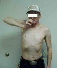
Reverse total shoulder arthroplasty combined with latissimus dorsi transfer would be
most appropriate for which of the following patients?
year-old male with post-traumatic shoulder arthritis after a four-part proximal humerus fracture with no motor dysfunction

year-old male with grade 4 shoulder arthritis with severe deltoid muscle dysfunction secondary to a stroke

year-old female with significant rotator cuff arthropathy, a negative Hornblower sign and less than 5 degrees of external rotation lag

year-old female with pseudoparesis of anterior elevation and external rotation, narrowing of gleno-humeral joint and acetabularization of the acromion

year-old male with grade 4 shoulder arthritis and an isolated supraspinatus tear
Reverse total shoulder arthroplasty combined with latissimus dorsi transfer would be most appropriate in a patient with pseudoparesis of anterior elevation and external rotation, in the setting of shoulder arthritis (narrowing of glenohumeral joint and acetabularization of the acromion).
Combining a latissimus dorsi tendon transfers with reverse total shoulder arthroplasty (R-TSA) helps to restore control of active external rotation. Dysfunction with external rotation can be determined clinically with external rotation lag sign, a positive Hornblower's sign, and radiographically with fatty degeneration of the teres minor classified as stage 2 or greater according to the system of Goutallier et al. or Fuchs et al.
Gerber et al. found that R-TSA with combined lat dorsi transfer yielded minimal improvements in external rotation ROM (13 deg to 19 deg) compared to increases in shoulder ROM in flexion (94 deg to 137 deg) and abduction (87 deg to 145 deg), with this procedure.
Boileau et al. examined 17 consecutive patients treated with reverse shoulder arthroplasty and latissimus dorsi and teres major transfer (L'Episcopo). They found that external rotation increased from -21 degrees to 13 degrees (+34 degrees ). They recommend transferring both the LD and TM, rather than the LD alone as it results in better active external rotation.
Illustration A is a radiograph showing a right reverse total shoulder replacement. Illustration B shows a cadaveric image of the positioning of the latissimus dorsi tendon transfer prior to implantation of the reverse total shoulder components.
Incorrect Answers
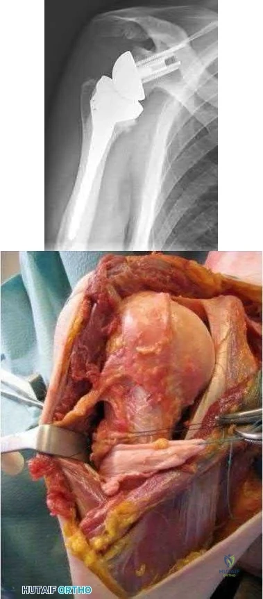
Figure A and B are MRI images of a 42-year-old male with symptoms of right shoulder neuropathy. If this patient has an abnormality detected on EMG and nerve conduction testing, which of the following nerves is most likely to be involved?
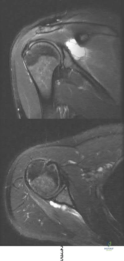
Subscapular nerve Axillary nerve Musculocutaneous nerve Suprascapular nerve Long thoracic nerve
This patient is presenting with suprascapular nerve compression secondary to a spinoglenoid cyst. Injuries of the posterior shoulder joint capsule or posterior-superior labrum can result in spinoglenoid cysts. They may lead to suprascapular nerve palsy.
Patients will present with characteristic findings of external rotation
( infraspinatus) weakness when the cyst is isolated in the spinoglenoid notch. If the cyst is located in the suprascapular notch, both external rotation weakness and abduction (supraspinatus) weakness will be present. Electromyography and MRI are the investigations of choice in depicting the etiology of this mononeuropathy.
Piatt et al. found posterosuperior labral tears in 65/73 patients who had spinoglenoid notch cysts. All patients presented with should pain and weakness. Patients undergoing surgical intervention by drainage or excision +/- arthroscopic labral repair had a better outcome than non-operative care.
Westerheide et al. reported fourteen patients who underwent arthroscopic decompression of ganglion cysts associated with suprascapular neuropathy. All patients had a labral tear intraoperatively with arthroscopic drainage and labral repair. There was not recurrence at an average of 51 months of followup.
Piasecki et al. reviewed suprascapular neuropathy. Causes include:nerve entrapment along this path, particularly at the vulnerable suprascapular and spinoglenoid notch, as well as extrinsic compression by soft-tissue masses.
Figures A is a coronal MRI showing a large hyperintense mass medial to the glenoid articulation. Figure B shows an axial MRI of the lesion posterior to the glenoid. Illustration A shows a diagram of the posterior right shoulder. The suprascapular nerve can be seen traveling through the spinoglenoid notch. Incorrect Answers:
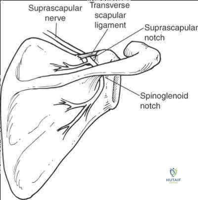
A 12-year-old right-hand-dominant pitcher presents with progressive right shoulder pain. He is now unable to pitch. He is tender to palpation over the lateral shoulder and has pain with rotation. An AP radiograph of the affected shoulder is shown in Figures A and a contralateral radiograph is shown in Figure B. What is the most likely diagnosis?
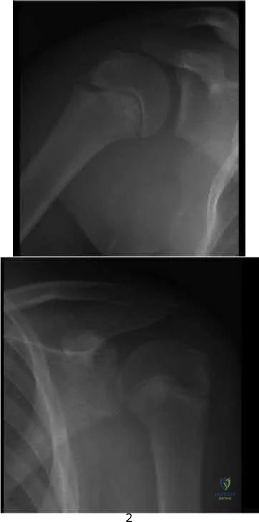
Septic arthritis of the shoulder SLAP tear

External impingement Internal impingement

proximal humerus. Patients may report a recent increase in pitching regimen. On examination, there is focal tenderness at the level of the physis. Treatment focuses on rest, physical therapy and a progressive throwing program. Pitching is often stopped for 2-3 months during rehabilitation.
Chen et al. review shoulder and elbow injuries in the young athlete. Little
Leaguer's shoulder results from epiphyseal lysis secondary to microtrauma. Pain over the anterolateral shoulder may be elicited on examination. The mainstay of treatment is 2-3 months of rest and return to pitching via a progressive throwing program.
Mcfarland et al. review techniques to prevent injuries in the throwing athletes. They note that overuse injures can be avoided when appropriate throwing mechanics are enforced and pitch counts are li
physeal widening noted especially when compared to the contralateral normal pediatric shoulder view seen in Figure B.
Incorrect Answers:
A 35-year-old carpenter has pain in the antecubital fossa that is worse with turning a screwdriver. He has undergone non-operative treatment for 6 months without relief. On physical examination his hook test is normal and there is pain and weakness with resisted supination. Radiographs are shown in Figures A-C. A MRI of the right elbow is shown in Figure D. The next most appropriate treatment is?
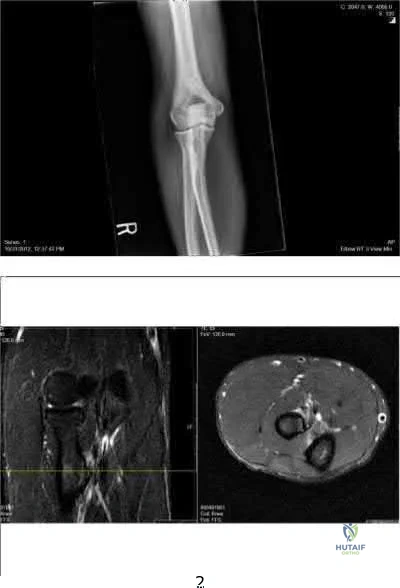
Exploration of the radial tunnel Superficial radial neurectomy

Detachment and repair of the biceps tendon Transfer of the biceps to the brachialis EMG with nerve conduction study
While complete trauamtic rupture of the distal biceps is more common, partial tears have been reported in the literature. The most common presentation is pain in the antecubital fossa worse with resisted supination.
Conservative management consists of NS

Transfer to the brachialis improves flexion strength but not supination.
Ramsey et al present a review article on distal biceps tendon injuries. They state that the most successful management of partial distal biceps tears that have failed conservative management is to surgically treat it like a complete rupture with release and surgical reattachment of the distal biceps to the radial tuberosity.
Figures A-C are normal radiographs of the elbow. Figure D is a crossreferenced axial and coronal T2 MRI that demonstrates increased signal and partial distal biceps tendon tearing. Illustration C shows the resected region of distal biceps tendon in the same patient and had an excellent functional outcome following distal biceps release and surgical reattachment with 2 double-loaded suture anchors.
Video V demonstrates The hook test for detecting complete distal biceps tendon avulsions.
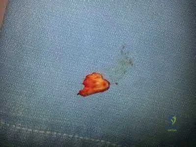
A 49-year-old man sustains a dislocation of his left elbow that is successfully reduced and splinted. He misses his scheduled follow-up appointments and returns 6 weeks later. He is immediately enrolled in a course of vigorous physical therapy. At a repeat visit at 6 months, examination reveals that he lacks 40 degrees of elbow extension, and has flexion to 80 degrees. He is taken to the operating room for surgical release. Figures A and B are diagrams depicting the ligamentous attachments about the elbow. To restore elbow flexion, in addition to releasing the articular capsule, which ligament should be released?
Ligament A Ligament B Ligament C Ligament D Ligament E
In addition to capsular release, the posterior band of the medial collateral ligament (MCL) should be released.
The posterior band of the MCL is attached dorsal to the axis of rotation and has greater variation in length. It increases in length by 9 mm between 60° and 120° of flexion. Posterior band contracture leads to loss of elbow flexion. In contrast, the anterior band of the MCL (AMCL) maintains a constant length ( isometric) throughout the entire arc of movement. Anterior capsule contracture leads to loss of extension.
Wada et al. treated 14 elbows with post traumatic contracture. Through a medial incision, the ulnar nerve was freed and the posterior band and posteromedial joint capsule were excised. Mean flexion increased from 89° preop to 127° postop. Anterior capsulectomy was performed for limited extension.
Morrey et al. studied structures providing stability about the elbow. They found that the anterior capsule stabilizes the elbow to varus-valgus stress in extension, not in flexion. The anterior band of the MCL is a primary stabilizer, especially in flexion.
Figures A and B are medial and lateral illustrations of the elbow, respectively, depicting the ligamentous attachments. Illustrations A and B are radiographs are 3D CT reconstruction images of the left elbow, respectively, showing heterotopic ossification around the posterior band of the MCL.
Incorrect Answers:
A 23-year-old male sustains a dislocation of his elbow that was successfully closed reduced in the emergency room. 3 months later, the patient presents with pain and a catching sensation in his elbow. On physical exam, he is noted to have a positive lateral pivot-shift test. Incompetence of which of the following ligaments in Figure A is most commonly associated with his condition?
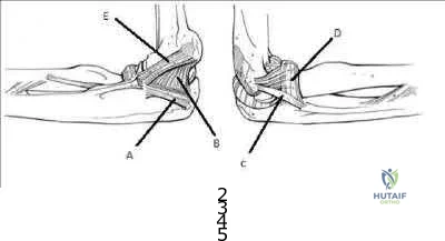
A B C D E
The patient is presenting with symptoms and physical exam consistent with posterolateral rotatory instability. Injury to the lateral ulnar collateral ligament
( LUCL), labeled C in Figure A, allows an abnormal external rotation
( supination) of the ulna on the humerus. This results in posterolateral rotatory instability. Posterolateral rotatory instability often presents as pain and recurrent clicking, snapping, clunking, or locking of the elbow. It should be noted that frank dislocations are not the most common presenting symptom. The physical exam is usually benign except for a positive lateral pivot-shift test or posterolateral rotatory drawer test. While injury to the LUCL is thought to be the primary pathology, other ligamentous stabilizers of the elbow may play a role.
Mehta et al. review posterolateral rotatory instability of the elbow. They state the instability usually results from an elbow dislocation with subsequent failure to heal of the ligamentous structures.
Patients with recurrent instability often require surgical intervention, as bracing is typically cumbersome and ineffective.
The video provided shows how to perform the lateral pivot-shift test. The patient is placed in the supine postion with forearm overhead and elbow extended. The elbow is then supinated with force and flexed to >40° while a valgus load applied. A positive result is palpable / visible clunk as the ulna and radius reduce suddenly. Illustration A shows the posterolateral rotatory drawer test.
External rotation and posterior forces are applied to the forearm attempting to sublux the radius posterior to the capitellum.
Incorrect Answers:
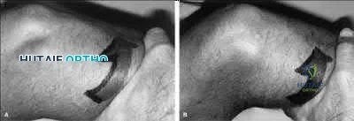
Figure A is the MR image of the left shoulder of an active 47year-old painter who has been experiencing shoulder pain for 9 months. In addition to the finding shown in Figure A, MRI examination of the intra-articular portion of the biceps tendon shows fraying greater than 50%. He has not obtained relief from an 8 month course of non-operative management including non-steroidal antiinflammatory medications, physical therapy and corticosteroid injection. What is the best next step in treatment?
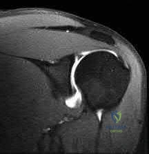
New course of physical therapy

Activity shutdown with 6 weeks sling immobilization

Arthroscopic superior labrum anterior to posterior (SLAP) tear repair Arthroscopic debridement and possible biceps tenotomy versus tenodesis

Arthroscopic rotator cuff repair and acromioplasty
This patient has a Type II SLAP lesion. These should only rarely be repaired in patients older than 40 years of age. If a source of pain refractory to nonoperative management, biceps tenotomy or tenodesis should be considered.
SLAP repair for Type II SLAP lesions is a procedure that has enjoyed a high success rate in young patients. These are generally not indicated for repair in patients greater than 40 years of age due to high rate of stiffness postoperatively. A subset of patients continue to do poorly after SLAP repair. Poor range of motion and the development of post-surgical adhesive capsulitis is often an etiology for poor results. Arthrofibrosis recalcitrant to diligent therapy over many months can be treated with arthroscopic capsular release. This is predicated on failure of a dedicated course of physical therapy as part of a non-operative management course lasting greater than six months. As the propensity for stiffness increases with age, consideration should be treated with SLAP tear debridement and biceps tenotomy or tenodesis in patients greater than 40 years old. Tenotomy or tenodesis, however, can be effective at providing pain relief in the presence of proximal biceps tendon pathology.
Katz et al. reviewed 34 patients who presented to their group for management of failed SLAP repair. 50% were Worker's Compensation cases. The mean age at the time of initial SLAP repair was 43 years. They treated these patients conservatively initially followed by revision surgery in 21 cases. All completed a course of physical therapy initially. They concluded that once a patient has failed SLAP repair, there is a high chance of further conservative treatment failing. Although revision surgery improves outcomes, 32% will continue to have a "suboptimal" result. Holloway et al. reviewed 50 patients who underwent arthroscopic capsular release for adhesive capsulitis, comparing three groups: (1) post-surgical; (2) post-fracture; and (3) idiopathic adhesive capsulitis. All patients had completed supervised physical therapy and a home exercise program for at least one year. They concluded that arthroscopic capsular release improved range of motion equally for all three groups but patients in the post-surgical group had poorer subjective pain, function and satisfaction scores.
Figure A is an MRI showing a Type II SLAP tear. Illustration A shows the classification of SLAP lesions.
Incorrect Answers:
A 30-year-old man undergoes arthroscopic Bankart repair for recurrent anterior dislocation. He continues to experience instability postoperatively. Examination reveals a positive apprehension test. Radiographs of both shoulders are seen in Figure A. CT scan of his left shoulder is seen in Figure B. What is the best treatment option?
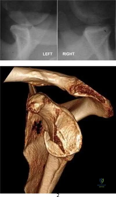
Bankart repair

Humeral head bone augmentation Remplissage

Coracoid autograft Connolly procedure
This patient has anterior glenoid bone deficiency (inverted pear glenoid) from a large bony Bankart lesion that was not adequately addressed in the index procedure. This is best treated with bony augmentation using the Latarjet vascularized coracoid transfer.
Patients with glenoid bone defects >20-30% have a high recurrence rate
(>60%) after Bankart repair alone. Bone grafting is necessary to offer containment. Autograft options include coracoid transfer (such as the Latarjet procedure which extends the articular arc and creates a conjoined tendon sling) and iliac crest bone grafting.
Burkhart et al. addressed glenohumeral bone defects. They advise that significant bone deficits cannot be adequately addressed via arthroscopic Bankart repair alone. The Latarjet transfer creates an extra-articular platform to extend the articular arc of the glenoid.
Hantes et al. assessed Latarjet repairs using CT. They found that there is almost complete repair of a 25% to 30% glenoid defect when using the Latarjet procedure.
Figure A comprises comparison Bernageau view glenoid profile radiographs of both shoulders.
Figure B is a 3D reconstruction CT with showing glenoid bone deficiency (inverted pear deformity) with a large bony Bankart lesion. Illustration A shows the method of obtaining a Bernageau glenoid profile view. Illustration B shows the "cliff sign" of anterior glenoid bone loss.
Illustration C depicts the Latarjet procedure. Illustration D depicts reduction in the articular arc with anterior glenoid loss.
Incorrect Answers:
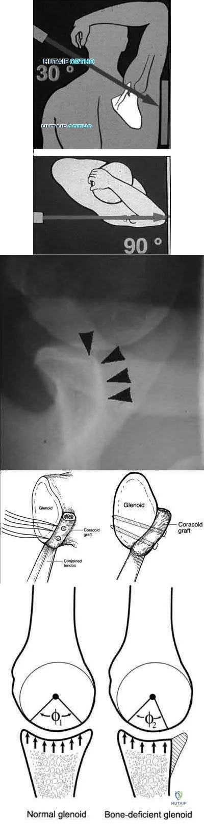
Figure A shows an arthroscopic picture of a 62-year-old male undergoing repair of a torn subscapularis tendon. In the image shown, G represents the glenoid, H represents the humeral head, and the dotted line represents the superolateral border of the subscapularis tendon. Which two ligaments form the structure marked with the asterisk?
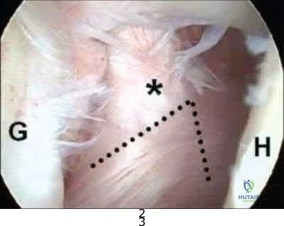
Inferior and middle glenohumeral ligaments Middle and superior glenohumeral ligaments Coracohumeral and coracoacromial ligaments

Coracohumeral and superior glenohumeral ligaments Superior and inferior glenohumeral ligaments
The coracohumeral and superior glenohumeral ligaments form a complex that marks the superolateral margin of the subscapularis tendon.
In chronic or degenerative tears, the subscapularis will often retract medially and become scarred to the deltoid fascia. This makes identification difficult during arthroscopic repair. The coracohumeral and superior glenohumeral ligaments form a complex that inserts on the superolateral margin of the subscapularis. This "comma sign" can usually be identified during arthroscopic repair making identification of the subscapularis tendon an easier task.
Burkhart and Brady present surgical pearls for arthroscopic repairs of the subscapularis. Amongst other things, they state the subscapularis is almost always repairable with proper mobilization, but an Achilles tendon allograft or a subcoracoid pectoralis major transfer may be used for a severely degenerated subscapularis.
Lo and Burkhart describe the comma sign for repair of chronic subscapularis tears. They describe how the superior glenohumeral ligament/coracohumeral ligament complex and subscapularis tendon are intimately associated, and often tear off the humerus while remaining attached to each other. This complex, when torn, forms a "comma sign," that marks the superior and lateral margins of the subscapularis tendon.
Illustration A shows why the convergence of the superior glenohumeral and coracohumeral ligaments on the superolateral border of the subscapularis is referred to as the "comma sign." Incorrect Answers:
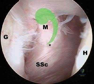
A 52-year-old man sustained the left elbow injury shown in Figure A while playing basketball 2.5 months ago. He underwent the procedure shown in Figure B. Post-operatively he was mobilized in a hinged brace. On examination today, his arc of elbow flexion is 75 degrees with loss of 45 degrees of full extension. His Disabilities of the Arm, Shoulder and Hand (DASH) Outcome Measure score is 45 points. What initial treatment option will likely provide the greatest improvement in this patients DASH score and functional range of motion?
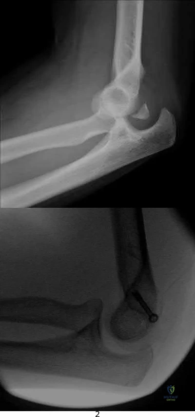
Self-directed exercise therapy Supervised exercise therapy

Supervised exercise therapy with static progressive elbow splinting Continuous passive motion device

Closed manipulation under anesthesia
The clinical presentation is consistent with post-traumatic elbow stiffness following an elbow fracture-dislocation. Supervised exercise therapy with static elbow splinting over a 6 month period
has shown to have a significant improvement on DASH scores and functional range of motion (ROM) in patients with post-traumatic elbow stiffness.
Post-traumatic elbow stiffness is often difficult to manage. The ultimate goal of treatment is to restore a functional range of elbow motion (30° to 130°). Nonoperative modalities are considered the first-line of treatment. Aggressive physical therapy has traditionally been advocated. However, the use of static progressive elbow splinting with a turnbuckle, alongside aggressive physical therapy, has shown to provide better functional outcomes. Treatment is usually maintained over a period of 6-12 months. Surgery is considered when nonoperative therapy fails.
Doornberg et al. looked at a retrospective case series of 29 patients with posttraumatic elbow stiffness. They showed that static progressive splinting can help gain additional motion when standard exercises fail to produce additional improvements.
Lindenhovius et al. randomized sixty-six patients with post-traumatic elbow stiffness into static progressive elbow splint therapy or dynamic elbow splinting over a 12 month period. There was no significant difference in outcomes between treatment modalities. ROM increased by 40° vs. 39° at six months, respectively. DASH scores improved from 50 vs 45 at enrollment to 32 vs. 25 at six months, respectively.
Figure A shows a posterior elbow dislocation with an associated medial epicondyle fracture. Figure B shows ORIF of the fracture seen in Figure A. Illustration A shows a static progressive turnbuckle elbow splint used for posttraumatic elbow stiffness.
Incorrect Answers:
tissues, causing hemarthrosis and additional fibrosis in the joint.
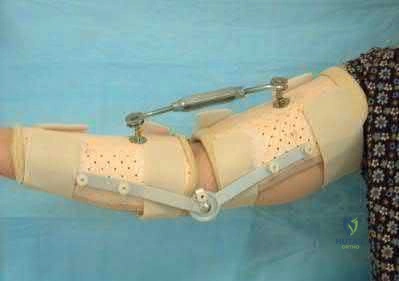
A 25-year-old right-hand baseball pitcher presents with persistent shoulder pain for the past several months in his dominant throwing arm. On physical examination, he is found to have full arc of motion with the exception of an internal rotation deficit of 30 degrees compared to his contralateral side. He is asked to complete the exercise shown in the video in Figure V. This form of rehabilitation is meant to address pathology in which anatomic structure?

Superior glenohumeral ligament Middle glenohumeral ligament

Anterior band of the inferior glenohumeral ligament Superior band of the inferior glenohumeral ligament

Posterior band of the inferior glenohumeral ligament

tissues in patients demonstrating symptoms of internal impingement. The sleeper stretch helps to address posterior tightness and the only posterior structure listed in the responses is the posterior band of the inferior glenohumeral ligament (posterior IGHL).
Internal impingement is a significant cause of pain in throwing athletes. It results from impingement of the articular undersurface of the posterior supraspinatus against the posteriorsuperior glenoid.
This is thought to be secondary to tightness in the posterior soft tissues including the capsule and posterior band of the IGHL. The mainstay of non-operative management is posterior capsular stretching with the sleeper stretches and cross-body adduction stretches. Heyworth et al. review the etiology, diagnosis and management of internal impingement of the shoulder. They note that repetitive contact between greater tuberosity and glenoid rim posterosuperiorly lead to impingement of the posterior rotator cuff and labrum. This occurs when the arm is externally rotated and abducted.
Tyler et al. reviewed the effects of posterior capsular stretching on alleviating symptoms in patients with internal impingement. Twenty-

Figure V is a video that demonstrates the sleeper stretch to address tightness of the posterior soft tissues. The arm is forward flexed 90 degrees and the patient lies on his side in order to stabilize the scapula while the arm is internally rotated. Illustration A depicts the sleeper stretch.
Incorrect Answers:
not the focus of the sleeper stretch.
A 28-year-old professional baseball pitcher sustains a complete rupture of his ulnar collateral ligament. He is neurovascularly intact on exam. Which of the following surgical reconstruction techniques has been shown to result in the lowest complication rate and best patient outcome?

Splitting of flexor-pronator mass, figure-of-8 graft fixation. Splitting of flexor-pronator mass, docking graft fixation.

Splitting of flexor-pronator mass, docking graft fixation, ulnar nerve transposition. Detachment of flexor-pronator mass, figure-of-8 graft fixation, ulnar nerve transposition.

Detachment of flexor-pronator mass, docking graft fixation, ulnar nerve transposition.
Ulnar collateral ligament (UCL) reconstruction using a flexor-pronator musclesplitting approach and a docking graft fixation technique are associated with the lowest complication rate and best patient outcomes.
Vitale et al. performed a systematic review of retrospective cohort studies evaluating UCL reconstruction techniques in overhead athletes. They demonstrated that the flexor-pronator musclesplitting approach was associated with better outcomes than detachment of the flexorpronator mass, had a lower rate of postoperative ulnar neuropathy, and a lower overal complication rate. They also found fixation of the graft utilizing the docking technique was associated with better outcomes than the figure-of-8 technique. Abandoning the obligatory ulnar nerve transposition was associated with improved patient outcomes (89% vs. 75%) and a lower rate of postoperative ulnar neuropathy (4% vs. 9%).
Rettig et al performed a case series review of 31 overhead throwing athletes with ulnar collateral ligament injuries managed nonoperatively with 3 months rest followed by rehabilitation exercises. They concluded that 42% of athletes were able to return to their previous level of competition at an average of 6 months from diagnosis (earlier than reconstruction). The authors were unable to identify any patient-specific factors (duration of symptoms, age, acuity of onset) that would predict the success of nonoperative treatment.
Illustration A shows the figure-of-8 (Jobe) graft fixation technique. It is performed by passing the tendon graft through two bone tunnels in the medial epicondyle of the humerus and through one tunnel in the ulnar sublime tubercle. The graft is then sutured to itself in a figure-of-8 configuration. Illustration B shows the docking graft fixation technique. The graft is placed in a triangular configuration through a single humeral tunnel. The suture limbs are then brought out through two separate bone holes and tied over a bony bridge on the superior aspect of the medial epicondyle.
Incorrect Answers:
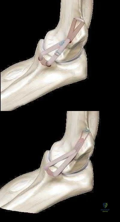
The right shoulder exercise seen in Figure A will put the LEAST amount of stretch on which structure?
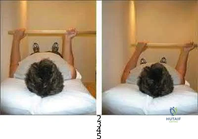
Inferior glenohumeral ligament Coracohumeral ligament Anterior-superior capsule Superior glenohumeral ligament Posterior capsule
Shoulder wand exercises, as shown in Figure A, are used to increase external range of motion of the shoulder. With the arm adducted and the elbow flexed, this exercise will put the LEAST amount of stretch on the posterior capsule.
External rotation shoulder wand exercises are commonly used for the treatment of adhesive capsulitis. Adhesive capsulitis is most commonly caused by contracture of the rotator interval. The rotator interval includes the anterior-superior capsule, superior glenohumeral ligament, coracohumeral ligament and long head biceps tendon. The structure most commonly contracted is the anterior-superior capsule, which limits external rotation when the arm is adducted. Kuhn et al. showed that in the neutral position, each ligament except the posterior capsule significantly affected the torque required for external rotation. The greatest effect on resisting external rotation at 0 degrees of abduction was the entire inferior glenohumeral ligament > coracohumeral ligament
> anterior band of the inferior glenohumeral ligament > superior and middle glenohumeral ligament.
Harryman et al. looked at the role of the rotator interval capsule in passive motion and stability of the shoulder. They found operative alteration of this capsular interval was found to affect flexion, extension, external rotation, and adduction of the humerus with respect to the scapula. Limitation of external motion was increased by operative imbrication of the rotator interval and decreased by sectioning of the rotator interval capsule.
Kim et al. reviewed shoulder MRIs to determine if abnormalities of the rotator interval were correlated with chronic shoulder instability. They found a significantly larger rotator interval height, rotator interval area, and rotator interval index in patients with chronic anterior shoulder instability compared to patients without instability.
Figure A shows a patient performing an exercise to increase right shoulder external rotation with a wand/stick. The right arm is fully adducted by her side, and her elbow flexed at 90 degrees.
Incorrect Answers:
) A 55-year-old male returns for followup 3 months after reverse shoulder arthroplasty. He reports limited function of his right shoulder but no antecedent trauma. A radiograph of his shoulder is shown in Figure A. All of the following variables are associated with this complication EXCEPT:
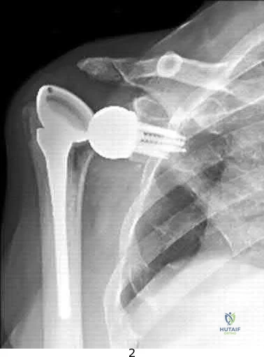
History of malunited proximal humerus fracture Proximal humeral bone loss

Failed primary arthroplasty Rheumatoid arthritis

Fixed preoperative glenohumeral dislocation
Rheumatoid arthritis is not associated with reverse shoulder arthroplasty (RSA) dislocation. RSA dislocation is a known complication of RSA. Risks include proximal humeral bone loss, chronic fracture sequelae with malunited/ununited tuberosities, failed previous arthroplasty, and fixed glenohumeral dislocation preoperatively. An irreparable subscapularis tears may be less of an issue with newer implant designs.
Trappey et al. studied instability and infection rates after RSA. They found that the rate of instability was similar in primary and revision surgery, but the rate of infection was higher in revision surgery. Instability was highest in the fracture sequelae group because of malunited tuberosities, contractures and proximal humeral bone loss.
Favre et al. examined the effect of component positioning on RSA stability.
They found that humeral version was more important than glenoid version. Stability is improved with the humerus in neutral or slight anterversion. They recommend avoiding retroversion >10deg. Edwards et al. examined subscapularis insufficiency and the risk of RSA dislocation. They found that of 138 RSA, all 7 dislocations occurred in patients with an irreparable subscapularis.
Dislocation was also more likely in patients with complex diagnoses, including proximal humeral nonunion, fixed dislocation, and failed prior arthroplasty.
Figure A shows reverse shoulder arthroplasty dislocation. Incorrect Answers:
) A 45-year-old man complains of chronic right shoulder pain. He has a history of chronic steroid use because of asthma. He recently completed a course of physical therapy
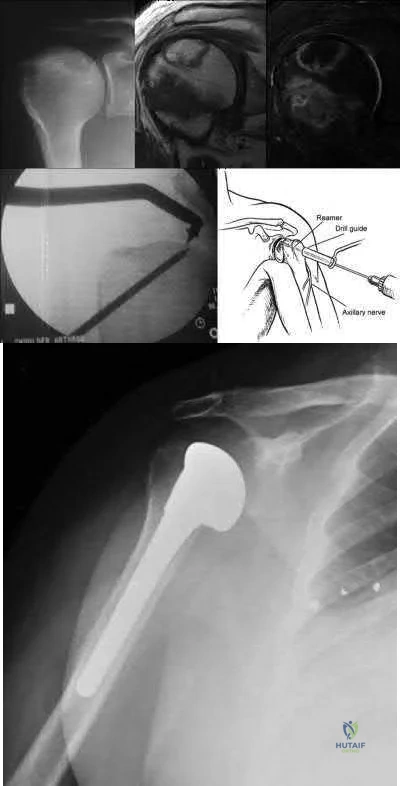
and has given up his job as a laborer in favor of a desk job. Examination reveals diminished shoulder abduction strength. A radiograph of his shoulder is shown in Figure A. Which of the following surgical treatment options (Figures B through F) is the most appropriate?
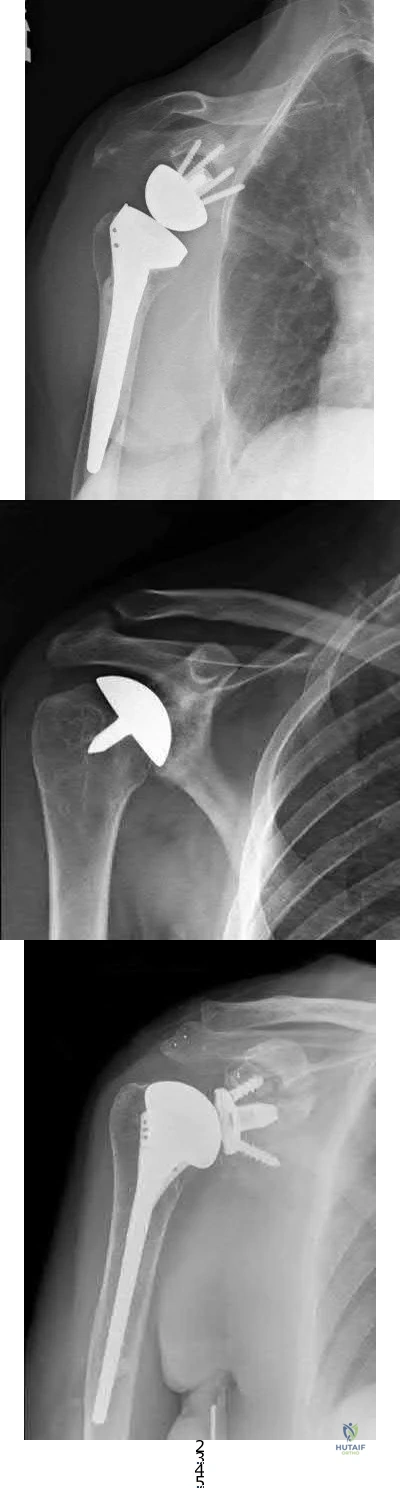
Figure B Figure C Figure D Figure E Figure F
This patient has early stage avascular necrosis (AVN) of the humeral head without subchondral collapse/flattening, likely related to chonic steroid use.
Core decompression is indicated.
Treatment of humeral head AVN is dependent on Cruess Stage. Precollapse stages (Stage I and II) may be treated by core decompression and joint preservation. Hemiarthroplasty is used for Stage III-IV disease. Total shoulder arthroplasty is used for Stage V disease. Resurfacing may be used for Stage III disease with focal chondral defects and sufficient remaining epiphyseal bone stock for fixation.
Harreld et al. reviewed humeral head AVN. They advocate attempting core decompression and arthroscopy for Stage III disease, and then tailoring resurfacing or replacement depending on defect size.
Smith et al. reviewed 31 hemiarthroplasties for steroid-related AVN (6 Stage III, 16 Stage IV, 5 Stage V). Unsatisfactory results were found in 45%. This was associated with glenoid cartilage wear over time. However, they still believed hemiarthroplasty was appropriate for younger active patients with stage III or stage IV disease.
LaPorte et al. performed core decompression for various stages of AVN.
Results were successful in 94%, 88%, 70% and 14% of Ficat-Arlet Stages I, II, III and IV humeral head AVN respectively, and more successful for nonsteroid related cases compared with steroidrelated cases. They recommend this treatment for Stages I-III.
Figure A comprises a radiograph showing Cruess Stage II disease ("snowcap" sign indicating sclerosis, preservation of the head contour and absence of subchondral collapse, left), a T1weighted
MRI (center) and T2-weighted fat saturated MRI (right) showing a variegated pattern of osteonecrosis, but with preservation of normal head contour. Figure B shows core decompression. Figure C shows hemiarthroplasty. Figure D shows reverse shoulder arthroplasty. Figure E shows resurfacing. Figure F shows total shoulder arthroplasty. Illustration A depicts the Cruess staging system. Illustration B shows a possible algorithm for management where they use the Ficat stages adapted from the hip.
Incorrect Answers:
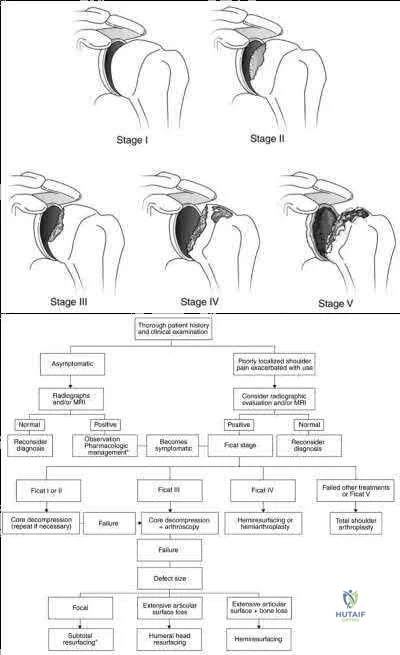
A 56-year-old otherwise healthy woman undergoes uncomplicated arthroscopic repair of a full-thickness rotator cuff tear. Prior to the procedure, the patient had attempted a long,
protracted course of physical therapy in an attempt to regain function without surgery. At her 10 day post-operative visit, she tearfully informs you that she cannot see her physical therapist because she has used up her 24-visit allotment for the year. She has 4 more weeks
until her insurance year turns over, and she does not have the financial means to pay out of pocket. What is the best next course of action?
Request a peer-to-peer review for authorization of additional visits or else the patient is likely to have a poor result

Provide a brief explanation and sheet of exercises for periscapular and cuff strengthening exercises using a resistance band

Test her range of motion and strength, and if limited, have her return weekly to your clinic for a guided home rehabilitation program

Explain to her that seeing a physical therapist during the first 6 weeks following cuff repair will not affect her range of motion one year removed from surgery

Give her a prophylactic corticosteroid injection to avoid early post-operative adhesive capsulitis
Early motion following rotator cuff repair has not been shown to impact range of motion and stiffness at one year post-operative clinical examinations.
Stiffness is a complication of protecting rotator cuff repairs from early re-tear with sling immobilization. However, with sling immobilization of up to 6 weeks, there is evidence of no increase in long-term stiffness. Accordingly, supervised physical therapy is not required in the first 6 weeks following arthroscopic rotator cuff repairs in order to obtain a good result.
Parsons et al. retrospectively reviewed 43 patients with full-thickness cuff tears who underwent a conservative early-postoperative protocol involving sling immobilization for 6 weeks with no formal therapy during that time. 10 /43 patient were characterized as stiff at their 6-week postoperative visit. At one year, there was no difference in the range of motion of the early stiff group compared to the non-stiff group.
Trenerry et al. collected prospective data on 209 consecutive patients undergoing primary rotator cuff repair. They found that patients in the stiffest quartile of range of motion testing at 6 weeks progressively regained range of motion by a post-operative visit at 72 weeks. The predictor of slowest recovery of early post-operative stiffness was found to be an internal rotation deficit with the patient reaching behind his or her back.
Incorrect Answers:
1: Early supervised physical therapy has not been shown to be essential to obtaining good motion post-operatively.
2 and 3: Strength testing and home strengthening programs in the early postoperative period would put the repair at risk for early failure.
5: Prophylactic corticosteroids are not indicated for prevention of postoperative stiffness, and furthermore, could theoretically interfere with healing.
A 62-year-old woman presents with chronic shoulder pain. On physical exam, she has anterior shoulder pain and her symptoms are reproduced with provocative testing of the biceps including supination against resistance and forward flexion of the shoulder against resistance. Internal and external rotation are painful, but her range of motion is intact. Shoulder radiograph and MRI images are shown in Figures A-E. Which of the following statements is true regarding the patient's condition?
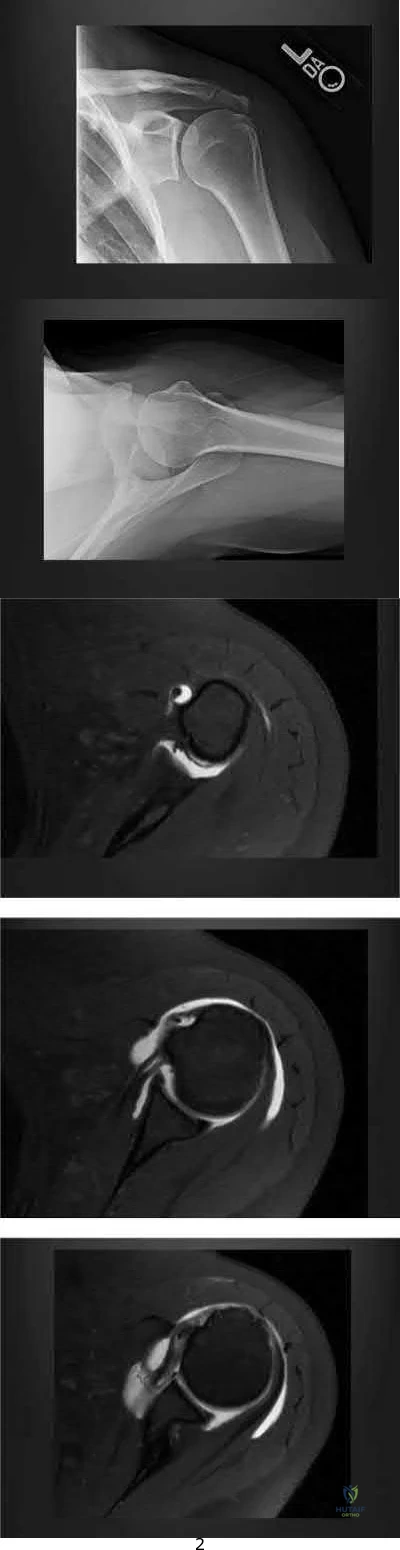
Her clinical examination is most consistent with a SLAP tear, which should be repaired. Her biceps pathology is due to her partial tearing of her subscapularis

She has isolated degenerative biceps tendonosis and an injection may cure her symptoms

She has end-stage rotator cuff arthropathy and should consider a reverse total shoulder arthroplasty

Her subacromial impingement is causing her biceps tendon sheath to be inflamed
The subscapularis tendon is the most important medial restraint to subluxation or dislocation of the long head of the biceps tendon (LHBT).
Anterior shoulder pain and positive provocative biceps clinical examination tests are common in patients with concomitant rotator cuff pathology. A consequence of subscapularis tendon tears -even partial tears - is that the LHBT can subluxate medially out of the intertubercular groove, as the subscapularis tendon is the most important restraint to medial instability of the LHBT. This instability can cause both pain and inflammation around the biceps tendon, leading to pain with resisted supination (Yergason's test) or resisted forward flexion (Speeds' test).
Walch et al. wrote a case series on 71 cases of biceps tendon instability. They found that dislocated LHBTs were associated with partial or complete subscapularis tears in 96% of cases.
Maier et al. published clinical results of treating acute traumatic medial LHBT instability with open repair of the subscapularis tendon and stabilization of the LHBT. They showed equivalent functional clinical outcomes to biceps tenotomy or tenodesis, with improved cosmesis and decreased muscle cramping.
Figures A and B show Grashey and axillary lateral views of the patient and demonstrate an os acromiale. Figures C, D, and E are axillary T2 MRI images. Figure C shows the tendon of the long head of the biceps in the distal aspect of the biceps grove. Moving proximally, Figure D shows the tendon subluxated onto the lesser tubercle and Figure E shows it fully dislocated medially.
Incorrect Answers:
A 78-year-old male presents to clinic 4 weeks after left total shoulder arthroplasty. He has not been wearing his sling and reports that he developed increased pain after slipping in the shower. He used the arm to catch himself from falling. On examination, he can flex the shoulder to 70 degrees, limited by pain. Active external rotation with arm at the side is 50 degrees and active internal rotation is 5 degrees. Passive external rotation is to 80 degrees. A radiograph of the left shoulder is shown below in Figure A. What other complaint is the patient most likely to have?

Pain with palpation of the bicipital groove Pain with palpation over the subdeltoid bursa Sensory loss over the lateral shoulder

Sensation of shoulder instability with external rotation Sensation of shoulder instability with internal rotation
The clinical presentation is consistent with a tear of the subscapularis, which is a well-described complication after total shoulder arthroplasty. The most likely additional complaint this patient will have is anterior shoulder instability, noticeable with external rotation of the shoulder.
Total shoulder arthroplasty is the preferred treatment for glenohumeral arthritis in patients with intact rotator cuff and good glenoid bone stock. The surgical approach involves detaching the subscapularis and capsule from the anterior humerus and dislocating the humeral head anteriorly. Post operatively, external rotation is limited to protect the subscapularis repair. If there is suspicion of a postoperative subscapularis tear, and ultrasound can be performed to confirm the diagnosis.
Miller et al. reported 7 cases of subscapularis tendon rupture after total shoulder arthroplasty, all of which were subsequently repaired. Decreased functional outcomes were observed in these patients, with lengthening techniques to address internal rotation contractures and prior surgery involving the subscapularis tendon as risk factors for rupture
Westoff et al. performed static and dynamic ultrasounds on 22 patients after total shoulder arthroplasty evaluating for numerous periarticular pathologies. The authors concluded that sonography is a useful tool for evaluation of periimplant tissues after TSA.
Figure A shows an intact left total shoulder arthroplasty without evidence of fracture, dislocation, or hardware loosening. Illustration A shows the incision for the subscapularis tendon during TSA. Incorrect Answers:
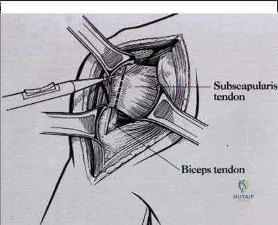
A 25-year-old lineman is referred to your office for a second opinion. 1 year ago, he underwent an arthroscopic procedure for shoulder instability. He complains of persistent sense of instability despite the surgery. Which of the following is a contraindication to revision arthroscopic labral repair for recurrent anterior glenohumeral instability?
Glenoid bone loss of 10%

Capsular attenuation from prior thermal capsulorraphy Anterior labral periosteal sleeve avulsion (ALSPA ) lesion Glenoid labral articular defect (GLAD) lesion

Combined Superior Labrum from Anterior to Posterior tear (SLAP) and recurrent Bankart lesion
Capsular attenuation or postthermal capsular necrosis from prior thermal capsulorraphy is a contraindicated to arthroscopic repair.
Thermal capsulorrhaphy utilizes heat generated by radiofrequency or laser ablation to cause capsular shrinkage in an effort to treat shoulder instability. However, high recurrence rates have been found, especially around two to three weeks after the index procedure, when the capsular tissue is the weakest. In the setting of recurrence following thermal capsulorrhaphy, open revision is recommended.
Creighton et al. reported on a series of 18 patients undergoing revision arthroscopic stabilization. Of the 18, 3 failed with recurrent instability, all with previous thermal capsulorrhaphy. Miniaci et al. reviewed the outcomes following thermal capsulorrhaphy noting high rates of recurrent instability, especially in the setting of initial treatment for multidirectional instability. Park et
al. reported on a series of 14 patients undergoing revision following thermal capsulorrhaphy. Ten out of 14 patients had signs of capsular thinning, insufficiency and attenuation.
Wong et al. surveyed 379 shoulder surgeons on the complications following thermal capsulorrhaphy. Capsular insufficiency and thinning were commonly associated with recurrent instability.
Hecht et al. performed thermal capsulorrhaphy and biomechanical analysis of the capsule in a sheep model. The authors found that the capsule was weakest at the 2-3 week post-operative timepoint, leading to the highest rate insufficiency, attenuation and mechanical failure at this time.
Incorrect answers:
A 47-year-old landscaper presents with worsening left shoulder pain and weakness. Three years ago, he injured the left shoulder in a fall and elected for nonoperative
management to minimize time off from work. Physical therapy was effective until 6 months ago when his shoulder function worsened to the point that he is now unable to work.
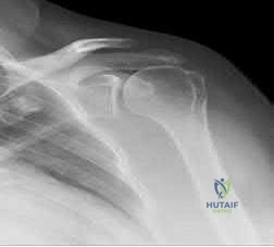
Examination of his active range of motion reveals forward elevation 120° with pain, abduction 100°, IR at neutral to T8 and ER at neutral 5°. He has a positive ER lag sign and Hornblower's sign. Belly press and lift-off tests are normal. A recent radiograph is shown in Figures A. MRI images are shown in Figures B and C. Which of the following is the best treatment option?
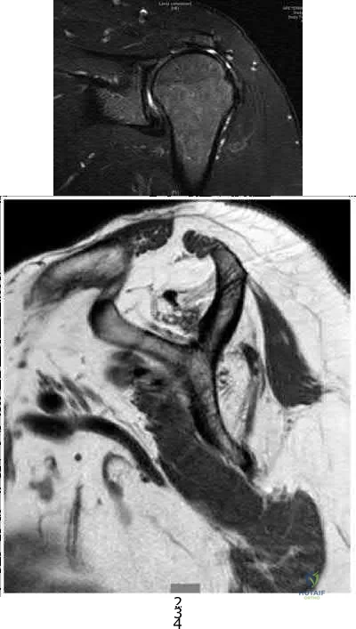
Continue physical therapy Latissimus dorsi transfer Arthroscopic rotator cuff repair Pectoralis major transfer

Reverse total shoulder arthroplasty
This patient has a chronic massive posterosuperior rotator cuff tear with marked atrophy, tendon retraction and loss of external rotation strength that is impacting his daily life. The best treatment option for this middle-aged laborer with an irreparable posterosuperior rotator cuff tear is a latissimus dorsi transfer to restore external rotation strength and motion.
Irreparable rotator cuff tears are marked by: (1) Superior displacement of the humeral head (AHI < 5-7mm), (2) Fatty infiltration of the rotator cuff muscles ( Goutallier stage 3-4), (3) Increased duration of the tendon tear and (4) Profound external rotation weakness. These findings are predictive of poorquality tissue and stiffness of the muscle-tendon unit, not amenable to primary repair. In this setting, a latissimus dorsi transfer can be utilized to restore shoulder strength, function and improve pain. Relative contraindications include subscapularis deficiency, deltoid deficiency, pseudoparalysis of the shoulder and advanced glenohumeral arthritis.
Gerber et al. performed a case series analysis of 67 patients with irreparable rotator cuff tears managed with latissimus dorsi transfer. Patients with an intact subscapularis demonstrated improvement in pain, range of motion and strength postoperatively, while no improvement was noted in patients with subscapularis deficiency. The authors conclude that latissimus dorsi transfer should not be performed in the setting of poor subscapularis function.
Iannotti et al. found that better clinical results following latissimus dorsi transfer were associated with: preserved active shoulder range of motion and strength (specifically forward elevation > 90° and external rotation > 20°), synchronous firing of the transferred latissimus dorsi muscle and male gender.
Figure A is an AP radiograph of the left shoulder with superior migration of the humeral head (AHI
< 5mm) and no evidence of glenohumeral arthritis. Figures B and C show a retracted posterosuperior rotator cuff tear and Goutallier stage 4 atrophy (more fat than muscle) of the supraspinatus, infraspinatus and teres minor, rendering this tear irreparable. Illustration A shows a latissimus dorsi transfer. The latissimus dorsi tendon is positioned over the top of the humeral head, covering most of the rotator cuff defect. The tendon is then secured to the subscapularis tendon edge and lesser tuberosity anteriorly, the remnant supraspinatus and infraspinatus tendons medially, and the greater tuberosity laterally.
Incorrect Answers:
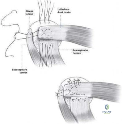
A collegiate swimmer develops medial winging of the scapula. If the EMG and nerve conduction studies are abnormal, the most likely nerve roots to be involved are?

C7, C8, T 1 C6, C7, C 8 C5, C6, C 7 C4, C5, C 6 C3, C4, C 5
Classic medial winging of the scapula is due to paralysis of the serratus anterior muscle which is supplied by the long thoracic nerve which holds the scapula to the chest wall and prevents the inferior angle of the scapula from migrating medially. It is innervated by the long thoracic nerve (C5, 6, 7).
Surgical treatment may include partial pec major transfer. Lateral winging may be caused by spinal accessory nerve palsy (CN XI, also ventral ramus C2,3,4). The nerve may be injured during neck surgery. This causes trapezius weakness, allowing the inferior pole of the scapula to migrate laterally. The modified Eden-Lange procedure may be used for this type of winging.
Gregg et al. describes paralysis of the serratus muscle in young athletes which they felt was due to repetitive traction. Full recovery usually occurs in an average of 9 months, and they recommend that surgical methods of treatment should be reserved for patients in whom function fails to return after a twoyear period.
Foo et al. describes a larger cohort of 20 patients again treated expectantly with observation and physical therapy. They reported consistent recovery but that it can take up to 2 years.
Illustration A shows a clinical photo of medial scapular winging. Illustration V is an instructional video of scapular winging. It begins with a clinical video of the condition.
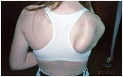
A 62-year-old man complains of shoulder pain for 2 years. He has had 1 course of intra articular sodium hyaluronate and 6 weeks of physical therapy with little relief. Examination reveals diminished arm flexion and abduction secondary to pain. Radiographs of his shoulder are shown in Figures A and B. According to the American Academy of Orthopaedic Surgeons Clinical Practice Guidelines, what is the next best step?
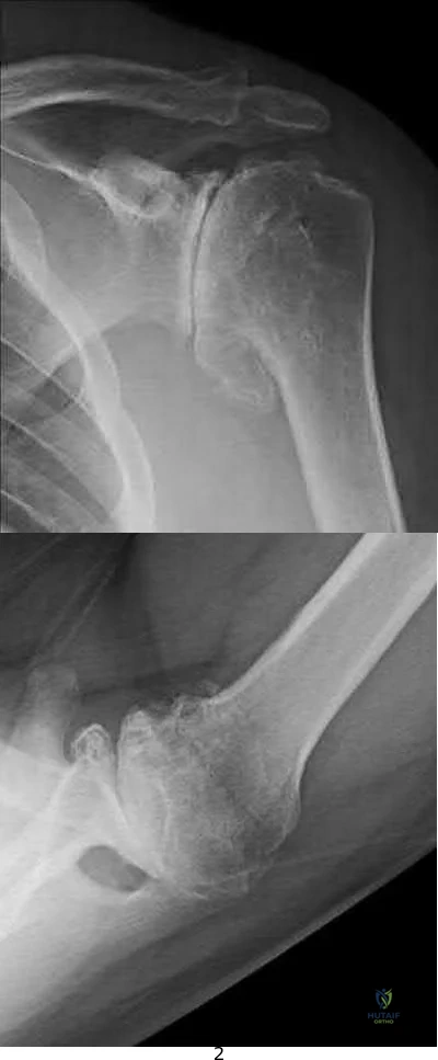
Humeral head replacement arthroplasty Hemiarthroplasty and ream-and-run glenoid procedure

Cuff tear arthropathy (CTA) prosthesis

Total shoulder arthroplasty with a metal-backed cemented glenoid component Total shoulder arthroplasty with an all-polyethylene cemented glenoid component
This patient has end-stage glenohumeral osteoarthritis (GH OA). According to the AAOS CPG, total shoulder arthroplasty (TSA) is recommended using an allpolyethylene cemented glenoid component.
TSA is indicated for cases of end-stage GH OA. It is preferred to hemiarthroplasty. It is contraindicated in cases with insufficient glenoid bone stock (glenoid wear to the level of the coracoid), rotator cuff arthropathy or irreparable cuff tears and deltoid dysfunction. It provides good pain relief and has good survival at 10 years (>90%).
Radnay et al. performed a systematic review involving 1952 patients comparing TSA with humeral head replacement (HHR). They found that TSR provided greater pain relief, range of motion, patient satisfaction, and had lower revision rates. They recommend TSA over HHR for GH OA. Izquierdo et al. described the AAOS Clinical Practice Guidelines (CPG) regarding treatment of GH OA. This is summarized in Illustration A.
Figures A and B show end-stage GH OA with large osteophytes and subchondral sclerosis. There is significant glenoid wear and posterior subluxation (Walch B glenoid deformity). Illustration A is a table summarizing the AAOS CPG on treatment of GH OA. Illustration B shows a CTA humeral component. It is not paired with a glenoid component.
Incorrect Answers:
Metal-backed glenoids have higher rates of revision than all-polyethylene glenoids.
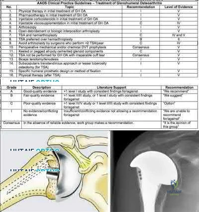
Posterior glenohumeral dislocations are as common as anterior dislocations in which of the following patient groups?
Football players

Marfan's syndrome patients Renal failure patients Epilepsy patients

Women
Millett et al and Robinson et al provide review articles on posterior shoulder dislocations, which are rare clinical entities that occur during seizures and electrocution (due to tetanic muscle contraction) or as a result of high energy trauma. Robinson et al noted that poor prognostic factors associated with posterior shoulder dislocation include late diagnosis, large bony defect of humeral head, associated proximal humerus fracture, and need for arthroplasty. In Gerber's series, posterior dislocations occurred with equal frequency to anterior in a cohort of epilepsy patients.
What nerve is injured most commonly during the superficial dissection when repairing a distal biceps rupture through a single incision anterior approach?

Medial antebrachial cutaneous nerve Lateral antebrachial cutaneous nerve Superficial radial nerve

Ulnar nerve

Posterior interosseous nerve
The lateral antebrachial cutaneous nerve (LABCN) is at risk during the superficial dissection when repairing a distal biceps rupture through a single incision anterior approach.
The LABCN is the terminal cutaneous branch of the musculocutaneous nerve, which supplies sensation to the volar-lateral aspect of the forearm. The LABCN pierces the deep fascia of the arm lateral to the musculotendinous junction of the distal biceps tendon after lying on top or piercing through the brachialis muscle. It exits the arm and lies in the subcutaneous tissues of the antecubital fossa. It is important to retract this nerve laterally during the approach to the distal biceps tendon.
Cohen describes the importance of identifying the LABCN during the superficial dissection as injury to this nerve is not uncommon (5-7%). Injury to the PIN (5 %) is devastating and occurs with retractor placement during the deep dissection and the use of suspensory fixation on the posterior cortex of the radius.
The review article by Ramsey et al covers the surgical anatomy and complications of biceps surgery, including injury to the LABCN.
Illustration A shows the LABCN relative to the anterolateral approach to the elbow which is commonly used to repair distal biceps avulsions. Illustration B shows the close proximity of the LABCN to the distal biceps in a human specimen.
Incorrect Responses:
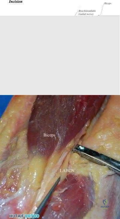
An MRI of the shoulder in a patient with chronic quadrilateral space syndrome is most likely to show which of the following?

Increased intra-capsular volume Loss of intra-capsular volume Fatty atrophy of the infraspinatus Fatty atrophy of the teres minor

Fatty atrophy of the latissimus dorsi
Quadrilateral space syndrome involves dysfunction of the axillary nerve, perhaps by entrapment or compression, resulting in the functional denervation of the teres minor.
The quadrilateral space is a potential space formed by the long head of the triceps medially, the humerus laterally, the teres minor above, and the teres major below. The axillary nerve and posterior circumflex humeral artery travel through this space.
The Sanders article describes the MRI appearance, which is that the muscle appears streaked with white on MRI and atrophied (See illustration A) consistent with fatty atrophy. Sanders group report this finding in 3% of shoulder MRIs. The posterior circumflex humeral artery also travels with the axillary nerve as it travels through this space. Loss of capsular volume on an arthrogram study is suggestive of adhesive capsulitis.
Illustration B is a diagram which shows the borders of the quadrilateral (or quadrangular) space.
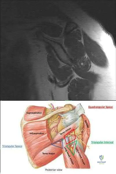
A 21-year-old collegiate volleyball player is noted to have weakness in external rotation and isolated atrophy of the infraspinatus on physical examination as seen in Figure A. An axial MRI image is shown in Figure B. This clinical condition is most likely caused by compression of the:
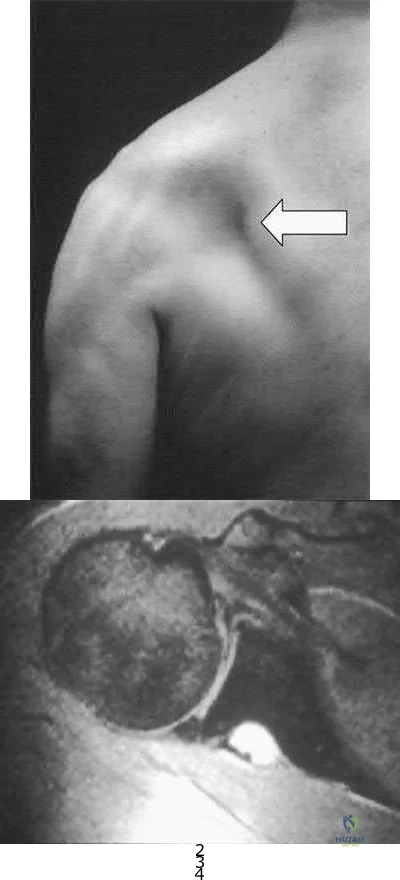
Axillary nerve at the triangular space Suprascapular nerve in the suprascapular notch Axillary nerve in the quadrangular space Suprascapular nerve in the spinoglenoid notch

Long thoracic nerve anterior to the scalenus and the first rib and posterior to the clavicle
The clinical presentation is consistent for a suprascapular neuropathy caused by compression of the suprascapular nerve by a cyst in the spinoglenoid notch.
The suprascapular nerve arises from the upper trunk of the brachial plexus with contributions from C5-6. It travels through the suprascapular notch of the scapula where it gives motor branches to the supraspinatus then around the spinoglenoid notch where it innervates the infraspinatus.
Compression of the nerve at the suprascapular notch will cause denervation and atrophy of both the supraspinatus and infraspinatus while compression at the spinoglenoid notch affects the infraspinatus in isolation. This is commonly seen in overhead athletes who sustain a SLAP tear and resultant spinoglenoid notch cyst as seen in the MRI. This will cause weakness and atrophy of the infraspinatus and can be noted both clinically and radiographically. Appropriate operative management is still not clear in the literature with some authors reporting a need for labral repair + cyst decompression and others reporting good outcomes with labral repair alone. Other cases have been treated with needle aspiration.
The cited reference by Cummins et al reviews the various causes, diagnosis, and treatment of suprascapular neuropathy.
The reference by Martin et al is a retrospective study of the results of nonoperative treatment of suprascapular neuropathy in which 5 had excellent results and 7 had good results.
Which of the following provocative tests would most likely be positive in a patient with medial epicondylitis?

Resisted forearm pronation and wrist flexion with a clenched fist Resisted forearm supination and wrist extension with a clenched fist Dynamic valgus stress test

Milking maneuver Pinch grip test
A provocative test for medial epicondylitis can be elicited by applying resistance to a patient with their fist clenched, wrist flexed and pronated.
Medial epicondylitis is an overuse syndrome of the flexor-pronator mass. The pronator teres (PT) and flexor carpi radialis (FCR) are thought to be most affected with this condition. It is most common in the dominant arm and occurs with activities that require repetitive wrist flexion/forearm pronation. Patients are most tender over the origin of PT and FCR at the medial epicondyle.
Resisting a patient with their fist clenched, wrist flexed and pronated can cause worsening of their pain. This maneuver can be used as a provocative test for this condition.
Cain et al. reviewed elbow injuries in throwing athletes. They comment that the common flexorpronator muscle origin provides dynamic support to valgus stress in the throwing elbow, especially during early arm acceleration and help produce wrist flexion during ball release.
Amin et al. reviewed the evaluation and management of medial epicondylitis. They report that medial epicondylitis typically occurs in the fourth through sixth decades of life, the peak working years, and equally affects men and women. Physical therapy and rehabilitation is the main aspect of recovery from medial epicondylitis, once acute symptoms have been alleviated.
Illustration A shows a video of this provocative test for medial epicondylitis. Incorrect Answers:
A 72-year-old woman presents for follow-up after elbow surgery. Her radiographs are shown in Figures A and B. Which of the following pre-operative diagnoses is a relative contraindication to the use of this prosthesis design?

Acute intra-articular distal humerus fracture Malunited intra-articular distal humerus fracture Late-stage rheumatoid arthritis

Post-traumatic bony ankylosis Osteoarthritis
This patient has had an unconstrained total elbow arthroplasty (TEA).
Unconstrained TEA is least preferred for late-stage rheumatoid arthritis where there is significant capsuloligamentous instability and bony erosion.
Unconstrained (unlinked or resurfacing prosthesis) TEA depend on intact bony and ligamentous constraints for stability. These are appropriate for humeroulnar conditions with intact collateral ligaments and radiocapitellar articulation e.g. osteoarthritis, post-traumatic arthritis, intra-articular distal humerus fracture, and malunion of the distal humerus. Conditions with increased risk of
instability (ligamentous injury, rheumatoid arthritis) will benefit from a linked or semiconstrained prosthesis.
Mansat et al. reviewed the Coonrad-Morrey linked (semi-constrained) TEA implant in 70 patients after 5 years. They found that patients with inflammatory arthritis had higher function than those with traumatic conditions
( fractures, nonunions and post-traumatic arthritis). Survival rate was 98% and 91 % at 5 and 10 years, respectively. They concluded that this implant provided satisfactory treatment for different indications although radiolucent lines and bushing wear were a concern.
Hildebrand et al. reviewed the functional outcome of the Coonrad-Moorey prosthesis in 51 elbows after 50 months. The inflammatory arthritis group had higher performance scores than the traumatic/post-traumatic conditions group. Isometric extensor torque was found to be less than the nonoperated side. Radiolucency was noted in 11 elbows.
Figures A and B show an unconstrained TEA with radial head replacement. Illustration A shows more examples of unconstrained TEA. Illustration B shows a semiconstrained TEA. The arrow points to the anterior flange. Illustration C shows radiolucent lines around the stems. Illustration D shows severe bushing wear leading to locking mechanism failure. Illustration E is a table comparing linked and unlinked implants.
Incorrect Answers:
A professional baseball team has several pitchers with complaints of velocity loss with their pitches and shoulder pain of their dominant shoulders during spring training. Pitch counts are properly monitored. The average glenohumeral internal rotation deficit on the pitching staff is 45 degrees. The best intervention would be:
Pitchers throwing less fastballs and more changeups

Evaluate the pitchers elbows for ulnar collateral ligament acute ruptures. Increasing the weight training for the deltoid and latissimus dorsi muscles Focused stretches and therapies that address posterior capsular tightness Firing the general manager for finding pitchers that "lose their stuff"
Glenohumeral Internal Rotation Deficit (GIRD) is a phenomenon that occurs in baseball pitchers and is due to posterior capsular tightness. Treatment should begin with a therapy program addressing the pathologic posterior capsule.
GIRD is a phenomenon that is frequently found in high-level overhead throwing athletes, predominantly baseball pitchers. It is defined as the measured difference in internal rotation between the non-dominant arm and dominant arm. Worsening range of motion deficits are seen with increased repetitions, both over a single season and a career. GIRD > 25º is associated with development of shoulder pathologies or pain requiring periods of inactivity. Cessation of overhead throwing activities and initiation of a stretching program to address posterior capsular contractures is largely effective (90% in some series).
Burkhart et al. reviewed the conditions associated with high-level overhead throwing athletes shoulders, culminating in a theory of pathologic progression to "dead arm syndrome" (loss of velocity and effective pitching). Their theory attributes adaptive hyperexternal rotation (occurs during late-cocking / early acceleration phases of pitching) to lead to posterior-inferior capsular contracture and GIRD. Subsequent injuries to anterior structures - including SLAP lesions - would then occur.
Illustration A is a cartoon depiction of how to perform the sleeper stretch. This is a common component of a pitcher's maintenance stretching program.
Incorrect Answers:
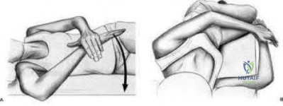
A 68-year-old man presents with severe right shoulder pain. He had a prolonged course of physical therapy and received several cortisone injections for his pain without improvement. Examination reveals pseudoparalysis of the right shoulder with a 20-degree external rotation lag with the shoulder adducted. With the shoulder placed in 90 degrees of abduction, he can actively externally rotate his shoulder. The patient was treated with a medialized reverse prosthesis shown in Figure A. Which of the following statement is true regarding this treatment option?
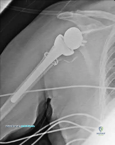
It is contraindicated in patients with shoulder pseudoparalysis

It can be used in patients with deltoid dysfunction when combined with latissimus dorsi transfer It shifts the center of rotation of the shoulder superior and lateral

The risk of scapular notching is increased with inferior placement of the glenoid component The risk of instability is increased with an irreparable subscapularis
The clinical presentation is consistent with a patient with pseudoparalysis that was treated with a reverse total shoulder arthroplasty (RTSA). The risk of postoperative instability is increased in patients with an irrepairable subscapularis when a medialized reverse prosthesis is used. Answers 1-4 are false statements.
RTSA is most commonly indicated for rotator cuff arthropathy. However, indications for use now include shoulder pseudoparalysis, anterosuperior escape of the humeral head, acute 3 or 4-part proximal humerus fractures, and greater tuberosity fracture nonunions. Contraindications to RTSA included deltoid dysfunction, insufficient glenoid bone stock, and bony deficiency of the acromion. Edwards et al. prospectively evaluated the risk of shoulder dislocation after reverse TSA. They found a significantly increased risk of dislocation (p=0.012) in patients with an irreparable subscapularis at time of surgery. There were no dislocations in the reparable group. Dislocations were more likely in patients with proximal humeral nonunions and failed prior arthroplasty. Mulieri et al. looked at the use of reverse TSA in patients with irreparable massive rotator cuff tears without evidence of glenohumeral arthritis. All outcomes were improved postoperatively, and they advocate
for reverse TSA in this subset of patients. Survivorship was over 90% at more than 4 years average follow up.
Boileau et al. evaluated the clinical outcomes of isolated biceps tenotomy/tenodesis in patients with massive rotator cuff tears and a biceps lesion. They found that the procedure can effectively treat pain and improve function in these patients. There was no difference in patients undergoing tenotomy versus tenodesis.
Figure A is a right shoulder radiograph status post RTSA with components in adequate position. Incorrect Answers:
Figure A shows immediate post-operative radiographs of a 75year-old patient with primary osteoarthritis. She presents 3 years later with increasing pain and weakness in the shoulder despite home physical therapy. Examination reveals limited active range of motion, with forward elevation of 80 degrees and external rotation of 50 degrees. Her deltoid function is intact. Repeat radiographs are seen in Figure B. Which treatment option would provide the best functional outcome for this patient?
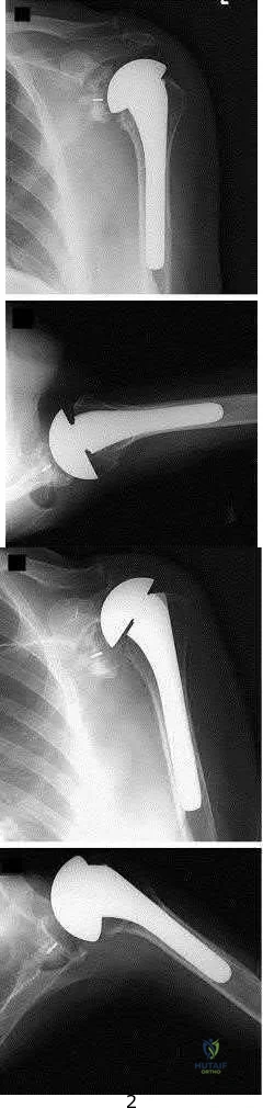
Open tendon transfer
Corticosteriod injection and supervised physical therapy

Open rotator cuff repair, subacromial decompression and distal clavical excision Revision to reverse shoulder arthroplasty

Revision to cuff arthropathy hemiarthroplasty
This patient presents with failed total shoulder arthroplasty. The best treatment option for functional outcome would be revision to reverse shoulder arthroplasty (rTSA).
RTSA is considered a viable treatment option for patients with failed shoulder arthroplasty. It allows for improved arm elevation and abduction in the setting of nonfunctional rotator cuff muscles, as seen in this example. Despite the expanding indications for rTSA, there are high complication rates in the revision setting. Complication rates for rTSA after failed shoulder arthroplasty have been reported to be between 11-36%. This procedure should, therefore, be performed by surgeons with extensive training in reconstructive shoulder arthroplasty.
Patel et al. retrospectively reviewed 31 patients (mean age, 68.7 years) who underwent rTSA for treatment of a failed shoulder arthroplasty. They found the greatest improvement with active forward elevation from 44° preoperatively to 108 ° postoperatively (P < .001). Complications occurred in 3 patients with periprosthetic fracture.
Hattrup et al. reviewed a series of 19 patients that underwent open rotator cuff repair after shoulder arthroplasty. Out of the 19 patients only 4 shoulders were successfully repaired. They concluded that successful rotator cuff repair after shoulder arthroplasty is possible but failure is more common. Figure A shows a left total shoulder arthroplasty that is well reduced in the glenoid. Figure B shows antero-superior escape of the prosthesis, indicative of a massive rotator cuff tear.
Incorrect Answers:
A 35-year-old man awoke following a night of heavy drinking with severe right shoulder pain and inability to raise his arm above his head. A radiograph from the emergency room is provided in Figure A. He was treated with a sling for a diagnosis of rotator cuff tear. Six weeks later, he complains of continued pain and difficulty using the arm. Which of the following is the next best step in management?
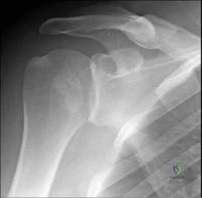
Physical therapy for range of motion followed by rotator cuff and deltoid strengthening exercises

Axillary radiograph of the shoulder

EMG to evaluate the suprascapular and axillary nerves Arthroscopic rotator cuff repair

Open subacromial decompression and latissimus dorsi transfer for massive cuff tear
The radiograph demonstrates overlap of the humeral head and glenoid suggesting shoulder dislocation. An Axillary radiograph is necessary to evaluate concentric reduction vs. dislocation of the shoulder. An example is provided in illustration A. Posterior shoulder dislocations can be easily be missed without the proper orthogonal views of the shoulder. Perron reviews the proper identification and emergency room care of posterior shoulder dislocation. Richardson found axillary radiographs to be more sensitive than trans-scapular radiographs for identifying posterior shoulder dislocations.
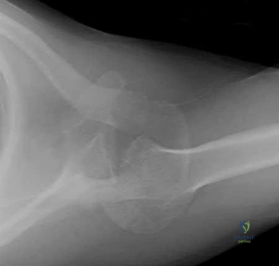
Recent randomized controlled trials comparing early passive range of motion to 6 weeks of immobilization after successful arthroscopic rotator cuff repair concluded that, compared to immobilization, early passive range of motion resulted in:
Higher Constant scores at 12 months

Increased rates of re-rupture as determined by ultrasound Equivalent functional outcomes

Less pain at 6 months

Inceased range of motion at 12 months
A series of high-quality RCTs have demonstrated that early passive range of motion has equivalent functional outcomes when compared to 6 weeks of immobilization after arthroscopic rotator cuff surgery.
Traditionally, most surgeons recommended early post-operative range of motion exercises for their patients in order to prevent adhesions and ultimately stiffness. However, recent evidence has found that there is no difference in the healing rate, range of motion or functional outcome between patients who undergo early versus delayed (i.e. initial 6 weeks of immobilization) passive range of motion exercises after arthroscopic rotator cuff repair.
Kim et al. conducted a randomized controlled trial comparing early passive range of motion vs. immobilization in 106 patients who underwent arthroscopic repair for full-thickness rotator cuff tears. They found that there was no clinically or statistically significant difference between the two groups in pain, healing or function.
Keener et al. also conducted a randomized controlled trial of 124 patients who were undergoing arthroscopic repair of a full-thickness rotator cuff tear and found no difference between early and delayed range of motion in healing and functional outcome.
Cuff & Pupello also compared early vs. delayed range of motion during the post-operative rehabilitation phase in a randomized controlled trial of 68 individuals undergoing arthroscopic rotator cuff repair and found no significant difference in range of motion or healing.
Incorrect Answers:
A 49-year-old male presents with right shoulder pain and weakness after undergoing open cervical lymph node biopsy approximately one year ago. A pertinent finding from the physical exam is seen in Figure A, with the patients arms by his side. Physical exam finding with the arms in a position of 90 degrees of forward elevation and 10 degrees of external rotation are shown in Figure B.
What nerve is most likely injured?
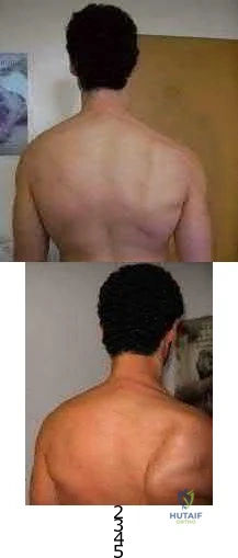
Long thoracic Suprascapular Spinal accessory Axillary Thoracodorsal
The patient is presenting with LATERAL scapular winging which is a result of injury to the spinal accessory nerve and resultant trapezius muscle palsy.
The spinal accessory nerve is fundamental to scapulothoracic function and essential for scapulohumeral rhythm. This nerve is vulnerable along its superficial course. The majority of injuries to the spinal accessory nerve are iatrogenic and occur secondary to head and neck surgery. There is often a marked delay in recognition and initiating treatment. Surgical treatment with the Eden-Lange transfer lateralizes the levator scapulae and rhomboids (transfer from medial border to lateral border)
Camp et al. reviewed the results of 111 patients who underwent operative management of a lesion to the spinal accessory nerve. They found that the majority (~80%) of injuries were sustained iatrogenically and that diagnosis was delayed for approximately 12 months.
Pikkarainen et al. reviewed the natural history of isolated serratus palsy. They found that symptoms mostly recover in 2 years, but at least one-fourth of the patients will have long-lasting symptoms, especially pain.
Figure A depicts a patient with lateral scapular winging. Figure B demonstrates physical exam of this patient with their arms in a position of 90 degrees of forward elevation and 10 degrees of external rotation. Illustration A highlights the difference between medial and lateral scapular winging. Illustration B depicts another example of a patient with lateral scapular winging.
Incorrect Answers:
An injury to the long thoracic nerve would result in serratus anterior palsy which would lead to MEDIAL scapular winging.

An injury to the suprascapular nerve would result in weakness and wasting of the supraspinatus and/or infraspinatus.
Question 59
A 16-year-old ice hockey player is struck on the chest by the puck. He skates a few strides and then collapses. What is the most likely diagnosis?
Explanation
REFERENCES: Maron BJ, Strasburger JF, Kugler JD, Bell BM, Brodkey FD, Poliac LC: Survival following blunt chest impact-induced cardiac arrest during sports activities in young athletes. Am J Cardiol 1997;79:840-841.
Link MS, Maron BJ, Estes NAM III: Commotio cordis, in Estes NAM III, Salem DN, Wang PJ (eds): Sudden Cardiac Death in the Athlete. Armonk, NY, Futura, 1998, pp 515-528.
Question 60
Which of the following enzymes is used to resorb bone by mature osteoclasts?
Explanation
Question 61
A patient who was involved in a motor vehicle accident 2 days ago now reports neck pain. He denies any other symptoms. Radiographs reveal a type II odontoid fracture that is 2 mm anteriorly displaced. Management consists of halo vest immobilization in extension, and repeat radiographs reveal that the fracture is completely reduced. The patient is discharged to home, but later that evening he notes difficulty swallowing while trying to eat dinner. What is the most likely cause of this difficulty?
Explanation
REFERENCES: Garfin SR, Botte MJ, Waters RL, Nickel VL: Complications in the use of halo fixation device. J Bone Joint Surg Am 1986;68:320-325.
Glaser JA, Whitehill R, Stamp WG, Jane JA: Complications associated with the halo-vest: A review of 245 cases. J Neurosurg 1986;65:762-769.
Question 62
Following its exit from the sciatic notch, the sciatic nerve passes between what two muscles?
Explanation
REFERENCES: Hoppenfeld S, deBoer P: Surgical Exposures in Orthopaedics: The Anatomic Approach. Philadelphia, PA, JB Lippincott, 1984, pp 335-348.
Anderson JE (ed): Grant’s Atlas of Anatomy, ed 7. Baltimore, MD, Williams & Williams,
1978, Figure 4-34.
FOR ALL MCQS CLICK THE LINK ORTHO MCQ BANK
Question 63
A 22-year-old professional ballet dancer reports a 3-month history of posterior ankle pain that occurs when she changes from a flat foot to pointe (hyperplantar flexed position). Examination does not elicit the pain with forced passive plantar flexion. A radiograph is shown in Figure 8. What is the most likely cause of the pain?
Explanation
REFERENCES: Hamilton WG, Geppert MJ, Thompson FM: Pain in the posterior aspect of the ankle in dancers: Differential diagnosis and operative treatment. J Bone Joint Surg Am 1996;78:1491-1500.
Khan K, Brown J, Way S, et al: Overuse injuries in classical ballet. Sports Med
1995;19:341-357.
Question 64
A 25-year-old man shot himself at the base of the right index finger while cleaning his handgun. Examination reveals that the finger is cool and cyanotic. A clinical photograph and radiograph are shown in Figures 44a and 44b. What is the recommended treatment?
Explanation
REFERENCES: Peimer CA, Wheeler DR, Barrett A, et al: Hand function following single ray amputation. J Hand Surg Am 1999;24:1245-1248.
Neumeister MW, Brown RE: Mutilating hand injuries: Principles and management. Hand Clin 2003;19:1-15.
Question 65
A 20-year-old unrestrained driver sustained a midshaft femur fracture in a high-speed motor vehicle accident. The femoral neck was evaluated with a CT scan with 2-mm cuts through the hip; no fracture was identified. What additional studies (if any) should be performed to minimize the risk of having an undiagnosed femoral neck fracture?
Explanation
Question 66
CLINICAL SITUATION Figures 1 through 3 are the radiographs of a 25-year-old man who is brought to the emergency department after a motorcycle collision. He is complaining of isolated knee pain. Examination reveals swelling, popliteal ecchymosis, joint line pain, and limited knee joint motion. His pulses and sensation are normal. Initial surgical management should consist of
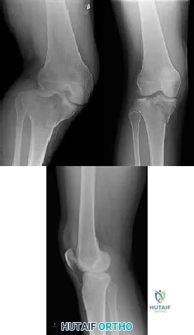
Explanation
Initial management of axially unstable tibial plateau fractures with soft tissue swelling should consist of spanning external fixation and closed manipulative realignment. This allows for soft tissue recovery with the knee joint provisionally stabilized in reduced station. It also provides time for pre-operative planning, which is typically empowered via a CT scan with reconstructions. If the pattern was initially misdiagnosed as a more typically bicondylar tibial plateau fracture, the CT scan will clarify the misconception and allow for better surgical decision making.
Supine positioning is preferred for definitive fixation, but surgical approaches vary. Attempting to stabilize a medial partial articular pattern in the supine position from a lateral utility approach is fraught with difficulties. Lateral locked plating is not designed for this indication. The lateral utility approach allows for visualization of the centrolateral impaction and lateral meniscal peripheral capsular avulsion repair,
but when used alone leads to biomechanically unsound implant placement. The primary plate should be on the medial side of the tibia rather than the intact lateral column.
Question 67
A 20-year-old girl reports a shoulder dislocation while reaching for a high shelf. Her history reveals multiple past dislocations with spontaneous reduction and no obvious traumatic event at onset. A photograph of her hand is shown in figure
Explanation
The first line of treatment for shoulder dislocations in patients with MDI should consist of physical therapy aimed at strengthening the rotator cuff and scapular stabilizers. For those who fail to respond to 3 to 6 months of nonsurgical treatment, surgical intervention can be considered with inferior capsular shift being the procedure of choice for multidirectional instability. Good results have been achieved with surgical procedures for posterior and multidirectional instability, but results have been less predictable than those achieved with procedures for traumatic anterior instability.
Ide et al. evaluated the results of an 8-week rehabilitation program with shoulder-strengthening exercises and a novel scapular-stabilizing shoulder orthosis in 46 patients with MDI. There was a significant increase in mean total scores on the modified Rowe grading system and mean torque of internal and external rotation with a normalization of mean external/internal torque ratios at the completion of the program. The authors concluded that shoulder strengthening exercises represent a
useful treatment option for patients with MDI.
Levine et al. reviewed treatments of multidirectional shoulder instability in athletes. Nonoperative management remains the initial treatment of choice. Open capsular shifts remain the operative treatment of choice, however, arthroscopic electrothermal capsulorrhaphy has become increasingly used as an alternative to an open approach.
Beasley et al. reviewed multidirectional instability in the shoulder of female athletes. The authors note that women tend to have greater ligamentous laxity than men and female athletes have a greater risk of converting laxity to symptomatic instability.
Figure A demonstrates metacarpophalangeal hyperextension, which is a sign of generalized ligamentous laxity.
Incorrect Answers:
Question 68
A 47-year-old man has left-sided motor weakness in the extensor digitorum longus and extensor hallucis longus, sensory loss in the lateral calf and dorsal foot, and no discernible reflex loss.
Explanation
Figures 72a and 72b are the T1-weighted MR images of a disk bulge. Disk bulges are common in asymptomatic people and are not predictive of pathology. Figures 72c and 72d show T1 MR images of a far lateral disk herniation at the L4-L5 level. This would affect the exiting or L4 nerve root. Radicular symptoms would occur at the L4 level. Figures 72e and 72f show an annular tear at the L4-L5 level. This pathology is associated with discogenic low-back pain. Figures 72g and 72h show the T1 MR images of a central lateral disk herniation at the L4-L5 level. This would affect the traversing or L5 nerve root. Radicular symptoms would occur at the L5 level.
RECOMMENDED READINGS
Jarvik JG, Hollingworth W, Heagerty PJ, Haynor DR, Boyko EJ, Deyo RA. Three-year incidence of low back pain in an initially asymptomatic cohort: clinical and imaging risk factors. Spine (Phila Pa 1976). 2005 Jul 1;30(13):1541-8; discussion 1549. PubMed PMID: 15990670.View Abstract at PubMed
Hoppenfeld S. Physical Exam of the Spine and Extremities. Stamford, CT: Appleton and Lange; 1995.
Madigan L, Vaccaro AR, Spector LR, Milam RA. Management of symptomatic lumbar degenerative disk disease. J Am Acad Orthop Surg. 2009 Feb;17(2):102-11. Review. PubMed PMID: 19202123.View Abstract at PubMed
Question 69
- A 12 month old infant has congenital complete absence of the tibia. Examination reveals that the femur in the abnormal limb is 3 cm short, with a normal ipsilateral hip. The patient has an intact fibula, an equinovarus foot with four rays, and moderate popliteal skin webbing. Management should consist of
Explanation
Question 70
Figure 53 is a coronal-cut CT scan of a 63-year-old woman who has a longstanding pes planus. She is seen for lateral ankle discomfort. Upon examination she is tender over the sinus tarsi and distal to the fibula. She has painless passive hindfoot eversion with 5/5 eversion strength. The most appropriate diagnosis is
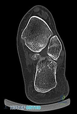
Explanation
With the use of CT scans, adults with symptomatic flatfoot deformity have been noted to develop subluxation of the talocalcaneal joint with resulting lateral hindfoot pain. Impingement of the talus and calcaneus in the sinus tarsi and/or between the tip of the fibula and the calcaneus may occur. This impingement is known as lateral impaction syndrome. Hindfoot motion is painless; therefore, this patient does not have symptomatic subtalar arthritis. Middle facet and calcaneonavicular coalitions are not present (hindfoot motion is present).
RECOMMENDED READINGS
Malicky ES, Crary JL, Houghton MJ, Agel J, Hansen ST Jr, Sangeorzan BJ. Talocalcaneal and subfibular impingement in symptomatic flatfoot in adults. J Bone Joint Surg Am. 2002 Nov;84-A(11):2005-9. PubMed PMID: 12429762. ? View Abstract at PubMed
Ananthakrisnan D, Ching R, Tencer A, Hansen ST Jr, Sangeorzan BJ. Subluxation of the talocalcaneal joint in adults who have symptomatic flatfoot. J Bone Joint Surg Am. 1999 Aug;81(8):1147-54. PubMed PMID: 10466647. ?View Abstract at PubMed
Question 71
Figure 33a shows a line drawing of a normal hemipelvis. The anterior acetabular rim is bold. Figure 33b illustrates a hemipelvis with a crossover sign, which is indicative of what acetabular pathology? Review Topic
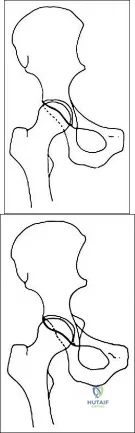
Explanation
Question 72
- A skeletal survey is more accurate than a bone scan for detecting skeletal involvement in which of the following neoplastic diseases?
Explanation
Question 73
Figures 113a and 113b are the radiographs of a 7-year-old girl who was evaluated for a visible elbow deformity by a foster parent. She thought the child fell, but her history was vague. On physical examination, a large prominence was seen over the posterolateral elbow, and the girl lacks the terminal 20 degrees of elbow extension. She has 75 degrees of elbow pronation and supination. She was nontender on examination. What is the most appropriate next treatment step? Review Topic
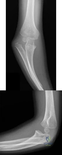
Explanation
Question 74
When performing a Kocher approach to the radial head for open reduction internal fixation the forearm is held in pronation. What structure is this maneuver attempting to protect?

Explanation
Question 75
During preparation for the NCAA wrestling championships, a participant reports the development of vesicular lesions on his right chest wall that are mildly painful; however, they have not affected his ability to wrestle. How should this athlete be managed? Review Topic
Explanation
Question 76
What is the most common clinically significant preventable complication secondary to the treatment of a displaced talar neck fracture?
Explanation
REFERENCES: Tile M: Fractures of the talus, in Schatzker J, Tile M (eds): Rationale of Operative Fracture Care, ed 2. Berlin, Springer-Verlag, 1996, pp 563-588.
Daniels TR, Smith JW, Ross TI: Varus malalignment of the talar neck: Its effect on the position of the foot and on subtalar motion. J Bone Joint Surg Am 1996;78:1559-1567.
Raaymakers EL: Complications of talar fractures, in Tscherne H, Schatzker J (eds): Major Fractures of the Pilon, the Talus, and Calcaneus: Current Concepts of Treatment. Berlin, Springer-Verlag, 1993, pp 137-142.
Question 77
Figure 31 shows the radiograph of a 64-year-old woman who is seen in the emergency department following a motor vehicle accident. She has no
Explanation
Question 78
A 52-year-old woman reports nagging shoulder pain that has been present for months and is slowly progressive in nature. The patient also reports nocturnal pain and notes that the pain is not activity related. Figures 27a and 27b show the radiograph and bone scan, and Figures 27c through 27e show T 1 -weighted, T 2 -weighted, and gadolinium MRI scans, respectively. Based on these findings, what is the most likely diagnosis?
Explanation
REFERENCES: Menendez LR (ed): Orthopaedic Knowledge Update: Musculoskeletal Tumors. Rosemont, IL, American Academy of Orthopaedic Surgeons, 2002, pp 187-194.
Resnick D (ed): Diagnosis of Bone and Joint Disorders. Philadelphia, PA, WB Saunders, 2002, pp 3897-3904.
Question 79
Figures 26a and 26b show the radiograph and MRI scan of an 18-year-old man who fell from a trampoline. Examination reveals exquisite local tenderness at the thoracolumbar junction, but he is neurologically intact. Management should consist of
Explanation
REFERENCES: Carl AL: Adult spine trauma, in DeWald RL (ed): Spinal Deformities: A Comprehensive Text. New York, NY, Thieme, 2003, pp 406-423.
Lewandrowski KU, McLain RF: Thoracolumbar fractures: Evaluation, classification, and treatment, in Frymoyer JW, Wiesel SW (eds): The Adult and Pediatric Spine. Philadelphia, PA, Lippincott Williams and Wilkins, 2004, pp 817-843.
Question 80
A 58-year-old woman with rheumatoid arthritis has progressive neck pain, upper extremity and lower extremity weakness, and difficulty with fine motor movements. Examination reveals hyperreflexia with mild to moderate objective weakness but the patient has no difficulty with ambulation for short distances. What is the most important preoperative imaging finding that predicts full neurologic recovery with surgical stabilization?
Explanation
REFERENCES: Boden SD, Dodge LD, Bohlman HH, et al: Rheumatoid arthritis of the cervical spine: A long-term analysis with predictors of paralysis and recovery. J Bone Joint Surg Am 1993;75:1282-1297.
Boden SD, Clark CR: Rheumatoid arthritis of the cervical spine, in Clark CR (ed): The Cervical Spine, ed 3. Philadelphia, PA, Lippincott Raven, 1998, pp 755-764.
Monsey RD: Rheumatoid arthritis of the cervical spine. J Am Acad Orthop Surg 1997;5:240-248.
Question 81
A 46-year-old man reports occasional squeaking of his hip 2 years after undergoing an uneventful total hip arthroplasty. History reveals no pain, physical examination cannot reproduce audible squeaking, and radiographs show appropriate implant position. What is the most appropriate management?
Explanation
Hopefully, with a better understanding of acoustic phenomena following ceramic total hip arthroplasty, this complication can be minimized.
REFERENCES: Yang CC, Kim RH, Dennis DA: The squeaking hip: A cause for concem-disagrees. Orthopedics
2007;30:739-742.
Walter WL, O’Toole GC, Walter WK, et al: Squeaking in ceramic-on-ceramic hips: The importance of acetabular component orientation. J Arthroplasty 2007;22:496-503.
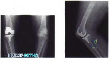
Figure 80a Figure 80b
Question 82
Following an acute dislocation of the patella, the risk of a recurrent dislocation is greater if the patient has which of the following findings?
Explanation
REFERENCES: Cash JD, Hughston JC: Treatment of acute patellar dislocation. Am J Sports Med 1988;16:244-249.
Larsen E, Lauridsen F: Conservative treatment of patellar dislocations: Influence of evident factors on the tendency to redislocation and the theraputic result. Clin Orthop
1982;171:131-136.
Question 83
During revision total knee arthroplasty (TKA), there is significant laxity in 90° of flexion and 10° short of full extension. Correcting the gap imbalance is best achieved by
Explanation
A. Nonoperative treatment with close radiographic follow-up
B. Revision THA with ceramic- on-polyethylene with abductor reconstruction
C. Removal of components and placement of spacer as stage 1 of 2-stage revision
D. Revision THA with metal-on- polyethylene and trochanteric slide
Question 84
A 37-year-old recreational tennis player undergoes surgery for tennis elbow. Following surgery, she describes clicking and popping on the lateral aspect of the elbow. A lateral pivot shift test is positive. What is the most likely cause of her symptoms?
Explanation
REFERENCES: O’Driscoll SW, Morrey BF: Surgical reconstruction of the lateral collateral ligament, in Morrey BF (ed): The Elbow. Philadelphia, PA, Lippincott, Williams and Wilkins, 1994, pp 169-182.
O’Driscoll SW, Bell DF, Morrey BF: Posterolateral rotatory instability of the elbow. J Bone Joint Surg Am 1991;73:440-446.
Question 85
A 45-year-old tennis player undergoes surgery for chronic lateral epicondylitis. After returning to play, he notes increasing lateral elbow pain with mechanical catching and locking. Examination shows positive supine posterolateral rotatory instability. What ligament has been injured?
Explanation
REFERENCES: O’Driscoll SW, Bell DF, Morrey BF: Posterolateral rotatory instability of the elbow. J Bone Joint Surg Am 1991;73:440-446.
O’Driscoll SW, Morrey BF: Surgical reconstruction of the lateral collateral ligament, in Morrey BF (ed): The Elbow. Philadelphia, PA, Lippincott, Williams and Wilkins, 1994, pp 169-182.
Question 86
Medial dislocation of the long head of the biceps tendon in the shoulder is most commonly caused by a
Explanation
REFERENCES: Werner A, Mueller T, Boehm D, et al: The stabilizing sling for the long head of the biceps tendon in the rotator cuff interval: A histoanatomic study. Am J Sports Med 2000;28:28-31.
Arendt EA (ed): Orthopaedic Knowledge Update: Sports 2. Rosemont, IL, American Academy of Orthopaedic Surgeons, 1999, pp 165-189.
Question 87
The MRI scan of a patient with symptomatic shoulder pain reveals subacromial bursitis. What markers have been shown to be significant contributors to this pain? Review Topic
Explanation
Question 88
Figures below show the radiographs, and the CT obtained from a 58-year-old woman who underwent cementless left total hip arthroplasty. Nine months after surgery, she continued to have groin pain when actively flexing her hip. She has trouble walking up stairs and getting out of her car. What is the most likely diagnosis?
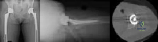
Explanation
Groin pain after total hip replacement has a number of possible causes, and an exact diagnosis may remain elusive in some patients. Infection should be ruled out with laboratory studies and, if indicated, diagnostic aspiration of the hip joint. Implant loosening should be evaluated by plain radiograph and bone scan, if indicated. Synovitis resulting from wear debris should be considered in patients with polyethylene liners who experience late-onset symptoms or in any patient with a metal-on-metal bearing. This patient's symptoms are classic for iliopsoas tendonitis. Physical examination usually reveals pain and weakness with resisted hip flexion. A cross-table lateral radiograph and CT show that the anterior edge of the acetabulum protrudes beyond the anterior wall, thereby acting as a source of iliopsoas tendon irritation. In such cases, acetabular component revision and repositioning are indicated. Fluoroscope-guided iliopsoas cortisone injection can help to establish the diagnosis and relieve groin pain. If the acetabular component is well positioned, then iliopsoas tenotomy should be considered.
Question 89
What neurovascular structure is in closest proximity to the probe in the arthroscopic view of the elbow shown in Figure 50?
Explanation
REFERENCES: Field LD, Altchek DW, Warren RF, et al: Arthroscopic anatomy of the lateral elbow: A comparison of three portals. Arthroscopy 1994;10:602-607.
Andrews JR, Carson WG: Arthroscopy of the elbow. Arthroscopy 1985;1:97-107.
Question 90
Intradiskal electrothermal therapy (IDET) uses an intradiskal catheter to deliver controlled thermal energy to the inner periphery of the annulus fibrosis of a chronically painful intervertebral disk. Lumbar diskography is used diagnostically to identify the presumed pain generator to be targeted with IDET. Based on the medical literature, what can be said about the current status of IDET?
Explanation
REFERENCES: Freeman BJ, Fraser RD, Cain CM, et al: A randomized, double-blind, controlled trial: Intradiscal electrothermal therapy versus placebo for the treatment of chronic discogenic low back pain. Spine 2005;30:2369-2377.
Pauza KJ, Howell S, Dreyfuss P, et al: A randomized, placebo-controlled trial of intradiscal electrothermal therapy for the treatment of discogenic low back pain. Spine J 2004;4:27-35.
Wetzel FT, McNally TA: Treatment of chronic discogenic low back pain with intradiskal electrothermal therapy. J Am Acad Orthop Surg 2003;11:6-11.
Question 91
Figure 1 is the MR image of a 36-year-old athlete who is tackled from behind and falls forward onto his left knee. He has pain, swelling, and stiffness. Examination includes a moderate effusion, positive quadriceps active test, and normal Lachman test finding. The injured structure is composed of an
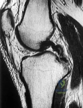
Explanation
Numerous strategies have been described to reduce the risk, including use of a posteromedial accessory incision to allow finger retraction of the popliteal neurovascular bundle, oscillating drills to prevent excessive soft-tissue entanglement, and tapered (rather than square) drill bits that may minimize cut-out of sharp edges as drilling reaches the posterior tibial cortex. Knee extension lessens, rather than increases, the distance between the posterior tibia and the neurovascular bundle and increases, not lessens, risk for vascular injury.
Question 92
Intrinsic muscles of the foot act on the toes by
Explanation
REFERENCES: Myerson MS, Shereff MJ: The pathologic anatomy of claw and hammertoes.
J Bone Joint Surg Am 1989;71:45-49.
Richardson EG (ed): Orthopaedic Knowledge Update: Foot and Ankle 3. Rosemont, IL, American Academy of Orthopaedic Surgeons, 2004, pp 71-80.
Question 93
Figures 15a and 15b are intrasurgical photographs from the posterolateral viewing portal that were taken at the beginning and end of a right shoulder arthroscopic procedure performed on a 54-year-old man. This technique demonstrates superior results compared to traditional arthroscopic techniques when evaluating which outcome?
Explanation
The images reveal a medium-sized tear of the rotator cuff. As more clinical studies are published comparing double-row with single-row rotator cuff repair, it has become clear that the retear rate is lower with a double-row construct for small and medium-sized tears. This may be attributable to the stronger time-zero repair construct that double-row repair provides. No study to date has demonstrated a significant difference in clinical outcomes (functional and pain scores at any time) or time to healing between the 2 techniques.
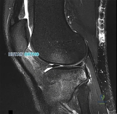
CLINICAL SITUATION FOR QUESTIONS 16 THROUGH 17
Figure 16 is the MR image of a 14-year-old football player who injured his right knee during a game. He describes feeling a "pop," and then he needed help walking off the field. His knee is visibly swollen.
Question 94
- A 25-yo man sustains the injury shown in Figures 29a and 29b as a result of high-speed trauma. Examination reveals diffuse weakness in the lower extremities that is slightly worse on the right side, and decreased rectal tone and sensation. A CT scan is shown in figures 29c and 29d. Definitive treatment of the injury to the spine is delayed because of a severe pulmonary contusion. At 15 days after the injury, the patient’s neurological status remains unchanged. Management should now consist of
Explanation
Question 95
Pedicle subtraction osteotomies (PSO) are commonly performed in the lumbar spine to treat sagittal imbalance. What is the most common complication following a PSO in the lumbar spine? Review Topic
Explanation
Question 96
A 33-year old man sustains a posterior elbow dislocation after a fall. Attempts at closed reduction result in recurrent instability. What is the most common ligamentous injury found at the time of surgical stabilization?
Explanation
with and without associated fractures at the time of surgery. Injury to the lateral collateral ligament complex was seen in every case, with avulsion from the distal humerus as the most common finding. Midsubstance
tears, proximal avulsions, and distal bony avulsions of the ulnar collateral ligament are less common.
Question 97
A 71-year-old woman with coronary artery disease underwent an uncomplicated right total hip arthroplasty for osteoarthritis 12 years ago. Her hip has functioned well until approximately 18 months ago when she noted the spontaneous onset of groin, buttock, and proximal thigh pain that is present at rest and made worse with activity. A radiograph is shown in Figure 15. What is the recommended management at this point?
Explanation
Reassurance and follow-up if symptoms worsen places the patient at risk for further bone loss and periprosthetic fracture. Emergent surgery is not required because the symptoms have been present for more than a year; however, urgent revision hip arthroplasty is recommended when the patient is medically cleared. While there is data to suggest that bisphosphonates may slow the progression of osteolysis in animal modes, there is no clear evidence that bisphosphonate treatment prevents the progression of osteolysis in humans. Additionally, this patient has a loose symptomatic femoral component.
REFERENCES: Chiang PP, Burke DW, Freiberg AA, et al: Osteolysis of the pelvis: Evaluation and treatment. Clin Orthop Relat Res 2003;417:164-174.
Dunbar MJ, Blackley HR, Bourne RB: Osteolysis of the femur: Principles of management. Instr Course Lect 2001;50:197-209.
Rubash HE, Dorr LD, Jacobs JJ, et al: Does alendronate inhibit the progression of periprosthetic osteolysis? Trans
Orthop Res Soc 2004;29:1888.
)
Figure 16a Figure 16b
Question 98
Figures 40a and 40b show the pre- and postoperative radiographs of an 82-year-old woman with bilateral hip pain who has had staged total hip arthroplasties. To minimize potential injury to the sciatic nerve at the time of surgery, the surgeon should
Explanation
limb lengthening.
REFERENCES: Koval KJ (ed): Orthopaedic Knowledge Update 7. Rosemont, IL, American Academy of Orthopaedic Surgeons, 2002, pp 430-431.
Sanchez-Sotelo J, Berry DJ, Trousdale RT, et al: Surgical treatment of developmental dysplasia of the hip in adults: II. Arthroplasty options. J Am Acad Orthop Surg 2002;10:334-344.
Question 99
Figure 31 is the abdominal radiograph of a 70-year-old woman who experiences nausea and abdominal tightness 48 hours following left total knee arthroplasty performed under general anesthesia. She received 24 hours of cefazolin antibiotic prophylaxis and a patient-controlled analgesia narcotic pump for pain management. She has been receiving warfarin for thromboembolic prophylaxis. Her severe abdominal distension and markedly decreased bowel sounds are most likely secondary to
Explanation
The radiograph reveals severe intestinal dilatation, which has occurred as the result of acute colonic pseudo-obstruction and is associated with excessive narcotic administration following total joint arthroplasty. Anesthetic type, antibiotic administration, and warfarin have not been associated with this obstruction. Electrolyte imbalances such as hypokalemia have been associated with postsurgical acute colonic pseudo-obstruction.
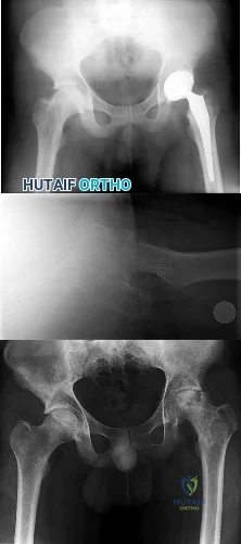
CLINICAL SITUATION FOR QUESTIONS 32 THROUGH 35
Figures 32a through 32c are the radiographs of a 30-year old man who is experiencing right hip pain. He has no current medical problems, but, with a body mass index of 41, he is morbidly obese. He was previously treated for leukemia with chemotherapy that included
high-dose steroids. He undergoes total hip arthroplasty (THA) with a ceramic-on-ceramic bearing.
Question 100
A 30-year-old elite marathon runner reports chronic pain over the lateral aspect of the distal right leg and dysesthesia over the dorsum of the foot with active plantar flexion and inversion of the foot. Examination reveals a tender soft-tissue fullness approximately 10 cm proximal to the lateral malleolus. The pain is exacerbated by passive plantar flexion and inversion of the ankle. There is also a positive Tinel’s sign over the site of maximal tenderness. There is no motor weakness, and deep tendon reflexes are normal. Radiographs and MRI of the leg are normal. What is the next most appropriate step in management?
Explanation
REFERENCES: Styf J: Diagnosis of exercise-induced pain in the anterior aspect of the lower leg. Am J Sports Med 1988;16:165-169.
Sridhara CR, Izzo KL: Terminal sensory branches of the superficial peroneal nerve: An entrapment syndrome. Arch Phys Med Rehabil 1985;66:789-791.
Styf J: Entrapment of the superficial peroneal nerve: Diagnosis and results of decompression.
J Bone Joint Surg Br 1989;71:131-135.
