Orthopedic Board Review MCQs: Trauma, Shoulder & Knee | Part 130
Key Takeaway
This page is Part 130 of a comprehensive OITE/AAOS Orthopedic Board Review series, offering 100 high-yield, verified MCQs. Designed for orthopedic residents and surgeons, these questions are precisely modeled on ABOS and AAOS examinations. This resource provides essential practice to master clinical scenarios and achieve successful board certification.
About This Board Review Set
This is Part 130 of the comprehensive OITE and AAOS Orthopedic Surgery Board Review series authored by Dr. Mohammed Hutaif, Consultant Orthopedic & Spine Surgeon.
This set has been strictly audited and contains 100 100% verified, high-yield multiple-choice questions (MCQs) modelled on the exact format of the Orthopaedic In-Training Examination (OITE) and the American Academy of Orthopaedic Surgeons (AAOS) board examinations.
How to Use the Interactive Quiz
Two distinct learning modes are available:
- Study Mode — After selecting an answer, you immediately see whether you are correct or incorrect, together with a full clinical explanation and literature references.
- Exam Mode — All feedback is hidden until you click Submit & See Results. A live timer tracks elapsed time. A percentage score and detailed breakdown are displayed upon submission.
Pro Tip: Use keyboard shortcuts A–E to select options, F to flag a question for review, and Enter to jump to the next unanswered question.
Topics Covered in Part 130
This module focuses heavily on: Ankle, Deformity, Elbow, Infection, Knee, Ligament, Nerve, Shoulder, Trauma.
Sample Questions from This Set
Sample Question 1: Botulinum toxin is used to treat vasospastic disorders of the hand such as the Raynaud phenomenon to improve digital perfusion and reduce pain. Botulinum toxin enables which transmitter to be unopposed, resulting in vasodilation?...
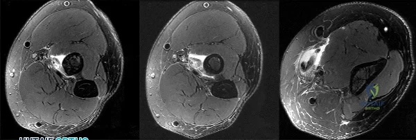
Sample Question 2: Figures 1 through 3 demonstrate the MRIs obtained from a 36-year-old man with an injury to the elbow. The expected result of nonsurgical treatment would be weakness of...
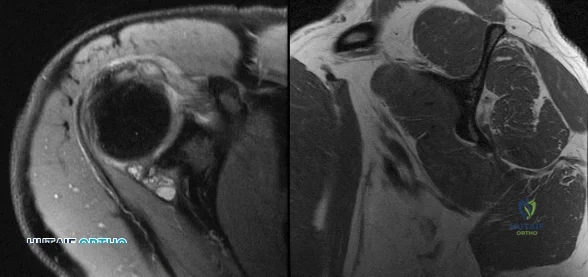
Sample Question 3: Figures 1 and 2 are the right shoulder MRI scans of a 22-year-old right-handed professional male volleyball player with 4 months of right shoulder pain. The pain began insidiously and is exacerbated by overhead activities and hitting during...
Sample Question 4: In the anterior cruciate ligament-deficient knee, what structure provides an important secondary restraint to anterior tibial translation? Review Topic...
Sample Question 5: Figure 14 shows the clinical photographs and radiograph of an 8-year-old girl who has a progressive equinus deformity of the right ankle. There is no history of trauma or infection. What is the most likely diagnosis?...
Why Active MCQ Practice Works
Evidence consistently demonstrates that active recall through spaced MCQ practice yields substantially greater long-term retention than passive reading alone (Roediger & Karpicke, 2006). All questions in this specific module have been algorithmically verified for clinical integrity and complete explanations.
Comprehensive 100-Question Exam
00:00
Start Quiz
Question 1
Botulinum toxin is used to treat vasospastic disorders of the hand such as the Raynaud phenomenon to improve digital perfusion and reduce pain. Botulinum toxin enables which transmitter to be unopposed, resulting in vasodilation?
Explanation
Nitric oxide is the only transmitter listed that is not inhibited by botulinum toxin. Substance P and glutamate are inhibited by botulinum toxin from release by pain nociceptors, thus reducing pain. Fonseca and associates have postulated that botulinum toxin inhibits the RhoA kinase pathway by blocking reactive oxygen species, which in turn does not allow actin/myosin to activate, thus preventing vasoconstriction of smooth muscle. Blocking the RhoA kinase pathway allows the action of nitric oxide to be unopposed, causing vasodilation. Nitric oxide is a potent vasodilator. Thus, botulinum toxin promotes nitric oxide activity to increase vasodilation.
Question 2
Figures 1 through 3 demonstrate the MRIs obtained from a 36-year-old man with an injury to the elbow. The expected result of nonsurgical treatment would be weakness of
Explanation
The axial T2-weighted MRIs demonstrate a distal biceps rupture. The increased signal is noted superficial to the brachialis muscle and adjacent to the biceps tuberosity. The distal biceps tendon is not seen in the distal cuts and has retracted proximally. The physical examination of patients with these injuries show abnormal contour of the arm and tenderness in the antecubital fossa. The hook test is a provocative maneuver that documents biceps integrity. When performing the maneuver, the examiner attempts to hook a finger around the distal biceps tendon while the patient actively supinates with the elbow held in flexion. Nonsurgical treatment has been documented to result in an average loss of 40% of supination strength and 30% of elbow flexion strength. Repair is optimal within several weeks of injury. The alternative options would not occur with a distal biceps rupture.
Question 3
Figures 1 and 2 are the right shoulder MRI scans of a 22-year-old right-handed professional male volleyball player with 4 months of right shoulder pain. The pain began insidiously and is exacerbated by overhead activities and hitting during games. He has maintained a daily program of shoulder stretching and strengthening exercises but has experienced a steady decline in function to the point of not being able to participate in volleyball. Examination reveals some mild atrophy at the posterior shoulder, full forward elevation, mild weakness of external rotation on the right shoulder, negative empty-can testing, positive O’Brien’s and negative apprehension. Surgical intervention would aim to resolve pathology related to which nerve?
Explanation
Question 4
In the anterior cruciate ligament-deficient knee, what structure provides an important secondary restraint to anterior tibial translation? Review Topic
Explanation
Question 5
Figure 14 shows the clinical photographs and radiograph of an 8-year-old girl who has a progressive equinus deformity of the right ankle. There is no history of trauma or infection. What is the most likely diagnosis?
Explanation
REFERENCE: Bottoni CR, Reinker KA, Gardner RD, Person DA: Scleroderma in childhood: A 35-year history of cases and review of the literature. J Pediatr Orthop 2000;20:442-449.
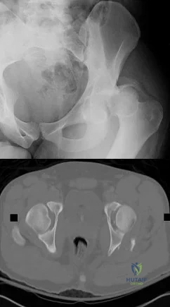
Question 6
A 15-year-old girl who swims the breaststroke has had hip pain after training excessively for a national level competition. Based on the MRI scans shown in Figures 5a through 5c, what is the most likely diagnosis?
Explanation
REFERENCES: Grote K, Lincoln TL, Gamble JG: Hip adductor injury in competitive swimmers. Am J Sports Med 2004;32:104-108.
Clemente C: Anatomy: A Regional Atlas of the Human Body, ed 3. Baltimore-Munich, Urban and Schwarzenberg, 1987, Figures 429, 430.
Question 7
Complications following a reverse shoulder prosthesis occur most frequently when performed for what diagnosis? Review Topic
Explanation
Question 8
Figure 11 shows the radiograph of an otherwise healthy 22-year-old man who sustained a midfoot injury in a motor vehicle accident 9 days ago. Treatment should consist of
Explanation
REFERENCES: Teng AL, Pinzur MS, Lomasney L, et al: Functional outcome following anatomic restoration of the tarsal-metatarsal fracture dislocation. Foot Ankle Int
2002;23:922-926.
Kuo RS, Tejwani NC, DiGiovanni CW, et al: Outcome after open reduction and internal fixation of Lisfranc joint injuries. J Bone Joint Surg Am 2000;82:1609-1618.
Question 9
Figure 1 shows the radiograph of a 60-year-old woman who underwent a previous operation for great toe pain 20 years ago. She has had increasing pain over the past 5 years and now reports pain with any motion, swelling, and clicking. She also reports pain under the ball of foot. What is the most appropriate management to alleviate her metatarsalgia and great toe pain?
Explanation
REFERENCES: Hecht PJ, Gibbons MJ, Wapner KL, et al: Arthrodesis of the first metatarsophalangeal joint to salvage failed silicone implant arthroplasty. Foot Ankle Int 1997;18:383-390.
Myerson MS, Schon LC, McGuigan FX, et al: Results of arthrodesis of the hallux metatarsophalangeal joint using bone graft for restoration of length. Foot Ankle Int
2000;21:297-306.
Question 10
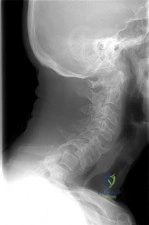
A 45-year-old man undergoes an anterior cervical diskectomy and fusion at C5-6 and C6-7 with instrumentation. During the first postoperative visit at 1 week, the patient reports difficulty swallowing and mild anterior cervical tightness. The anterior wound is benign and the patient denies any dyspnea or shortness of breath. A postoperative radiograph is seen in Figure 25. What is the most appropriate management at this time?
Explanation
6 months with nonsurgical management. A minority of patients experience moderate or severe symptoms by 6 months after the procedure. Female gender and multiple surgical levels have been identified as risk factors for the development of postoperative dysphagia.
REFERENCES: Lee MJ, Bazaz R, Furey CG, et al: Risk factors for dysphagia after anterior cervical spine surgery: A two-year prospective cohort study. Spine J 2007;7:141-147.
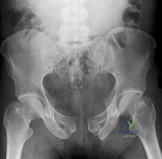
Bazaz R, Lee MJ, Yoo JU: Incidence of dysphagia after anterior cervical spine surgery:
A prospective study. Spine 2002;27:2453-2458.
Question 11
A 51-year-old woman with shoulder pain responds transiently to a subacromial injection and physical therapy exercise program. When her symptoms recur, an arthroscopic subacromial decompression is recommended. During the surgery, a partial-thickness articular-sided supraspinatus tear is noted. The supraspinatus footprint is exposed for 3 mm from the articular margin. The remaining intra-articular structures are normal. Inspection from the bursal surface reveals the tendon to be intact. What is the most appropriate course of management? Review Topic
Explanation
Question 12
An active 66-year-old man who underwent total shoulder arthroplasty 3 years ago now reports pain. Laboratory studies reveal an elevated erythrocyte sedimentation rate and C-reactive protein. Intraoperative frozen section reveals greater than 10 white blood cells per high power field on two slides and the Gram stain reveals gram-positive cocci in clusters. What is the most appropriate surgical treatment to eradicate the infection and maintain function? Review Topic
Explanation
Question 13
Figure 9 shows the AP radiograph of a 65-year-old man who has knee pain and swelling. What is the most likely diagnosis?
Explanation
REFERENCES: Barrack RL, Booth RE Jr, Lonner JH, et al (eds): Orthopaedic Knowledge Update: Hip and Knee Reconstruction 3. Rosemont, IL, American Academy of Orthopaedic Surgeons, 2006, p 188.
Berkow R (ed): The Merck Manual, ed 14. Rathway, NJ, Merck, 1984, pp 910, 1176, 1200.
Question 14
Which of the following lesions most closely resembles Ewing’s sarcoma histologically?
Explanation
REFERENCE: Wold LE, McLeod RA, Sim FH, Unni KK: Atlas of Orthop Pathology. Philadelphia, PA, WB Saunders, 1990.
Question 15
In recurrent posterior shoulder instability, what is the recommended approach to the posterior capsule?
Explanation
REFERENCES: Dreese J, D’Alessandro D: Posterior capsulorrhaphy through infraspinatus split for posterior instability. Tech Shoulder Elbow Surg 2005;6:199-207.
Shaffer BS, Conway J, Jobe FW, et al: Infraspinatus muscle-splitting incision in posterior shoulder surgery: An anatomic and electromyographic study. Am J Sports Med 1994;22:113-120.
Fuchs B, Jost B, Gerber C: Posterior-inferior capsular shift for the treatment of recurrent voluntary posterior subluxation of the shoulder. J Bone Joint Surg Am 2000;82:16-25.
Question 16
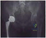
The failure of the acetabular component shown in Figure 15 is most likely the result of the use of a 32-mm head and
Explanation
REFERENCE: Astion DJ, Saluan P, Stulberg BN, Rimnae CM, Li S: The porous-coated anatomic total hip prosthesis: Failure of the metal-backed acetabular component. J Bone Joint Surg Am 1996;78:755-766.
Question 17
Which of the following anatomic structures is labeled 6 in Figure 27?
Explanation
REFERENCES: Hollinshead WH: Anatomy for Surgeons: The Back and Limbs, ed 3. Philadelphia, PA, Harper and Row, 1982, p 467.
Tubiana R, McCullough CJ, Masquelet AC: An Atlas of Surgical Exposures of the Upper Extremity. Philadelphia, PA, JB Lippincott, 1990, p 309.
Question 18
A 12-year-old boy has a head-on head collision while playing soccer. He had no loss of consciousness but has persistent headaches for 2 weeks. The patient is now back to school and has no headaches. What is the best next step?
Explanation
Question 19
A healthy 64-year-old man just underwent an uncomplicated shoulder arthroplasty for severe glenohumeral osteoarthritis. Intraoperatively, 60 degrees of external rotation was obtained. Postoperatively, he starts on a range-of-motion program. What limitations are recommended?
Explanation
REFERENCES: Boardman ND III, Cofield RH, Bengston KA, et al: Rehabilitation after total shoulder arthroplasty. J Arthroplasty 2001;16:483-486.
Matsen FA III, Lippitt SB, Sidles JA, et al: Practical Evaluation and Management of the Shoulder. Philadelphia, PA, WB Saunders, 1994, pp 215-218.
Question 20
What is the most common cause of early failure for patellofemoral arthroplasty?
Explanation
REFERENCES: Lonner JH: Patellofemoral arthroplasty. J Am Acad Orthop Surg 2007;15:495-506. Argenson JN, Flecher X, Parratte S, et al: Patellofemoral arthroplasty: An update. Clin Orthop Relat Res 2005;440:50-53.
Question 21
A 37-year-old electrician is diagnosed with a frozen shoulder after sustaining an electrical injury at work 2 weeks ago. Examination reveals that he cannot actively or passively externally rotate or abduct the arm. The glenohumeral joint and scapula move in a 1:1 ratio. Radiographs are shown in Figures 15a and 15b. The best course of action should be
Explanation
REFERENCES: Hawkins RJ, Neer CS II, Pianta RM, Mendoza FX: Locked posterior dislocation of the shoulder. J Bone Joint Surg Am 1987;69:9-18.
Norris TR (ed): Orthopaedic Knowledge Update: Shoulder and Elbow. Rosement, IL, American Academy of Orthopaedic Surgeons, 1997, pp 181-189.
Rockwood CA Jr, Thomas SC, Matsen FA III: Subluxations and dislocations about the glenohumeral joint, in Rockwood CA Jr, Green DP, Bucholz RW (eds): Fractures in Adults, ed 3. Philadelphia, PA, JB Lippincott, 1991, vol 1, pp 1058-1065.
Question 22
When harvesting iliac crest bone graft during a posterior spinal decompression and fusion, injury to what structure can result in painful neuromas or numbness over the skin of the buttocks?
Explanation
REFERENCES: An HS: Principles and Techniques of Spine Surgery. Baltimore, MD, Williams and Wilkins 1998, pp 770-773.
Kurz LT, Garfin SR, Booth RE Jr: Harvesting autogenous iliac bone grafts: A review of complications and techniques. Spine 1989;14:1324-1331.
Mrazik J, Amato C, Leban S, et al: The ilium as a source of autogenous bone grafting: Clinical considerations. J Oral Surg 1980;38:29-32.
Question 23
Figure 7 shows the MRI scan of a 23-year-old competitive rugby player who has anterior ankle pain and swelling. He states that he has been playing for many years and has sprained his ankle several times. Examination will reveal what specific hallmark feature?
Explanation
REFERENCES: Ogilvie-Harris DJ, Mahomed N, Demaziere A: Anterior impingement of the ankle of the ankle treated by arthroscopic removal of bony spurs. J Bone Joint Surg Br 1993;75:437-440.
Cannon LB, Hackney RG: Anterior tibiotalar impingement associated with chronic ankle instability. J Foot Ankle Surg 2000;39:383-386.
Question 24
Which of the following patients is considered the most appropriate candidate for an isolated split posterior tendon transfer?
Explanation
REFERENCES: Drennan JC (ed): The Child’s Foot and Ankle. New York, NY, Raven Press, 1992, pp 291-294.
Green NE, Griffin PP, Shiavi R: Split posterior tibial-tendon transfer in cerebral palsy. J Bone Joint Surg Am 1983;65:748-754.
Kling TF Jr, Kaufer H, Hensinger RN: Split posterior tibial-tendon transfers in children with cerebral spastic paralysis and equinovarus deformity. J Bone Joint Surg Am 1985;67:186-194.
Question 25
Radiographs of an 80-year-old woman with back pain reveal a compression fracture. Which of the following imaging studies best evaluates the acuity of the fracture?
Explanation
REFERENCES: Phillips FM: Minimally invasive treatments of osteoporotic vertebral compression fractures. Spine 2003;28:S45-S53.
Rao RD, Singrakhia MD: Painful osteoporotic vertebral fracture: Pathogenesis, evaluation, and roles of vertebroplasty and kyphoplasty in its management. J Bone Joint Surg Am 2003;85:2010-2022.
Question 26
A 20-year-old man reports painless snapping about the lateral aspect of the right hip. He denies any history of trauma. Examination reveals no limp or tenderness. Hip range of motion is full, and there is good strength. Radiographs are normal. What anatomic structure is most likely causing these symptoms?
Explanation
REFERENCES: White RA, Hughes MS, Burd T, et al: A new operative approach in the correction of external coxa saltans: The snapping hip. Am J Sports Med 2004;32:1504-1508.
Faraj AA, Moulton A, Sirivastava VM: Snapping iliotibial band: Report of ten cases and review of the literature. Acta Orthop Belg 2001;67:19-23.
Choi YS, Lee SM, Song BY, et al: Dynamic sonography of external snapping hip syndrome.
J Ultrasound Med 2002;21:753-758.
Question 27
A 14-year-old Little League pitcher who plays in 2 leagues concurrently has pain in his throwing shoulder while pitching but not at rest.
Explanation
Multidirectional shoulder instability can be diagnosed by demonstrating instability in at least 2 planes. The sulcus sign is often present with a prominent depression below the acromion when traction is applied to the arm. The mechanism of anterior shoulder dislocation or subluxation is most commonly a combination of abduction, external rotation, and a posteriorly directed force applied to the arm. Among baseball players, the lead shoulder is susceptible to posterior capsulolabral lesions termed “batter’s shoulder.” SLAP tears are common among overhead
athletes and can cause symptoms similar to impingement as well as a glenohumeral internal rotation deficit, which may predispose players to labral tears. Little League shoulder is an overuse injury typically seen in baseball pitchers who are around 14 years of age. It is an osteochondrosis of the proximal humeral epiphysis attributable to overuse from throwing.
RECOMMENDED READINGS
Kang RW, Mahony GT, Harris TC, Dines JS. Posterior instability caused by batter's shoulder. Clin Sports Med. 2013 Oct;32(4):797-802. doi: 10.1016/j.csm.2013.07.012. Epub 2013 Aug 22. Review. PubMed PMID: 24079435. View Abstract at PubMed
Carson WG Jr, Gasser SI. Little Leaguer's shoulder. A report of 23 cases. Am J Sports Med. 1998 Jul-Aug;26(4):575-80. PubMed PMID: 9689382. View Abstract at PubMed
Ren H, Bicknell RT. From the unstable painful shoulder to multidirectional instability in the young athlete. Clin Sports Med. 2013 Oct;32(4):815-23. doi: 10.1016/j.csm.2013.07.014. Review. PubMed PMID: 24079437. View Abstract at PubMed
Werner BC, Brockmeier SF, Miller MD. Etiology, Diagnosis, and Management of Failed SLAP Repair. J Am Acad Orthop Surg. 2014 Sep;22(9):554-565. Review. View Abstract at PubMed
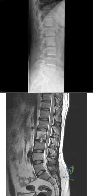
Question 28
A B C Figures 62a through 62c are the MR images and CT scan of a 65-year-old man with a history of diabetes mellitus, hypertension, and smoking. He has a 6-week history of increasing midback pain, lower extremity pain, and weakness. What is the most likely diagnosis, and how should this diagnosis be confirmed?
Explanation
The sagittal T2-weighted and axial T2-weighted images show a lesion within the T8 vertebral body that involves the posterior elements. There is an associated epidural component that results in compression of the spinal cord. The sagittal reconstructed CT image shows a lytic lesion within the T8 vertebral body. This pattern of vertebral body involvement with preservation
of the adjacent disks and endplates in a 65-year-old patient is most compatible with a diagnosis of a tumor. The most likely tumor is a metastatic lesion. A CT-guided biopsy will confirm this diagnosis. Although thoracic tuberculosis does not typically cross the disk space, the lack of an anterior soft-tissue component decreases the likelihood of this diagnosis.
RECOMMENDED READINGS
Khanna AJ, Shindle MK, Wasserman BA, Gokaslan ZL, Gonzales RA, Buchowski JM, Riley LH 3rd. Use of magnetic resonance imaging in differentiating compartmental location of spinal tumors. Am J Orthop (Belle Mead NJ). 2005 Oct;34(10):472-6. Review. PubMed PMID: 16304794. View Abstract at PubMed
White AP, Kwon BK, Lindskog DM, Friedlaender GE, Grauer JN. Metastatic disease of the spine. J Am Acad Orthop Surg. 2006 Oct;14(11):587-98. Review. PubMed PMID: 17030592. View Abstract at PubMed
Question 29
Figure 89a is the radiograph of a 24-year-old man who was involved in a motor vehicle accident. A closed reduction is performed and a post-reduction CT scan is shown in Figure 89b. What is the next most appropriate step in management?
Explanation
Question 30
Compared with retention of the native patella in primary total knee arthroplasty, routine patellar resurfacing is associated with
Explanation
Despite concerns regarding fracture, osteonecrosis, and patellar clunk, the routine retention of the native patella during primary total knee replacement is associated with a 20% to 30% increased revision risk in
large joint registries. In addition, the retention of the native patella results in a 5.7% revision surgery rate in patients with anterior knee pain.
Question 31
The use of a screw between the clavicle and the coracoid process to maintain the clavicle and acromioclavicular (AC) joint in a reduced position is a treatment option for AC joint separations. Screw removal is generally recommended after soft-tissue healing. What effect does this rigid coracoclavicular fixation have on shoulder kinematics?
Explanation
REFERENCES: Flatow EL: The biomechanics of the acromioclavicular, sternoclavicular, and scapulothoracic joints. Instr Course Lect 1993;42:237-245.
Kenedy JC, Cameron H: Complete dislocation of the acromioclavicular joint. J Bone Joint Surg Br 1954;36:202-208.
Rockwood CA Jr, Williams GR, Young CD: Disorders of the acromioclavicular joint, in Rockwood CA Jr, Matsen FA III (eds): The Shoulder. Philadelphia, PA, WB Saunders, 1998, vol 1, pp 483-553.
Inman VT, Saunders JB, Abbott LC: Observations of the function of the shoulder joint. Clin Orthop 1996;330:3-12.
Question 32
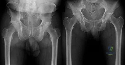
Figures below demonstrate the radiographs obtained from a 56-year-old man with a 3-year history of right groin pain. A comprehensive nonsurgical program has failed, and the patient would like to proceed with total hip arthroplasty. He is seen by a pain management specialist and is currently taking 40 mg of sustained-release morphine twice daily with oxycodone 10 mg 2 to 3 times a day for severe pain. What is the recommended course of action regarding his chronic narcotic use?
Explanation
Chronic opioid consumption prior to total joint arthroplasty has been associated with increased pain after surgery, increased opioid requirements, a slower recovery and longer hospital stay, and higher 90-day postoperative complications compared with patients not on chronic opioids preoperatively. Based on this information, Nguyen and associates performed a study in three patient groups that included 1) chronic opioid users who underwent no preoperative intervention, 2) chronic opioid users who were weaned down to 50% of their prior opioid regimen, and 3) patients who were not chronic opioid users. The authors found that the reduction of preoperative opioid use improved postoperative function, pain, and recovery and that the weaned group performed more like the opioid naive group than the chronic opioid user
group. Increasing opioid use prior to surgery in this patient would make it more difficult to control pain after surgery. Stopping all of his opioids just prior to surgery would place the patient at substantial risk for opioid withdrawal and is not recommended. Avoiding the use of all narcotics and using only acetaminophen postoperatively is very unlikely to provide appropriate pain relief in a chronic opioid user. The recommendation based on the provided literature is to decrease the patient's narcotic use prior to
surgery.
Question 33
Ganglion cysts about the wrist most commonly arise from what structure?
Explanation
REFERENCE: Thornburg LE: Ganglions of the hand and wrist. J Am Acad Orthop Surg 1999;7:231-238.
Question 34
A patient reports persistent anterior shoulder pain following a forceful external rotation injury to the shoulder. An MRI scan is shown in Figure 4. The patient remains symptomatic despite 3 months of nonsurgical management. Treatment should now consist of Review Topic
Explanation
Question 35
A 72-year-old man was involved in an automobile accident 4 weeks ago. Initially he noted pain about his nondominant left shoulder, which resolved within a few weeks after the accident. He now describes trouble with gripping and carrying items in his left hand. Radiographs are shown in Figures 20a through 20c. His signs and symptoms are the result of injury to which of the following ligaments?
Explanation
REFERENCES: Berger RA: The ligaments of the wrist: A current overview of anatomy with considerations of their potential functions. Hand Clin 1997;13:63-82.
Cohen MS, Taleisnik J: Direct ligamentous repair of scapholunate dissociation with capsulodesis augmentation. Tech Hand Up Extrem Surg 1998;2:18-24.
Question 36
The mother of a 5-year-old child reports that he has had a fever of 103°F (39.4°C), leg swelling, and has been unwilling to bear weight on his right lower leg for the past 7 days. Examination reveals point tenderness at the distal femur. Aspiration at the metaphysis yields 10 mL of purulent fluid, and a Gram stain reveals gram-positive cocci. In addition to hospital admission, management should include
Explanation
REFERENCE: Kasser JR (ed): Orthopaedic Knowledge Update 5. Rosemont, Ill, American Academy of Orthopaedic Surgeons, 1996, pp 149-161.
Question 37
Based on the type of articulation shown in Figure 32, wear is not affected by which of the following factors?
Explanation
REFERENCES: Amstutz HC, Grigoris P: Metal on metal bearings in hip arthroplasty. Clin Orthop 1996;329:S11-S34.
Amstutz HC, Campbell P, McKellop H, et al: Metal on metal total hip replacement workshop consensus document. Clin Orthop 1996;329:S297-S303.
McKellop H, Park SH, Chiesa R, et al: In vivo wear of three types of metal on metal hip prostheses during two decades of use. Clin Orthop 1996;329:S128-S140.
Question 38
A 17-year-old African American high school football player is in the afternoon session of an August “2-a-day” practice. He tells his trainer he is experiencing weakness, dizziness, and nausea. The ambient temperature is 31°C with a relative humidity of 70%. An examination by the team trainer reveals a body temperature of 39°C and headache, chills, confusion, and disorientation. What is the most likely diagnosis?
Explanation
Heat exhaustion and heatstroke are both forms of heat illness during which the body is unable to self-regulate internal temperature. The hallmarks of heatstroke are altered mental status and/or core temperature higher than 40°C. Heat exhaustion may be marked by nausea, vomiting, headache, dizziness, chills, and excessive sweating, but there are no mental status
changes. In heatstroke, sweating can often slow or cease as dysregulation worsens. Simple dehydration would not result in mental status changes or elevated core temperature. Sickle-cell crisis is marked by extreme pain, with location depending on the site of crisis. Four main patterns are common: bone, chest, abdominal, or joint crises. Sickle-cell crisis can be precipitated by dehydration, although it also can occur as a result of cold exposure.
Question 39
A 21-year-old professional ballet dancer reports a painful popping sensation over her right hip joint. Examination reveals that symptoms are reproduced with hip flexion and external rotation. Which of the following studies will best confirm the diagnosis?
Explanation
REFERENCES: Gruen GS, Scioscia TN, Lowenstein JE: The surgical treatment of internal snapping hip. Am J Sports Med 2002;30:607-613.
Garrick JG (ed): Orthopaedic Knowledge Update: Sports Medicine 3. Rosemont, IL, American Academy of Orthopaedic Surgeons, 2004, pp 139-153.
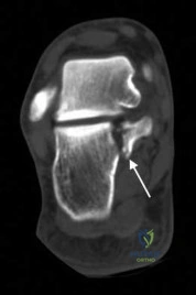
Question 40
Following a fall from a height of 5 feet, a patient reports pain along the lateral border of the foot. The CT scan shown in Figure 54 indicates what pathology?
Explanation
REFERENCE: Robbins MI, Wilson MG, Sella EJ: MR imaging of anterosuperior calcaneal process fractures. Am J Roentgenol 1999;172:475-479.
Question 41
Treatment of adhesive capsulitis has a high failure rate when the underlying cause is
Explanation
REFERENCES: Fisher L, Kurtz A, Shipley M: Association between cheiroarthropathy and frozen shoulder in patients with insulin-dependent diabetes mellitus. Br J Rheumatol 1986;25:141-146.
Janda DH, Hawkins RJ: Shoulder manipulation in patients with adhesive capsulitis and diabetes mellitus: A clinical note. J Shoulder Elbow Surg 1993;2:36-38.
Pollock RG, Duralde XA, Flatow EL, Bigliani LU: The use of arthroscopy in the treatment of resistant frozen shoulder. Clin Orthop 1994;304:30-36.
Question 42
A 42-year-old woman who has had an 18-month history of severe low back pain is referred to your office for surgical evaluation. She reports that the pain initially began with right lower extremity pain and management consisted of oral analgesics, nonsteroidal anti-inflammatory drugs, and muscle relaxants. She has seen a chiropractor as well as a pain management specialist and she is status-post epidural steroid injections. She has also completed exhaustive physical therapy, as she is a certified athletic trainer and runs a health fitness program at a community hospital. Currently, she denies lower extremity pain and her pain is isolated to her low back and is subjectively graded as 8/10, with 10 being the worst pain she has ever experienced. The pain is interfering with her activities of daily living and she is seeking definitive treatment. Figures 32a through 32c show current MRI scans. Based on the current available medical literature, what is the most appropriate treatment? Review Topic
Explanation
In a recent updated Cochrane Review of surgery for degenerative lumbar spondylosis, it was noted that while Fritzell and associates appeared to provide strong evidence in favor of fusion, a more recent trial by Brox and associates demonstrated no difference between those patients undergoing lumbar fusion compared to those receiving cognitive intervention and exercise. The Cochrane Review suggests that this may reflect a difference between the control groups. Fritzell and associates compared lumbar fusion to standard 1990s “usual care,” whereas Brox and associates compared lumbar fusion to a “modern rehabilitation program.” Bear in mind that this patient is a certified athletic trainer and runs a hospital health fitness department; therefore, at least for purposes of this question, it can be assumed that she has participated in a “modern rehabilitation program.” The Cochrane Review goes on to state that preliminary results of three small trials of intradiskal electrotherapy suggest that it is ineffective and that preliminary data from three trials of disk arthroplasty do not permit firm conclusions.
Question 43
Figures 21a through 21c show the MRI scans of a 21-year-old football player who sustained a valgus knee injury while changing direction. Examination reveals swelling and tenderness along the medial aspect of the knee. There is a positive Lachman test, 3+ valgus laxity at 30 degrees, and 1+ valgus laxity at 0 degrees extension. The anterior drawer test is increased with the tibia in external rotation. The increase in the anterior drawer test with the tibia in external rotation is most likely the result of
Explanation
REFERENCES: Warren LA, Marshall JL, Girgis F: The prime static stabilizer of the medial side of the knee. J Bone Joint Surg Am 1974;56:665-674.
Indelicato PA: Injury to the medial capsuloligamentous complex of the knee, in Feagin J (ed): The Crucial Ligaments, ed 2. 1994, pp 351-360.
Question 44
A minimally invasive plate osteosynthesis is seen in Figure 15. The resultant fracture healing can best be attributed to a fixation construct that was
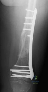
Explanation
Question 45
A 38-year-old man reports a 2-week history of acute lower back pain with radiation into the left lower extremity. There is no history of trauma and no systemic signs are noted. Examination reveals a positive straight leg test at 35 degrees on the left side and a contralateral straight leg raise on the right side. Motor testing demonstrates mild weakness of the gluteus medius and weakness of the extensor hallucis longus of 3+/5. Sensory examination demonstrates decreased sensation along the lateral aspect of the calf and top of the foot. Knee and ankle reflexes are intact and symmetrical. Radiographs demonstrate no obvious abnormality. MRI scans show a posterolateral disk hernation. The diagnosis at this time is consistent with a herniated nucleus pulposus at
Explanation
REFERENCES: Hoppenfeld S: Orthopedic Neurology. Philadelphia, PA, JB Lippincott, 1977, pp 45-74.
Spivak JM, Connolly PJ (eds): Orthopaedic Knowledge Update: Spine 3. Rosemont, IL, American Academy of Orthopaedic Surgeons, 2006, pp 43-56.
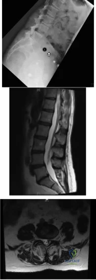
Question 46
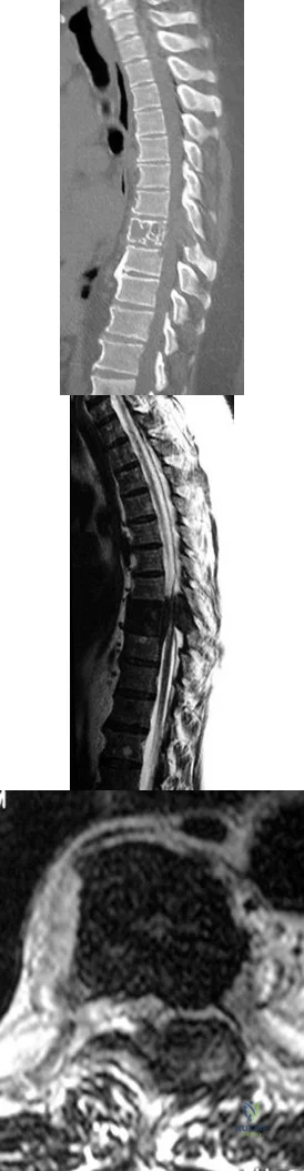
A 42-year-old man with a history of renal cell carcinoma has progressive weakness in the lower extremities for the past 3 weeks. The patient desires intervention. A sagittal T 2 -weighted MRI scan is shown in Figure 9a, and a sagittal contrast enhanced T 1 -weighted MRI scan is shown in Figure 9b. He currently ambulates minimal distances with a walker. His life expectancy is 8 months. Treatment of the spine lesion should consist of
Explanation
REFERENCES: Spivak JM, Connolly PJ (eds): Orthopaedic Knowledge Update: Spine 3. Rosemont, IL, American Academy of Orthopaedic Surgeons, 2006, pp 351-366.
White AP, Kwon BK, Lindskog DM, et al: Metastatic disease of the spine. J Am Acad Orthop Surg 2006;14:587-598.
Question 47
Figures 5a and 5b show the radiograph and MRI scan of a patient who has severe mechanical neck pain but no neurologic problems. Biopsy and work-up show the lesion to be a solitary plasmacytoma. Treatment should consist of
Explanation
REFERENCES: Corwin J, Lindberg RD: Solitary plasmacytoma of bone vs. extramedullary plasmacytoma and their relationship to multiple myeloma. Cancer 1979;43:1007-1013.
Durr HR, Wegener B, Krodel A, et al: Multiple myeloma: Surgery of the spine. Retrospective analysis of 27 patients. Spine 2002;27:320-324.
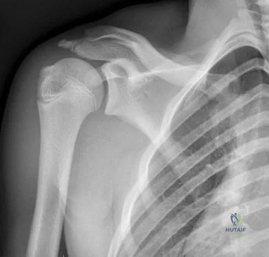
Question 48
A 72-year-old woman who fell on her right shoulder while using a treadmill is now unable to elevate her right arm. An MRI scan is shown in Figure 7. What is the most likely diagnosis?
Explanation
REFERENCE: Gerber C, Myer DC, Schneeberger AG, et al: Effect of tendon release and delayed repair on the structure of the muscles of the rotator cuff: An experimental study in sheep. J Bone Joint Surg Am 2004;86:1973-1982.
Question 49
The Lisfranc ligament connects the base of the
Explanation
REFERENCES: Sarrafian SK: Anatomy of the Foot and Ankle: Descriptive, Topographic, Functional, ed 2. Philadelphia, PA, JB Lippincott, 1993.
Solan MC, Moorman CT III, Miyamoto RG, et al: Ligamentous restraints of the second tarsometatarsal joint: A biomechanical evaluation. Foot Ankle Int 2001;22:637-641.
Question 50
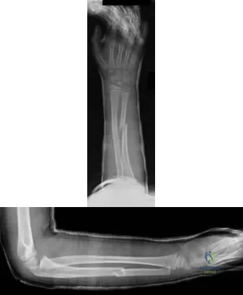
A 45-year-old male falls off his motorcycle and injures his arm. AP and lateral radiographs reveal an ulnar shaft fracture, 30 degrees apex anterior, and a radial head dislocation. Which direction is the radial head most likely dislocated?

Explanation
Question 51
Smoking has been associated with lower fusion rates in both cervical and lumbar fusion. Which of the following statements best describes an explanation for these findings?
Explanation
have been performed to offer an explanation of the mechanism mediating this effect. Whereas all of the above have been postulated as explanations, more recent studies have demonstrated that nicotine delivered via a transdermal patch significantly enhanced posterior spinal fusion in rabbits. Thus it appears that the effects of smoking on fracture healing are multifactorial and not yet fully understood.
Question 52
Posterior sternoclavicular dislocations are most commonly associated with which of the following complications?
Explanation
REFERENCES: Brooks AL, Henning GD: Injury to the proximal clavicular epiphysis, abstracted. J Bone Joint Surg Am 1972;54:1347-1348.
Beaty JH (ed): Orthopaedic Knowledge Update 6. Rosemont, IL, American Academy of Orthopaedic Surgeons, 1999, pp 255-260.
Question 53
Which of the following is the best method of initial pelvic stabilization for a patient with hemodynamic instability and the pelvic ring injury seen in Figure 199?
Explanation
Question 54
Intramedullary nailing of proximal tibial shaft fractures are technically demanding, and use of an extended medial parapatellar incision with a semiextended technique can prevent what common deformity at the fracture site?

Explanation
Lang et al. reported in their study of 32 proximal third tibia fractures that 56% of the fractures had 5 degrees or more valgus angulation and 28% had 10 degrees or more valgus angulation. Angulation in the AP plane ranged from 0 degrees to 20 degrees, all of which was apex anterior. Nineteen (59%) fractures demonstrated 5 degrees or more angulation, and 7 (22%) fractures demonstrated 10 degrees of more angulation.
Tornetta advocates use of extended medial parapatellar incision with the leg in a semiextended position to allow for a more proximal and lateral starting point. This modified starting point forces the nail to overcome the tendency of the fracture to flex (apex anterior) and go into valgus.
Question 55
A patient falls off a roof and sustains the fracture shown in Figure 29. What is the most likely complication that results from injury to the structure that is located at the arrow?
Explanation
Question 56
During total hip arthroplasty, what characteristic of irradiated (10 Mrad) and subsequently melted highly cross-linked polyethylene should provide a more wear-resistant construct than traditional gamma-irradiated (2.5-4 Mrad)-in-air polyethylene mated with the same head?
Explanation
Highly cross-linked polyethylene makes material resistant to adhesive wear. Abrasive wear from third bodies does not decrease wear. The fatigue strength of such material is inferior to that of traditional polyethylene, and its resistance to creep is the same, if not lower, than that of traditional polyethylene.
Question 57
A 51-year-old woman with no preoperative neurologic deficit is undergoing elective anterior cervical diskectomy and fusion (ACDF) with plating and fusion for a C5-6 disk herniation with right-sided neck pain. Thirty minutes into the surgery the neurophysiologic monitoring shows a rapid drop and then loss of amplitude in the right cortical somatosensory-evoked potential waveform. All other waveforms remained normal and unchanged, including right-sided cervical (subcortical) and peripheral (Erb’s point), and those from the left-sided upper extremity and both lower extremities. What is the most likely cause of the change? Review Topic
Explanation
Question 58
- What is the most common nerve injury following a Monteggia fracture-dislocation of the forearm in adults?
Explanation
Question 59
Which of the following findings is most prognostic for the ability of a young child with cerebral palsy to walk? Review Topic
Explanation
(SBQ13PE.27) A 15-year-old male patient presents requesting clearance to perform in the Special Olympics. He has had abnormal facies, has had mental developmental delay and cytogenetic analysis confirmed abnormalities on chromosome 21. Orthopaedically, he has been treated in the past for an elbow dislocation as well as bilateral patellar dislocation. He has already visited his cardiologist and endocrinologist and has been cleared. He has no complaints, denies any pain, difficulties with walking and reports that his training has been going well. What is the next best step? Review Topic
With a normal physical exam, patient can be cleared for participation
MRI bilateral knees and elbow to ensure no ligamentous injury
Referral to a neurologist for clearance
Lateral cervical spine flexion and extension radiographs
AP pelvis radiograph
Ruling-out C1-C2 instability with flexion/extension radiographs is necessary prior to any spine surgery or participation in sports in patients with Down's Syndrome.
Patients with Down's Syndrome typically present with generalized ligamentous laxity and decreased tone. Thus, dislocations (elbow or patella) along with asymptomatic instability in the cervical spine can commonly occur. Imaging analysis is necessary prior to sports participation.
McKay et al. performed a systematic review to summarize all congenital causes of cervical instability. They found in Down's patients, cervical instability due to ligamentous laxity is found mostly at C1-2. They recommend asymptomatic patients
with an ADI <4.5mm can resume unrestricted activities, while asymptomatic patients between 4.5-10mm should not participate in contact sports. With ADI >10mm OR symptoms/cord changes on MRI, surgery is recommended. Symptomatic patients with ADI between 4.5-10mm should be observed with activity restriction.
Dedlow et al. outlines the most recent 2011 update and guidelines for cervical instability in Down's syndrome patients. One of the major highlighted changes is the emphasis placed on radiographic re-examination, regardless of prior clearance. Re-examination should occur prior to participation in sports and/or the onset of new symptoms.
Illustrations A, B and C highlight the C1-2 instability on flexion-extension lateral radiographs. Careful attention can be placed on the relationship of the anterior arch of C1 and the dens (Illustration C). This allows for the measurements of ADI and the space available for the cord (SAC), which is highlighted in Illustration D.
Incorrect answers:
Question 60
Figure 31 shows the radiograph of a 64-year-old woman who is seen in the emergency department following a motor vehicle accident. She has no
Explanation
Question 61
A 27-year-old runner training for his first marathon reports lateral knee pain after an unusually long training run. He states that the most significant pain occurs while running downhill. Examination of the patient while he is laying on the unaffected side reveals increased pain when manual pressure is applied to the lateral femoral epicondylar area during knee range of motion of 30° to 45°. What is the most likely diagnosis?
Explanation
REFERENCES: Noble CA: The treatment of iliotibial band friction syndrome. Br J Sports Med 1979;13:51-54.
James SL: Running injuries to the knee. J Am Acad Orthop Surg 1995;3:309-318.
James SL, Jones DV: Biomechanical aspects of distance running, in Cavanagh PR (ed): Biomechanics of Distance Running. Champaign, IL, Human Kinetic Books, 1990, pp 249-269.
Question 62
Which of the following patients requires preoperative noninvasive cardiac testing?
Explanation
REFERENCES: Bushnell BD, Horton JK, McDonald MF, et al: Perioperative medical comorbidities in the orthopaedic patient. J Am Acad Orthop Surg 2008;16:216-227.
Auerback A, Goldman L: Assessing and reducing the cardiac risk of noncardiac surgery. Circulation 2006;113:1361-1376.
Fischgrund JS (ed): Orthopaedic Knowledge Update 9. Rosemont, IL, American Academy of Orthopaedic Surgeons, 2008, pp 105-113.
Question 63
Figures 59a and 59b are the radiographs of a 7-year-old boy who was seen 1 week after he underwent a closed reduction and casting in the emergency department after a fall on an outstretched arm. What is the most appropriate next step for this patient? Review Topic
Explanation
Question 64
A 78-year-old man with ankylosing spondylitis sustains a minor fall. Shortly afterward he experiences sudden worsening of his chronic back pain and is brought to the emergency department by his caregiver. Radiographs and a CT scan of the spine do not show a clear fracture. What is the most appropriate next step?
Explanation
Patients with ankylosing spondylitis are at high risk for occult fractures after low-energy injuries. Although radiographs and a CT scan do not demonstrate a spinal fracture in this patient, high risk for an unstable occult fracture necessitates further imaging with MRI to ensure that no fractures are missed. Although a CT scan is typically the primary imaging modality for workup of spine injuries in similar patients, CT and MRI complement each other and each detects fractures that are missed using the other modality. A CT myelogram might detect cord or root compression but would not aid in the diagnosis of an occult fracture. Nonsteroidal anti-inflammatory drugs are first-line treatment for idiopathic low-back pain. In a patient with ankylosing spondylitis at high risk for fracture, further workup is needed to rule out an occult fracture. Flexion and extension radiographs of the spine are inferior to MRI for evaluating occult fractures and ligamentous injuries. The primary concern for this patient remains an unstable spinal fracture, which necessitates an MRI of the spine before initiating a workup for other possible causes of his back pain.
RECOMMENDED READINGS
Duane TM, Cross J, Scarcella N, Wolfe LG, Mayglothling J, Aboutanos MB, Whelan JF, Malhotra AK, Ivatury RR. Flexion-extension cervical spine plain films compared with MRI in the diagnosis of ligamentous injury. Am Surg. 2010 Jun;76(6):595-8. PubMed PMID: 20583514. View Abstract at PubMed
Hitchon PW, From AM, Brenton MD, Glaser JA, Torner JC. Fractures of the thoracolumbar spine complicating ankylosing spondylitis. J Neurosurg. 2002 Sep;97(2 Suppl):218-22. PubMed PMID: 12296682. View Abstract at PubMed
Koivikko MP, Koskinen SK. MRI of cervical spine injuries complicating ankylosing spondylitis. Skeletal Radiol. 2008 Sep;37(9):813-9. doi: 10.1007/s00256-008-0484-x. Epub 2008 Apr
Question 65
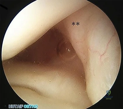
A 56-year-old woman presents with left hip pain and diminishing range of motion. Examination reveals pain with range of motion of the hip. Radiographs reveal multiple calcific lesions within the hip and well-preserved joint space. MRI scan shows thickened synovium nodular loose bodies with decreased signal on T1 and T2. What is the best next step?
Explanation
Question 66
The main blood supply to the lateral two thirds of the talar body is provided by the
Explanation
REFERENCES: Sanders R: Fractures and fracture-dislocations of the talus, in Coughlin MJ, Mann RA (eds): Surgery of the Foot and Ankle, ed 7. St Louis, MO, Mosby, 1999,
pp 1465-1518.
Haliburton RA, Sullivan CR, Kelly PJ, et al: The extra-osseous and intra-osseous blood supply of the talus. J Bone Joint Surg Am 1958;40:1115-1120.
Question 67
Figures 1 and 2 are the radiographs of a 20-year-old college multisport athlete who has had longstanding pain in his left hip. He denies any specific event that initiated his pain, but he notes that he had hip problems when he was an infant. He denies pain with activities of daily living, but he believes his pain is increasingly limiting his ability to exercise. He localizes the pain to his groin. He denies low-back or buttock pain or pain that radiates down his leg. What examination findings are most consistent with the pathology seen in the radiographs?
Explanation
trochanter renders trochanteric bursitis unlikely. Although athletic pubalgia can be a source of longstanding groin pain, he lacks the pain with a resisted sit-up and tenderness along the pubic ramus that is frequently noted in patients with pubalgia. His radiographs reveal a focal femoral neck prominence consistent with cam impingement, although pistol grip deformities and flattening of the lateral femoral head are often present as well. His MRI scan shows a labral tear, which is common in cam impingement. Surgical treatment for cam impingement can be effective for symptomatic patients. Even among high-level athletes, open surgical dislocation of the hip has been shown to have good results. Most patients with cam impingement can be treated with arthroscopic osteoplasty and achieve results comparable with those realized with open surgical dislocation. The literature describes success in terms of athletes returning to sports (even professional athletes) to be approximately 90% after arthroscopic treatment. Byrd and Jones described five patients who developed transient neurapraxias that resolved uneventfully. The patients in his series who had concomitant microfracture had a 92% return to sports within the follow-up period. Cam impingement has long been thought to be associated with a history of a slipped capital femoral epiphysis. The capitis in these patients is displaced posteriorly, resulting in a prominent anterior femoral neck and decreased hip internal rotation. Pincer impingement is associated with a deep acetabulum, such as protrusion acetabula and acetabular retroversion. A patient who underwent a periacetabular osteotomy can develop a more retroverted acetabulum as well.
Question 68
A 46-year-old male construction worker has right hip pain that has failed to respond to nonsurgical management. His body mass index (BMI) is 32, he is 6’2” tall, and he has no other medical comorbidities. AP and lateral radiographs of the right hip are shown in Figures 23a and 23b. The patient inquires about his suitability for metal-on-metal hip resurfacing. The patient should be educated that he is at higher risk for failure secondary to which of the following?
Explanation
The results of hip resurfacing arthroplasty have been reported to be best in young, male patients who are younger than 55 years of age with a diagnosis of osteoarthritis. Although some authors advocate metal- on-metal hip resurfacing as an option for patients with osteonecrosis of the femoral head, in this particular patient, given the size of the necrotic segment, he would be at higher risk for failure and a conventional total hip arthroplasty would be a more conservative option. As the acetabulum is resurfaced in metal- on-metal hip resurfacing, the secondary changes of the acetabulum are not an issue and his BMI is in an acceptable range for the procedure.
REFERENCES: Mont MA, Ragland PS, Etienne G, et al: Hip resurfacing arthroplasty. J Am Acad Orthop Surg 2006;14:454-463.
Revell MP, McBryde CW, Bhatnagar S, et al: Metal-on-metal hip resurfacing in osteonecrosis of the femoral head. J Bone Joint Surg Am 2006;88:98-103.
Buergi ML, Walter WL: Hip resurfacing arthroplasty: The Australian experience. J Arthroplasty 2007;22:61-65. Question 24
A 31-year-old woman had disabling right knee pain. An arthroscopic assessment reveals chondromalacia of both the lateral femoral condyle and tibial plateau. The standing femorotibial axis measures 10 degrees of valgus. The
optimum treatment of this condition should include
distal femoral varus osteotomy.
osteoarticular transplant to the lateral femoral condyle.
unicondylar arthroplasty.
high tibial osteotomy.
Fulkerson tibial tubercle transfer. PREFERRED RESPONSE: 1
DISCUSSION: The long-term outcome of a distal femoral varus osteotomy has been quite favorable and should remain the primary choice for this young active woman. Sharma and associates have shown that a 5-degree valgus malalignment has a five-fold chance of progressing at least one grade within 18 months, making a corrective osteotomy the most important surgical maneuver.
REFERENCES: Sharma L, Song J, Felson DT, et al: The role of knee alignment in disease progression and function decline in knee osteoarthritis. JAMA 2001 ;286:188-195.
Murray PB, Rand JA: Symptomatic valgus knee: The surgical options. J Am Acad Orthop Surg 1993; 1:19.
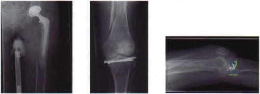
Figure 25a Figure 25b Figure 25c
Question 69
A 27-year-old man has recurrent right shoulder instability. He first dislocated his shoulder in college while playing rugby and was treated nonsurgically. Since then, he has sustained nearly 1 dozen dislocations and says that his shoulder always feels “loose.” The shoulder recently dislocated in his sleep and while he was putting on clothes. Which factor is a contraindication to an arthroscopic soft-tissue repair?
Explanation
There is much debate in the literature regarding optimal techniques for treatment of shoulder instability. Although some studies suggest that open stabilization may result in lower recurrence rates in contact athletes, this approach is now under scrutiny. Extensive labral involvement (posterior labral involvement in this scenario) is likely more accessible via arthroscopic methods. Although HAGL lesions may be more easily accessible via an open approach (particularly for inexperienced arthroscopists), numerous authors describe successful repair via arthroscopic techniques. Among these responses, the strongest indication for an open approach, including possible bony transfer, is high-grade glenoid bone loss. Although the critical amount of bone loss is a topic of debate, most surgeons and authors suggest a cutoff of 20% to 25%.
CLINICAL SITUATION FOR QUESTIONS 90 THROUGH 92
Figure 90 is the radiograph of a 14-year-old pitcher who plays in a year-round baseball program and has vague pain in his dominant shoulder. The pain occurs with throwing, and it has been worsening for 2 months. Pain typically occurs during the late cocking phase of throwing. He has no tenderness of the rotator cuff and 5/5 rotator cuff strength. His arc of motion is symmetric between his dominant and nondominant arms. The sulcus sign is negative.
Question 70
During percutaneous iliosacral screw placement for an unstable pelvic ring injury, use of the lateral sacral fluoroscopic image is critical to help avoid iatrogenic injury to what structure?

Explanation
In the 2000 reference by Routt et al, they state "a thorough knowledge of pelvic osseous anatomy, injury patterns, deformities, and their fluoroscopic correlations are mandatory for percutaneous pelvic fixation to be effective."
Illustration A shows a representative lateral sacral radiograph, with the major anatomic landmarks labeled. Safe SI screw insertion in the S1 body should be underneath the sacral ala line to minimize risk of a "in-out-in" screw that would come out in the area of the ala and injure the L5 nerve root that sits directly on top of this structure. Dysmorphic pelvic rings will often have a more vertical sacral line, or one that starts more inferiorly.
Question 71
A year-old woman has severe anterior knee pain. Her radiographs indicate end-stage patellofemoral compartment osteoarthritis. The tibiofemoral compartments are preserved. Extensive nonsurgical treatment has failed to provide relief, and she is offered patellofemoral arthroplasty (PFA). What is the most common long-term mode of failure for PFA using an implant with an onlay prosthesis design?
Explanation
Contemporary onlay-design trochlear prostheses in PFA replace the entire anterior trochlear surface. Previous inlay designs were inset within the native trochlea and carried a higher risk of catching and patellar instability, particularly in patients with trochlear dysplasia; they also generally have higher failure rates. The current most common mode of failure is progression of arthritis throughout the knee, in some series as high as 25% at 15 years. Aseptic loosening, particularly of cemented implants, is less common. Infection is an uncommon long-term complication. Patients considering PFA should be advised of the risk of arthritis progression. Many authors routinely obtain a preoperative MRI to assess the status of the tibiofemoral compartments.
Question 72
- Which of the following medicolegal relationships between an attending surgeon and a resident assistant applies when a patient files a malpractice suit relating to surgical complications following a total knee arthroplasty?
Explanation
Question 73
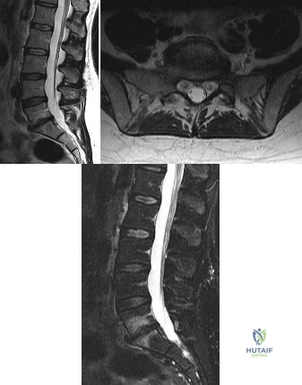
A 56-year-old male presents to your office with a primary complaint of pain in his lower back that extends down his left leg when he walks. He states he rides a stationary bike without pain, but he has severe pain walking more than two blocks. On exam he has 5/5 strength in all major muscle groups, and his sensation is intact to light touch in all dermatomes. He has no upper motor neuron signs. The pain has been going on for about a year, and he has had no improvement with physical therapy or anti-inflammatory medication. Figure A is an upright lateral radiograph of his lumbar spine. Figures B is his sagittal MRI, and Figure C is an axial image through L4/5. Assuming this patient is going to undergo surgery, what is most important in ensuring longterm symptomatic relief? Review Topic
Explanation
Degenerative spondylolisthesis occurs when there is anterior translation of one vertebral body (most commonly L4) on another vertebral body (most commonly L5). When this occurs, there can be compression of the traversing/caudal (L5) nerve root in the lateral recess. Because of the instability associated with this diagnosis, a fusion is needed to preserve longterm outcomes.
Kornblum et al., reported on the outcomes of 47 patients with degenerative spondylolisthesis at an average follow-up of 7 years and 8 months. Excellent to good results were reported in 86% of patients with a solid arthrodesis compared to 56% of patients with a pseudarthrosis.
Weinstein et al., reported the 4-year data from the SPORT study and found that patients with degenerative spondylolisthesis treated with surgery had statistically significant improvements in health related quality of life scores compared to those treated non-operatively.
Figure A is an upright lateral radiograph of the lumbar spine in which a degenerative spondylolisthesis of L4 on L5 is identified. Figure B is a sagittal T2 MRI re-demonstrating the spondylolisthesis as well as spinal stenosis. FIgure C is the axial image through L4/5 demonstrating spinal stenosis; additionally, a left sided facet cyst can be seen.
Illustration A and B are the postoperative films from the same patient after he underwent an L4/5 posterior decompression and instrumented fusion.
Incorrect
(SBQ13PE.7) A 45-year-old HIV-positive homeless man presents with increasing low back pain for the last three weeks. He now reports difficulty ambulating, fever, and loss of appetite. He denies bowel and bladder symptoms. He denies any symptoms radiating into his buttock or legs. On physical exam he has in obvious discomfort with standing which worsens in the forward flexion position. He has a normal motor and sensory exam in his lower extremities. Blood cultures are performed which come back negative. What would be the most next appropriate step in treatment? Review Topic
Broad spectrum antibiotics
Isoniazid, rifampin, and pyrazinamide therapy
CT guided biopsy with cultures
Technetium bone scan
Anterior corpectomy with a retroperitoneal approach, strut grafting and instrumentation
The clinical presentation is consistent with spondylodiscitis. Although the patient has risk factors for spinal tuberculosis, a CT guided biopsy should be performed to establish a diagnosis.
There is an increasing incidence of TB in United States due to increasing immunocompromised population from HIV. 15% of patients with TB will have extrapulmonary involvement. 5% of all TB patients have spine involvement. With any type of spondylodiscitis the infectious organism must be identified with blood cultures or a biopsy prior to initiating treatment.
Khoo et al. emphasize with the recent global pandemic of human immunodeficiency virus, the number of tuberculosis and secondary spondylitis cases is again increasing at an alarming rate. They report that medical treatment alone remains the cornerstone of therapy for the majority of Pott disease cases. Surgical intervention should be limited primarily to cases of severe or progressive deformity and/or neurological deficit.
Hadjipavlou et al. performed a retrospective study of 101 cases of spondylodiscitis. They found Staphylococcus aureus was the main organism. Infection elsewhere was the most common predisposing factor. Leukocyte counts were elevated in 42.6% of spondylodiscitis cases. The erythrocyte sedimentation rate was elevated in all cases of epidural abscess.
Figure A shows lumbar radiograph with a radiolucent lesion in L2 with mild loss of disc height of the L2/3 level. Figure B shows a T2-weighted MRI showing a lesion involving the L2/3 disc spaced and extending into the L2 vertebral body.
Incorrect Answers:
Question 74
A 22-year-old ballet dancer undergoes hip arthroscopy for increasing hip pain and popping with activity. She experiences complete resolution of signs and symptoms post-operatively. Her pre- and post-operative magnetic resonance sagittal images shown in Figure A (left, pre-operative; right, post-operative). Which of the following pre-operative physical examination findings may have been positive? Review Topic
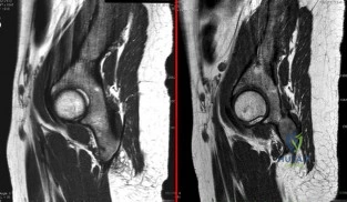
Explanation
Snapping hip exists in 3 forms: (1) external snapping hip, which is caused by the iliotibial band (ITB) sliding over the greater trochanter, (2) internal snapping hip, and
(3) intraarticular snapping hip, which is caused by loose bodies (traumatic, or from synovial chondromatoses) or labral tears. While painless snapping hip requires no treatment, painful snapping hip may be addressed with activity modification, physical therapy, steroid injections. Surgical release (ITB z-plasty or psoas tenotomy) is indicated if nonoperative management is unsuccessful.
Ilizaliturri et al. evaluated the results of endoscopic iliopsoas tendon release at the lesser trochanter (10 patients) vs endoscopic transcapsular psoas release from the peripheral compartment (9 patients). There were improvements in WOMAC scores in both groups, and no difference between groups. They conclude that both techniques are equally effective.
Marquez Arabia et al. evaluated if the psoas tendon regenerates after tenotomy in 27 patients. At 23 months, they found that tendon regeneration occurred in all patients, to
a mean circumference of 84% of the original. One patient had persistent pain, but all had 5/5 hip flexion strength. They hypothesize that the bulk of iliopsoas muscle fibers attaches directly to the proximal femoral shaft without a tendon, preventing retraction and allowing regeneration to occur easily.
Figure A shows pre- and post-operative arthroscopic psoas tenotomy magnetic resonance sagittal images. Illustration A shows the psoas tendon (white arrows) prior to transection. Illustration B shows the psoas tendon after transection (green arrows, proximal tendon segment; yellow arrows, distal segment). Illustration C and D are diagrams showing release at the level of the lesser trochanter and hip joint respectively.
Incorrect
90 degrees, but full external rotation. Answer 2: Decreased internal rotation and a positive impingement test (forced
flexion, adduction, femoroacetabular Answer 4: These
and internal
rotation) are classic findings
findings may
be found with intra-articular
for cam-type impingement loose bodies.
Question 75
When converting the knee shown in Figure 20 to a total knee arthroplasty, satisfactory outcome can be expected in what percent of patients?
Explanation
REFERENCE: Naranja RJ Jr, Lotke PA, Pagnano MW, Hanssen AD: Total knee arthroplasty in a previously ankylosed or arthrodesed knee. Clin Orthop 1996;331:234-237.
Question 76
What is the best approach to reduce and stabilize a displaced volar lunate facet fracture of the wrist?
Explanation
REFERENCES: Hanel DP, Jones MD, Trumble TE: Wrist fractures. Orthop Clin North Am 2002;33:35-57.
Trumble TE, Culp RW, Hanel DP, et al: Intra-articular fractures of the distal aspect of the radius. Instr Course Lect 1999;48:465-480.
Question 77
An elderly woman with radiographic evidence of spinal stenosis reports difficulty walking and calf pain that is relieved by rest and a change of position. The most likely cause of pain is ischemia of the
Explanation
Question 78
Which of the following pieces of equipment currently offers the greatest opportunity for lowering the number of equestrian injuries? Review Topic
Explanation
> or = 12). Injuries included the chest (54%), head (48%), abdomen (22%), and extremities (17%). Only 9% of riders wore helmets, and 64% believed the accident was preventable. The authors noted that "helmet and vest use will be targeted in future injury prevention strategies." In another study, Frankel and associates noted that helmet use was only documented in 34% of riders. Although orthopaedic injuries are common, knee pads, wrist guards, boots, and quick release stirrups would most likely have less impact on injury prevention.
Question 79
A 7-year-old boy has a swollen and deformed right arm after falling off his bicycle. Radiographs reveal a completely displaced posterolateral supracondylar humeral fracture. Examination reveals a warm, pink hand and forearm but absent pulses. What is the next most appropriate step in management?
Explanation
REFERENCES: Shaw BA: The role of angiography in assessing vascular injuries associated with supracondylar humerus fractures remains controversial. J Pediatr Orthop 1998;18:273.
Sabharwal S, Tredwell SJ, Beauchamp RD, et al: Management of pulseless pink hand in pediatric supracondylar fractures of humerus. J Pediatr Orthop 1997;17:303-310.
Schoenecker PL, Delgado E, Rotman M, et al: Pulseless arm in association with totally displaced supracondylar fracture. J Orthop Trauma 1996;10:410-415.
Question 80
What structure is most often injured in a volar proximal interphalangeal joint dislocation?
Explanation
REFERENCES: Doyle JR: Extensor tendons: Acute injuries, in Green DP, Hotchkiss RN (eds): Operative Hand Surgery, ed 3. New York, NY, Churchill Livingstone, 1993, p 1925.
Newport ML: Extensor tendon injuries in the hand. J Am Acad Orthop Surg 1997;5:59-66.
Question 81
A 79-year-old healthy male has 1 year of progressively worse left hip pain. He denies any significant weight loss but does complain of night pain. Radiograph and MRI are shown in Figures A & B. Bone scan and histology is shown in Figures C-E. What is the definitive treatment of this?
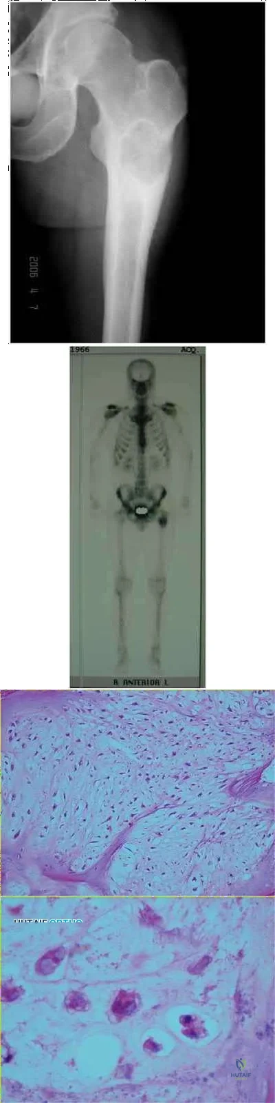
Explanation
Chondrosarcoma tends to occur in an older individuals and the most common sites of origin are the femur, tibia, humerus, ribs, scapula then pelvis.
Chondrosarcomas grow slowly and patients present with dull aching pain.
X-rays typically have a subtle, radiolucent, permeative lesion or may have hazy or speckled calcifications with either a diffuse "salt & pepper" pattern or a more discrete "popcorn" pattern. The MRI defines the amount of marrow and soft-tissue involvment, and typically has high T2 signal intensity. Bone scintigraphy will be positive.
In pathological sections, low-power imaging demonstrates lobulated clusters of chondrocytes, and high-power imaging demonstrates a bland cellular appearance, extensive basophilic cytoplasm, and no mitotic figures (low grade chondrosarcoma).
Question 82
- A patient undergoes an acute repair of a laceration of the median nerve in the antecubital fossa. A lack of functional recovery 6 months later is most likely due to
Explanation
Functional recovery is generally complete after a crush injury because the basement membrane and endoneurium are left intact, and the damaged axons can regenerate within their original endoneurial tubes and reinnervate their original target organ. After a complete lesion to the nerve, however, functional recovery of movement is often quite poor. The loss of functional recovery probably is related to the failure of the axons to regenerate and the misdirection of regenerating axons, which leads to inappropriate innervation of denervated muscles. Inappropriate innervation is thought to result in a loss in the ability to accurately recruit individual muscles and motor units within a muscle, resulting in the loss of motor control.
Question 83
Figure 91 is the radiograph of a 20-year-old man who kicked a door while intoxicated. At the emergency department, his leg is placed into a long-leg cast. After 2 hours, he reports increasing pain, numbness, and tingling in his toes. What is the most appropriate initial treatment?
Explanation
(SBQ12TR.88) When evaluating a fracture dislocation of the elbow, a varus and posteromedial rotation mechanism of injury typically results in what injury pattern?
A fracture of the radial head requiring ORIF
A highly comminuted radial head fracture requiring radial head arthroplasty or resection
An MCL injury requiring repair
A type I avulsion fracture of the coronoid
An anteromedial coronoid fracture
A varus and posteromedial rotation mechanism of injury typically results in a fracture of the anteromedial facet of the coronoid which frequently requires reduction and fixation to restore stability.
A varus and posteromedial mechanism of injury about the elbow presents with an injury pattern distinctly different from other injury patterns. A key part of treating this injury pattern is recognizing a fracture of the anteromedial facet of the coronoid, which often requires reduction and fixation to restore stability about the elbow. It is important to recognize this during preoperative planning since this injury typically requires a medial approach.
Steinman presents a review article describing coronoid fracture patterns and their mechanisms of injury.
Doornberg and Ring present a level 4 review showing that coronoid fracture patterns and their required treatments are predictable based on mechanism of injury. Varus and posteromedial mechanisms were found to reliably create a fracture of the anteromedial facet of the coronoid, and were associated with sparing of the MCL and radial head.
Doornberg and Ring also presented a Level 3 review of anteromedial facet cornoid fractures. They found that they could not be adequately visualized and treated from a lateral approach, and that they typically required reduction and fixation to restore adequate stability to the elbow. This stresses the importance of recognizing this injury pattern during preoperative planning.
Illustrations A and B are AP and lateral radiographs of an elbow following a varus/posteromedial injury with an anteromedial coronoid facet fracture. Illustration C is a diagram demonstrating fracture lines that create an anteromedial facet fracture fragment. This fracture can be subclassified into three subtypes [anteromedial rim (a), rim plus tip (b), and rim and tip plus the sublime tubercle (c)]
Incorrect answers:
(SBQ12TR.78) A 67-year-old female patient presents with increasing right hip/thigh pain over the past three months, which is now recalcitrant to anti-inflammatories. There is no history of trauma or constitutional symptoms. Her past medical history consists of hypertension, coronary artery disease, osteoporosis and gastric reflux. Physical examination reveals mild pain at the extremes of range of motion of the hip and a painful right sided limp. A radiograph of the right hip is seen in Figure A. What would be the most appropriate treatment for this patient at this time? Review Topic
Observation only
Referral to physiotherapy
MRI spine and hip
Total hip arthroplasty
Intramedullary femoral nail
This osteoporotic female patient is presenting with subtrochanteric lateral cortical thickening and hip pain. This is consistent with an insufficiency fracture of the femur secondary to use of bisphosphonate medication for treatment of osteoporosis. The most appropriate treatment would be intramedullary femoral nail fixation.
Bisphosphonate medications have been shown to be associated with atypical (subtrochanteric) femur fractures. These patients often have prodromal hip pain and lateral cortical thickening on radiographs prior to fracture. In addition, there has shown to be a significantly increased risk of fracture in the presence of the “dreaded black line” that occurs at the site of thickening.
Lenart et al. examined a case series of patients using bisphosphonates for the treatment of osteoporosis. They identified 15 postmenopausal women who had been receiving alendronate for a mean (±SD) of 5.4±2.7 years and who presented with atypical low-energy fractures. Cortical thickening was present in the contralateral femur in all the patients with this pattern.
Goh et al. retrospectively reviewed patients who had presented with a low-energy subtrochanteric fractures. They identified 13 women of whom nine were on long-term alendronate therapy. Five of these nine patients had prodromal pain in the affected hip in the months preceding the fall, and three demonstrated a stress reaction in the cortex in the contralateral femur.
Figure A shows a right hip radiograph with subtrochanteric lateral cortical thickening. There is mild arthritic changes in the hip. Illustration A shows a bone scan and radiographs of subtrochanteric lateral cortical thickening that resulted in fracture.
Incorrect Answers
Question 84
The husband of a 22-year-old woman has hypophosphatemic rickets. The woman has no orthopaedic abnormalities, but she is concerned about her chances of having a child with the same disease. What should they be told regarding this disorder?
Explanation
REFERENCES: Herring JA: Metabolic and endocrine bone diseases, in Tachdjian’s Pediatric Orthopaedics, ed 3. New York, NY, WB Saunders, 2002, pp 1685-1743.
Sillence DO: Disorders of bone density, volume, and mineralization, in Rimoin DL, Conner JM, Pyerite RE, et al (eds): Principles and Practice of Medical Genetics, ed 4. New York, NY, Churchill Livingstone, 2002.
Staheli LT: Practice of Pediatric Orthopedics. Philadelphia, PA, Lippincott Williams & Wilkins, 2001.
Question 85
A football lineman who sustained a traumatic injury while blocking during a game now reports that his shoulder is slipping while pass blocking. Examination reveals no apprehension in abduction and external rotation; however, he reports pain with posterior translation of the shoulder. He has full strength in external rotation, internal rotation, and supraspinatus testing. What is the pathology most likely responsible for his symptoms?
Explanation
A traumatic blow to the outstretched arm results in posterior glenohumeral forces. Labral detachment at the glenoid rim is common. Patients report slipping or pain with posteriorly directed pressure. Rarely do these patients have true dislocations that require reduction; however, recurrent episodes of subluxation or pain are not uncommon. Posterior repair has
been shown to be successful in the treatment of traumatic instability.
REFERENCES: Bottoni CR, Franks BR, Moore JH, et al: Operative stabilization of posterior shoulder instability. Am J Sports Med 2005;33:996-1002.
Williams RJ III, Strickland S, Cohen M, et al: Arthroscopic repair for traumatic posterior shoulder instability. Am J Sports Med 2003;31:203-209.
Kim SH, Ha KI, Park JH, et al: Arthroscopic posterior labral repair and capsular shift for traumatic unidirectional recurrent posterior subluxation of the shoulder. J Bone Joint Surg Am 2003;85:1479-1487.
Question 86
below show the radiographs, MRI, and MR arthrogram obtained from a year-old collegiate soccer player who has new-onset left groin pain. He played competitive soccer from a young age and has competed or practiced 5 to 6 times per week since the age of He denies any specific hip injury that necessitated treatment, but his trainer contends that he had a groin pull. He reports groin pain with passive flexion and internal rotation of the left hip, and his hip has less internal rotation than his asymptomatic right hip. He is otherwise healthy. What is the primary cause of a cam deformity?
Explanation
Question 87
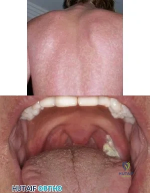
A 19-year-old football player is taken off the field because of fatigue. Examination reveals a rash shown in Figure A. Oral examination reveals findings shown in Figure B. Posterior cervical glands are palpable. A mass is palpable in the left upper quadrant. Which of the following is true regarding the most likely diagnosis? Review Topic
Explanation
IM is caused by the Epstein-Barr virus (EBV). Annual incidence is 1-3% in college freshmen. It is characterized by Hoagland's triad (fever, pharyngitis, lymphadenopathy). Some have rash and splenomegaly. Splenic rupture is rare (0.1-0.2% of patients). It is caused by sudden increase in portal venous pressure from a simple Valsalva maneuver or from external trauma. The risk of rupture is highest in the first 3 weeks of illness.
Putukian et al. reviewed IM and athletic participation. They recommend return to LIGHT activity after 3 weeks from symptom onset when the athlete is afebrile, has a good energy level, and does not have any significant associated abnormalities. They recommend returning to CONTACT sports after at least 3 weeks when the athlete has no remaining clinical symptoms, is afebrile, and has a normal energy level.
Jaworski et al. discussed infectious diseases in athletes. They state that splenic rupture occurs because of lymphocytic infiltration that distorts the support structure of the spleen, leading to fragility. They recommend return to light, non-contact activities once the athlete is afebrile and appropriately hydrated, fatigue has improved, and a minimum period of 3 weeks has passed from symptom onset.
Figure A shows a petechial rash, which can be seen in IM. Amoxicillin increases the risk of rash. Figure B shows unilateral exudative pharyngitis. The left tonsil is
covered
by
a
white
exudate/pseudomembrane.
Incorrect
Question 88
Dislocation following primary total hip arthroplasty is more likely to occur in which of the following situations?
Explanation
REFERENCE: Berry DJ: Dislocation, in Steinberg ME (ed): Revision Total Hip Arthroplasty. Philadelphia, PA, 1999, pp 463-482.
Question 89
01 (left). What is the most appropriate next step?
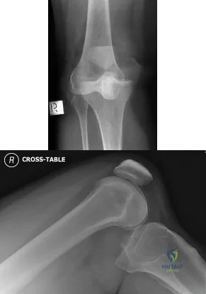
Explanation
Knee dislocations are associated with popliteal artery injury in 18-45% of cases and range from intimal tears to complete transection. Amputation rates of 85% have been reported if revascularization is delayed greater than 6 to 8 hours. Neurologic injury occurs in 15-40% of cases and is most common after posterolateral dislocation. The peroneal nerve is more commonly injured.
Rihn et al. reviewed the acutely dislocated knee. They recommend a vascular consult if pulses are weak, or ABI is compromised. They warn that in arterial injury, pulses, temperature and capillary refill can be normal. If the limb remains ischemic, surgical exploration and revascularization is indicated.
Medina et al. systematically reviewed neurovascular injury after knee dislocation in 862 patients. Vascular injury rate was 18%, and nerve injury rate was 25%. Repair was performed in 80% of vascular injuries, and amputation in 12%. The most vascular injury was seen in KDIIIL injuries (32%) and posterior dislocation (25%).
Figure A is an AP radiograph of a posterior knee dislocation. Figure B is a lateral showing the same injury.
Incorrect Answers:
Question 90
A 40-year-old man who is a manual laborer has had 3 years of worsening medial-sided left knee pain that has inhibited his ability to work. He reports undergoing a left subtotal medial meniscectomy 10 years ago. He has been treated with nonsteroidal anti-inflammatory drugs and two different corticosteroids, with the most recent injection given 1 month ago. Each injection provided him with a few weeks of pain control. His medical history is unremarkable, and he has smoked 20 cigarettes per day for the last 15 years. His BMI is 22. On examination, he has varus alignment of the involved leg and medial joint line tenderness and no lateral or patellofemoral pain. His knee range of motion is 3° shy of full extension to 130° of flexion. He has negative Lachman and posterior drawer test results. He demonstrates no lateral thrust with ambulation. What imaging study is most appropriate to determine treatment options for this patient?
Explanation
Question 91
A workers' compensation carrier for a local manufacturing company requests a second opinion on a 59-year-old man who sustained a crush injury to his foot and leg at work 6 months ago. His leg and foot were pinned between a forklift and a wall when an employee he was supervising lost control of the forklift. The employer suspects that the injured worker is malingering because the treating physician released him to work, but he has not returned to work. Which of the following elements of your history will best help you determine that the injured worker does not want to return to work out of fear of a confrontation with the employee he was supervising?
Explanation
Question 92
Figure 18a shows the initial lateral radiograph of a 6-year-old girl who sustained a fracture in a motor vehicle accident and was treated in a cast 1 year ago. She now has the valgus deformity seen in Figure 18b. Treatment should consist of
Explanation
REFERENCES: Cozen L: Knock-knee deformity in children: Congenital and acquired. Clin Orthop 1990;258:191-203.
Jackson DW, Cozen L: Genu valgum as a complication of proximal tibial metaphyseal fractures in children. J Bone Joint Surg Am 1971;53:1571-1578.
Brammar TJ, Rooker GD: Remodeling of valgus deformity secondary to proximal metaphyseal fracture of the tibia. Injury 1998;29:558-560.
Ogden JA, Ogden DA, Pugh L, et al: Tibia valga after proximal metaphyseal fractures in childhood: A normal biologic response. J Pediatr Orthop 1995;15:489-494.
Salter RB, Best TN: Pathogenesis of progressive valgus deformity following fractures of the proximal metaphyseal region of the tibia in young children. Instr Course Lect 1992;41:409-411.
Question 93
A 1-year-old infant has the hand deformities shown in Figure 40. What pathologic process is the most likely cause of these deformities?
Explanation
REFERENCE: Foulkes GD, Reinker K: Congenital constriction band syndrome: A seventy-year experience. J Pediatr Orthop 1994;14:242-248.
Question 94
Fixed hyperextension of the metatarsophalangeal joint is associated with
Explanation
REFERENCES: Marks RM: Anatomy and pathophysiology of lesser toe deformities. Foot Ankle Clin 1998;3:199-213.
Myerson MS, Shereff MJ: The pathological anatomy of claw and hammer toes. J Bone Joint Surg Am 1989;71:45-49.
Question 95
A 7-year-old girl who sustained a type III posteromedial extension supracondylar fracture underwent a closed reduction at the time of injury. Figure 27a shows the position of the fracture fragments prior to percutaneous medial and lateral pin fixation. Following surgery, healing was uneventful and the patient regained a full painless range of motion. Fifteen months after the injury, she now reports loss of elbow motion and moderate pain with activity. A current AP radiograph is shown in Figure 27b. What is the most likely cause of her symptoms?
Explanation
REFERENCES: Haraldsson S: The interosseous vasculature of the distal end of the humerus with special reference to the capitellum. Acta Orthop Scand 1957;27:81-93.
Morrissy RT, Wilkins KE: Deformities following distal humeral fracture in childhood. J Bone Joint Surg Am 1984;66:557-562.
Question 96
Clinical evidence suggests that grafts for replacing a torn anterior cruciate ligament often stretch after surgery. What is the most probable mechanism for this behavior?
Explanation
REFERENCES: Boorman RS, Thornton GM, Shrive NG, et al: Ligament grafts become more susceptible to creep within days after surgery. Acta Orthop Scand 2002;73:568-574.
Buckwalter JA, Einhorn TA, Simon SR (eds): Orthopaedic Basic Science: Biology and Biomechanics of the Musculoskeletal System, ed 2. Rosemont, IL, American Academy of Orthopaedic Surgeons, 2000, pp 596-609.
Lu L, Kaufman KR, Yaszemski MJ: Biomechanics, in Einhorn TA, O’Keefe RJ,
Buckwalter JA (eds): Orthopaedic Basic Science: Foundations of Clinical Practice, ed 3. Rosemont, IL, American Academy of Orthopaedic Surgeons, 2006, in press.
Question 97
Figure 11 shows the radiograph of a 3-year-old girl who sustained a proximal radius injury. Appropriate initial management should include
Explanation
REFERENCES: Leung AG, Peterson HA: Fractures of the proximal radial head and neck in children with emphasis on those that involve the articular cartilage. J Pediatr Orthop
2000;20:7-14.
Radomisli TE, Rosen AL: Controversies regarding radial neck fractures in children. Clin Orthop 1998;353:30-39.
Skaggs DL, Mirzayan R: The posterior fat pad sign in association with occult fracture of the elbow in children. J Bone Joint Surg Am 1999;81:1429-1433.
Gonzalez-Herranz P, Alvarez-Romera A, Burgos J, et al: Displaced radial neck fractures in children treated by closed intramedullary pinning (Metaizeau technique). J Pediatr Orthop 1997;17:325-331.
Question 98
Which of the following best characterizes the injury shown in Figure 53? Review Topic
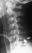
Explanation
Question 99
A 12-year-old girl with foot pain who has been diagnosed with hereditary motor sensory neuropathy is seen for the foot deformity shown in Figure 59a. A “block test” is performed and shown in Figure 59b. What is the most appropriate management for this patient?
Explanation
REFERENCES: Paulos L, Coleman SS, Samuelson KM: Pes cavovarus: Review of a surgical approach
using selective soft-tissue procedures. J Bone Joint Surg Am 1980;62:942-953.
McCluskey WP, Lovell WW, Cummings RJ: The cavovarus foot deformity: Etiology and management. Clin Orthop Relat Res 1989;247:27-37.
Ward CM, Dolan LA, Bennett DL, et al: Long-term results of reconstruction for treatment of a flexible
cavovarus foot in Charcot-Marie-Tooth disease. J Bone Joint Surg Am 2008;90:2631-2642.

Figure 60a Figure 60b Figure 60c
Question 100
5 standard deviations below young normals (< -2.5). The Z-score represents a comparison to age-matched normals.
Explanation
This patient has an impending subtrochanteric femur fracture and should be stabilized with cephalomedullary nailing.
Bisphosphonates have been shown to prevent osteoporotic fractures. They suppress osteoclastic recruitment and activity and induce apoptosis of osteoclasts. However, they have also been associated with subtrochanteric femur fractures. Cortical stress reactions in the form of lateral cortical thickening have been documented when radiographs were performed during the prodromal period preceding these fractures. If radiographs are obtained and demonstrate lateral cortical thickening in the presence of thigh pain, the entire femur should be stabilized with prophylactic cephalomedullary nailing to prevent fracture.
Shane et al. performed a review of atypical subtrochanteric and diaphyseal
femoral fractures. They report that long-term bisphosphonate use is associated with these injuries. Bisphosphonates localize in areas that are developing stress fractures and suppress intracortical remodeling. When bisphosphonate use has stopped, the risk of fracture decreases over time. They conclude that teriparatide may advance the healing of atypical femur fractures after surgical treatment.
Koh et al. studied the natural history of femoral stress lesions associated with bisphosphonate therapy. They determined certain features that predispose to complete stress fractures. They found all patients had thigh pain before fracture. They conclude that cortical stress reactions associated with prolonged antiresorptive therapy and the "dreaded black line" should be prophylactically stabilized to avoid a complete fracture.
Figure A is a radiograph of the proximal femur demonstrating lateral cortical thickening with the "dreaded black line." Illustration A is the same image with an arrow indicating the "dreaded black line."
Incorrect Answers:
Which of the following statements regarding bone mineral density (BMD) is true?
The 2017 American College of Rheumatology/American Association of Hip and Knee Surgeons Guideline for the Perioperative Management of Antirheumatic Medication states that hydroxychloroquine can be continued and etanercept
should be held for 2 weeks prior to undergoing total hip arthroplasty.
Patients with rheumatoid arthritis (RA) report high satisfaction following hip or knee replacement despite the higher rates of infection, dislocation, and readmission rates. Patients with RA may present on a variety of different biologic and nonbiologic medications used to control their systemic RA.
Optimal preoperative management of these immunosuppressant medications may help mitigate some of the risks of postoperative complications in RA patients.
Singh et al. reviewed the evidence surrounding the benefits and harms of various antirheumatic medications. They found evidence for traditional DMARDs, biologic agents, and nonbiologic agents in acute and established RA totaling 74 recommendations. They concluded that these recommendations are not prescriptions and that ultimate decision making should be patient- specific in a shared-decision
making process between the patient and physician.
Goodman et al. performed a multistep literature review on optimal antirheumatic medication management prior to joint replacement surgery. They were able to provide recommendations on when to continue, when to withhold, and when to restart these medications, and the optimal perioperative dosing of glucocorticoids. They concluded that these guidelines will help physicians manage antirheumatic medications at the time of elective THA or TKA.
Incorrect Answers:
An 83-year-old woman presents complaining of thigh pain. The pain has been progressing over the last few months. She denies any night chills or recent weight loss. She has smoked 1 pack per day for the last 40 years. Her current medications are alendronate and citalopram. Her current imaging is shown in Figure A. What is the next best step in treatment?
The patient is presenting with complex regional pain syndrome (CRPS) type 1 after a distal 1/3 tibial shaft fracture. The best diagnostic test for this is a thorough history and physical exam.
CRPS is a disorder of increased sympathetic activity in a region of prior trauma. Cases can be classified as type 1, where there is no demonstrable nerve damage, or type 2, where a specific nerve is affected. Patients will typically present with cool and mottled skin that atrophic and absent of hair. Many times the affected limb will be noticeably cooler than the contralateral side. In advanced stages, there will be joint contractures and extensive osteopenia on radiographs. Several diagnostic aids have been developed, but remain inadequate to diagnostic sensitivity compared to a thorough history and physical.
Shah et al. reviewed the diagnosis and treatment of CRPS. The authors reported that sweat quantification testing, skin thermography, and electromyography may be useful aids in diagnosis, but there is a lack of diagnostic sensitivity to make these tests reliable. The authors concluded that evidence suggests gabapentin, prazosin, propranolol, nifedipine, and mexiletine are the best medications for treatment.
Hogan et al. reviewed the diagnosis and treatment of CRPS. The authors reported that the most sensitive means for diagnosis is a good history and physical exam as there is no single test to confirm the diagnosis. The authors recommended a multidisciplinary team approach including pain specialists, physical therapists, and orthopedic surgeons as syndrome response to medications is variable.
Figures A and B demonstrate an AP and lateral radiograph of the right tibia and fibula with a distal 1/3 tibia fracture. Illustration A depicts that Lankford and Evans classification for CRPS.
Incorrect Answers:
A 72-year-old female with rheumatoid arthritis is scheduled to undergo total hip arthroplasty. She presents for her preoperative visit and asks about dosing of her antirheumatic medications. She currently takes etanercept weekly and hydroxychloroquine daily. Which of the following is the best dosing recommendation for her antirheumatic medications prior to surgery?
the entire right lower extremity, with sensitivity to cold temperatures. Physical exam demonstrates hyperesthesia of the extremity, thin and shiny appearing skin, cyanotic appearing with skin cool to the touch. What is the likely diagnosis and what is the best diagnostic test?
In a 5-year-old female without a history of trauma or rashes and with persistent oligoarthritis that improves during the day, the most likely diagnosis is juvenile idiopathic arthritis (JIA). Early-onset JIA is associated with chronic uveitis.
JIA is defined by the American College of Rheumatology as persistent arthritis and swelling in one or more joints for 6 weeks or longer in a patient younger than 16 years of age. It is a diagnosis of exclusion, usually entailing pattern recognition after a thorough history and physical exam. Serologic studies, including rheumatoid factor (RF), antinuclear antibody (ANA), and HLA-B27, may be helpful to rule out other etiologies (septic arthritis, systemic lupus erythematosus, rheumatic fever); however, these are neither sensitive nor specific. In patients with JIA, evaluation for possible uveitis by an ophthalmologist should be considered. Although this patient could have Lyme disease given the likely recent exposure to ticks during her camping trip, the lack of a rash, unresponsiveness to antibiotics, and polyarthritic nature make it less likely.
Arvikar et al. compared clinical features of systemic autoimmune arthritides to those of Lyme arthritis. They found that patients with Lyme arthritis usually had a clinical picture of monoarticular knee arthritis, whereas patients with systemic autoimmune arthritis manifested with polyarthritis. They concluded that systemic autoimmune arthritis with or without a history of Lyme disease should be treated with disease-modifying antirheumatic drugs (DMARDs).
Punaro et al. reviewed common rheumatologic conditions in children who may present to orthopaedic surgeons. For JIA, they reported a typical history of oligoarthritis for 6 weeks or more in a white female patient, with a peak onset between ages 1 and 3 years. Uveitis was typically chronic, bilateral, and asymptomatic. They concluded that while serologic tests were useful in
excluding other diagnoses, they were less useful in confirming JIA.
Incorrect Answers:
A 40-year-old patient sustains the injury in Figures A and B six months ago and underwent the appropriate fixation method. The patient is continuing to experience a tremendous amount of pain in
returning from summer camp. She denies any antecedent trauma, fevers, or rashes. Antibiotics prescribed by her primary care doctor have provided no significant relief, but she reports feeling better at
the end of the day. Labs reveal a negative rheumatoid factor. Which of the following is most commonly associated with her diagnosis?
The hardware shown in Figure A is a tension band plate. It is able to perform its function due to the Hueter-Volkmann Law.
Bones undergo continuous remodeling and turnover which are sensitive to the surrounding mechanical environment. Bone remodeling is governed by Wolff’s law, while the mechanical influence on longitudinal bone growth is controlled by the Hueter– Volkmann law. Wolff’s law relates to the adaptation of bone to its mechanical environment, and involves bone apposition stimulated by intermittent increased stress and bone resorption following reduced intermittent stress. The Hueter–Volkmann law relates to immature bone growth suppression through sustained compressive loading and growth acceleration by reduced loading or distraction.
Villemure et al. performed a review of growth plate mechanics and mechanobiology. They report that growth plates are sensitive to the surrounding mechanical environment. There are a number of clinical conditions of the skeleton that are thought to result from abnormal mechanical loading conditions influencing longitudinal growth prior to skeletal maturity, such as clubfoot (associated with limb position in utero), slipped capital femoral epiphysis, tibia vara, spondylolisthesis, and scoliosis.
Shabtai et al. performed a review of the limits of growth modulation using tension band plates in the lower extremities. Tension band plates have been found to be safe and effective at correcting pediatric frontal plane angular deformities. They found that the success rate for idiopathic cases nears 100%. The success rate for pathologic cases is lower and they have a higher complication rate. They conclude that tension band plates are a reasonable option for all but the most extreme frontal and sagittal plane deformities.
Figure A is a bilateral knee radiograph of a pediatric patient with tension band plates on the right tibia. Illustration A is a bilateral knee radiograph of the same pediatric patient.
The physis appears to have partially closed down and the angle of the screws has changed. One of the screws has broken which happens frequently.
Incorrect Answers:
A 5-year-old girl presents with an 8-week history of pain and swelling in the right knee, right shoulder, and left elbow after
Limb buds develop at 4 weeks and are first able to be visualized by transvaginal ultrasound at 8 weeks.
In a developing fetus, limb buds form at 26 days. The development of the limb is guided by a complex interaction of gene transcription factors and regulatory loops. The most important genes in limb development are sonic hedgehog (SHH), HOX genes, and WNT genes. Ultrasound evaluation is increasingly
being utilized to diagnose and guide treatment for developmental anomalies of a fetus. The limb buds of the fetus can be first seen at 8 weeks of gestation.
Krakow et al. reported on the prenatal diagnosis of fetal developmental dysplasias. They found that differentiating these disorders in the prenatal period can be challenging because they are rare and because many of the ultrasound findings are not necessarily pathognomic for a specific disorder.
Oetgen et al. authored a review on prenatal diagnosis of musculoskeletal conditions. They note that ultrasonography is a safe and cost-effective tool used to prenatally detect common musculoskeletal conditions such as clubfoot, skeletal dysplasias, limb-length discrepancies, spinal abnormalities, and hand and other upper extremity deformities.
Illustration A is a pictorial representation of limb bud formation Incorrect Answers:
The hardware shown in Figure A relies on which of the following principles to achieve its function?
Both Hemophilia A and B are inherited by X-linked recessive patterns. Hemophilia A is caused by factor VIII deficiency, whereas hemophilia B is caused by factor IX deficiency.
Factor VIII deficiency, also known as Hemophilia A, most commonly affects males due to the X-linked recessive inheritance pattern and occurs with a frequency of 1:5000 males. Factor IX deficiency, also known as hemophilia B, also only affects males due to X-linked recessive inheritance, with a frequency of 1:30000 males. Both disorders commonly present with recurrent spontaneous hemarthroses that affect large joints, typically the knee, leading to chronic synovitis and eventually joint destruction. Initial treatment involves factor replacement to within 60% normal, joint aspiration, and immobilization until the physical exam is normal. Treatment for chronic synovitis involves radiosynovectomy, open synovectomy, or arthroscopic synovectomy. End- stage joint destruction requires reconstructive surgery with aggressive factor replacement pre- and postoperatively.
Luck et al. performed a review on hemophilic arthropathy and recommended treatment options for hemophilic arthropathy. The authors recommend that infants get
primary prophylaxis with factor replacement prior to developing a "target" joint. In patients that experience a hemarthrosis, factor replacement with joint aspiration and immobilization until a normal physical exam are paramount for reducing chronic synovitis. Synovectomy, either arthroscopic or open, is recommended for chronic synovitis to reduce the progression of arthropathy. Then arthroplasty is reserved for end-stage joint destruction characteristic of recurrent synovitis.
Zingg et al. performed a retrospective review of 43 consecutive TKA in patients with hemophilic arthropathy. At 9.5 years follow-up there were two hematogenous infections, three revisions, 94% good-to-excellent patient- reported outcomes, and significantly increased ROM compared to preoperative examination. The authors concluded that TKA remains a successful option for end-stage arthropathy for hemophiliacs, but outcomes do not reach the level of non-hemophiliacs.
Journeycake et al. performed a retrospective review on 28 arthroscopic synovectomies performed on pediatric hemophiliac patients with chronic synovitis. At 5-years follow-up 76% of affected joints had stable or improved levels of function. The authors concluded that arthroscopic synovectomy provides a reliable means for limiting current hemorrhage in the affected joint and improving ROM.
Illustration A depicts a pedigree with an X-linked recessive inheritance pattern. Notice how only males are affected, but women can be carriers. Illustration B depicts the process by which recurrent hemarthroses leading to chronic synovitis and then arthropathy.
Incorrect Answers:
deficiency of von Willibrand factor; which assists in platelet adhesion. It is inherited in autosomal recessive pattern with the gene locus found on chromosome 12.
In terms of fetal limb bud development, which of the following is true?
Fluoroquinolones such as levofloxacin act by block DNA replication by inhibiting DNA gyrase.
Fluoroquinolone antibiotics are bactericidal, and their mechanism of action works through the inhibition of DNA gyrase. Side effects of fluoroquinolones include inhibition
of early fracture healing through toxic effects on chondrocytes and increased rates of tendinitis, with a special predilection for the Achilles tendon.
Levine et al. published a review on fluoroquinolones. They report that fluoroquinolones act by inhibiting DNA topoisomerases such as DNA gyrase (topoisomerase II). Due to increasing antibiotic resistance, their use is limited to specific clinical scenarios.
Additionally, their use in children is restricted due to a potential for growth disturbance and cartilage damage.
Perry et al. performed an experimental study on the inhibition of fracture healing by levofloxacin and trovafloxacin in rats. They found that experimental fractures systemically exposed to levofloxacin or trovafloxacin have diminished healing during the early stages of fracture repair. They, therefore, concluded that the administration of quinolones during early fracture repair may compromise fracture healing in humans.
Illustration A is an image illustrating the targets of the various antibiotic classes. Incorrect Answers:
Which of the following bleeding disorders is caused by an X- linked recessive mutation?
An isotonic muscle contraction is a muscle contraction with constant tension such as the upwards and downwards motions of a biceps curl.
The word “isotonic” is derived from two Greek words: “iso”, meaning “same”, and “tonikos” meaning “tension”. An isotonic muscle contraction is one during which the muscle maintains the same tension as it shortens. There are two types of isotonic contractions: concentric and eccentric. In a concentric isotonic contraction, the muscle shortens while contracting. In an eccentric isotonic contraction, the muscle lengthens during contraction.
Ashe et al. review exercise programs used in physical therapy. They report that muscle strengthening can be classified into isotonic, isometric, and isokinetic contractions.
Isotonic exercises involve the development of muscular force through range of motion or movement. Isokinetic exercises involve the force generation at a constant speed.
Isometric exercises involve the development of force without movement, as in tensing and holding a muscle
at a certain part of the range.
Illustration A is an image which illustrates the difference between isotonic and isometric contractions.
Incorrect Answers:
Which of the following correctly describes a class of antibiotics and its mechanism of action?
The third step in applying EBM is to appraise the evidence.
Evidence-based medicine (EBM) refers to an explicit process of using and evaluating information to make medical decisions. When applying EBM in practice, there are 5 steps that should be followed: 1) formulate an answerable question, 2) gather evidence, 3) appraise the evidence, 4)
implement the evidence, and 5) evaluate the process to determine the efficacy of the proposed treatment.
Bernstein published a review on EBM. He advocates for the use of a five-step process for sound decision making: formulate answerable questions, gather evidence, appraise the evidence, implement the valid evidence, and evaluate the process.
Spindler et al. published a review on reading and reviewing the orthopaedic literature. They focus on the third step of EBM: appraising the evidence. They report that systematic review assists the orthopaedic surgeon in interpreting study results and in understanding the relative validity of these results in the hierarchy of evidence.
Illustration A is a table listing the 5 steps of EBM.
Incorrect Answers:
4: Gathering evidence is the second step of EBM.
Which of the following activities describes an isotonic muscle contraction?
On average, physicians interrupt patients within 23 seconds of their interview.
The patient-physician interaction often begins with an initial "survey of problems" through an open-ended question designed to give the patient the freedom to speak and explain
their reason for seeking care. This is followed by a set of focused or closed-ended questions designed to elicit additional details and clarification. Unfortunately, physicians are often quick to interrupt or redirect patients prior to the completion of their reason for seeking care. This practice may lead to missed information, poor communication, and poor
patient satisfaction. Time constraints on physicians may contribute to this behavior. Marvel et al. looked at 264 patient-physician interviews by board-certified family practice physicians. They found patients' initial statement of concern were only complete 28% of the time with an average physician redirection
time of 23.1 seconds. They found patients only needed an additional 6 seconds to complete their statement of concern compared to those who were
redirected by the physician. They conclude that obtaining a complete patient agenda takes little time and can improve interview efficacy and increase data collection.
Incorrect Answers:
When applying evidence-based medicine (EBM) in practice, there are 5 steps that should be followed. Which of the following describes the third step?
For upper extremity surgery, the majority of narcotic pills prescribed by hand surgeons are not consumed by patients.
Patients in the United States are treated very aggressively for pain control. This is due, in part, to The Joint Commission, which has controversially identified pain as the "5th vital sign." An opioid epidemic has ensued which has been linked to a decreased the life expectancy in the United States for three consecutive years beginning in 2014. As a result, unused prescription pain medication following upper extremity surgery represents a liability for patients and surgeons. Following simple soft tissue surgeries (trigger finger, carpal tunnel, mass removal) patients typically only require pain
medication for 2-3 days, and there is no difference in pain control between narcotic or anti-inflammatory medication.
Stanek et al. implemented a standardized postoperative opioid prescription protocol for a group of academic hand surgeons. They found that the protocol decreased the opioid prescription size by 15%, prescription variability, and decreased refills. The authors recommend the development of specific prescription guidelines.
Rodgers et al. interviewed 250 patients after upper extremity surgery about opioid consumption. They reported that patients most frequently received 30 narcotic pills, which provided relief in 92% of cases. The authors found that patients undergoing bone procedures used on average 14 pills, most patients took medication for less than two days post-operatively, and the number of pills consumed on average was 10, with 19 pills unused. As a result, the authors advocated for more limited narcotic prescriptions post-operatively.
Weinheimer et al. randomized patients undergoing hand surgery to receive either Norco or acetaminophen/ibuprofen. They found no difference in time until patients were pain- free, average VAS scores, or the absolute number of those patients who were pain-free. They did find that the narcotic group experienced more adverse side effects (23% vs 3%), ultimately recommending for limiting narcotics post-operatively.
Incorrect answers:
During a new patient office visit, a physician asks an initial open- ended question to the patient. On average, how much time elapses before the physician redirects the patient's initial statement of concern?
The patient’s laboratory workup is likely to reveal hypovitaminosis D. Metabolic and endocrine abnormalities should be considered in patients who exhibit poor fracture healing, especially in those who lack history and exam findings suggestive of infection.
In addition to ruling out infection, a nonunion workup should include tests to identify metabolic and endocrine abnormalities. 25-hydroxyvitamin D3, synthesized from cholecalciferol in the liver, is the laboratory study of choice to determine vitamin D deficiency. Low vitamin D is a common, and easily treated, form of malnourishment in orthopedic trauma patients. Other important factors that can negatively impact fracture healing include protein malnourishment, diabetes mellitus, nicotine use, and HIV.
Warner et al. showed perioperative vitamin D deficiency correlated with
inferior clinical outcomes in patients who underwent operative fixation of ankle fractures. Of the 98 patients studied, 36 (37%) were found to be deficient in vitamin D (<20 ng/ml) and 31 (32%) were found to have a vitamin D insufficiency (> 20 ng/ml, <30 ng/ml). They concluded insufficient vitamin D may result in worse outcomes in orthopedic trauma patients recovering from fracture fixation.
Brinker et al. reviewed metabolic and endocrine abnormalities in 37 patients with nonunions. Inclusion criteria were: 1) an unexplained nonunion that occurred despite adequate reduction and stabilization; 2) history of multiple low-energy fractures with at
least one nonunion; or 3) a nonunion of a nondisplaced pubic rami or sacral ala fracture. Of the 37 patients who met screening criteria, 31 (84%) had metabolic or endocrine abnormalities. Vitamin D deficiency, discovered in 25 of 37 patients (68%), was the most common newly diagnosed abnormality. The authors conclude all patients with nonunion who meet their screening criteria should be referred to an endocrinologist.
Bishop et al. reviewed the assessment of compromised fracture healing and advocate for a metabolic and endocrine workup in the presence of nonunion. If an endocrinology consultation is unavailable, the initial laboratory screening should include 25- hydroxyvitamin D, calcium, thyroid-stimulation hormone, phosphorus, and alkaline phosphatase levels. They also emphasize that the presence of normal inflammatory markers does not exclude the possibility of infection, which should remain in consideration until fracture union and resolution of symptoms.
Incorrect Answers
However, the rate of infection is lower than hypovitaminosis D and both can occur simultaneously.
A hand surgeon plans on performing a carpal tunnel release on a healthy 45- year-old female. Which of the following is true regarding pain management for this case in the post-operative setting?
The ideal scenario to use the ANOVA test is when comparing parametric continuous data (i.e. BMI) for three or more groups.
In statistical analyses, data can be described as discrete (categorical, ordinal) or continuous. Discrete data are observations that can be expressed as categories such as gender, race, or disease status. Continuous data, such as age, are observations for which the difference between the numbers have meaning on a numerical scale. The ANOVA test is used to compare differences in mean values (i.e. continuous data) when there are more than two independent sample groups. When discrete data is compared in the setting of two or more independent sample groups, the chi-squared (parametric) or Fischer's exact test (non-parametric) may be utilized.
Kuhn et al. reviewed statistical tests when discrete data are analyzed. They reported that data may be either discrete (i.e. categorical) or continuous (i.e. age, BMI, height). They presented examples of tests used for discrete data including the chi-square test and Fischer's exact test. They emphasized the importance of scrutinizing the data presented prior to selecting a statistical test.
Greenfield et al. reviewed statistical tests when continuous data are analyzed. They reported that statistical tests for continuous data must be used if the outcome of interest is a comparison of sample means for data that are continuous (i.e. the height of populations). They discuss one-sample t-tests, independent two-sample t-tests, paired t- tests, and ANOVA.
Illustration A demonstrates an algorithm that is helpful in selecting the correct statistical test.
Incorrect Answers:
A 40-year-old Hispanic male presents with persistent pain seven months after open reduction internal fixation of a closed distal tibial fracture. His postoperative course was unremarkable and weight- bearing was resumed at six weeks. Exam reveals a well-healed incision with tenderness at the fracture site. There is no swelling or erythema. Radiographs demonstrate intact hardware and an oligotrophic nonunion. Laboratory workup is most likely to support which of the following interventions:
General anesthesia carries an increased risk of thromboembolism compared to spinal anesthesia. The remaining statements are false.
There have been multiple factors that demonstrate an increased risk of venous thromboembolism (VTE). Some of these risk factors include a previous VTE, obesity (BMI
> 30), surgery type (i.e. total joint arthroplasty),
hypercoagulable states (i.e. cancer, inheritable traits), myocardial infarction (MI), congestive heart failure, family history of VTE, hormone replacement therapy, elevated hormone conditions, varicose veins, and general anesthesia (increased risk compared to epidural/spinal anesthesia). Current AAOS guidelines recommend mechanical prophylaxis in all total hip and knee arthroplasty patients and chemoprophylaxis is recommended, but no optimal regimen is recommended. Chemical prophylaxis should be individualized for each patient and their risk factors.
Geerts et al. put forth their recommendations on the prevention of VTE from the American College of Chest Physicians in 2008. Some of the important points include aspirin not being recommended as a monotherapy (this recommendation was changed in 2012 and is now accepted as monotherapy), recommendation for mechanical prophylaxis, and recommendation for routine chemoprophylaxis for elective hip and knee arthroplasty for a minimum of 10 days.
Caprini et al. retrospectively reviewed 939 patients with either a DVT, PE, or PE and DVT and their treatment. They found that there was lower than anticipated use of low molecular weight heparin, insufficient bridging of patients to warfarin, and insufficient continuation of anticoagulation following hospitalization. They concluded that hospitals need to re-evaluate adherence to VTE treatment guidelines and develop strategies to optimize therapy.
Incorrect Answers:
An orthopedic surgery intern is preparing to perform statistical analyses for his research project. He presents to the department statistician inquiring about the Analysis of Variance (ANOVA) test. Which of the following below is the ideal scenario to use the ANOVA test?
While almost all patients undergoing major orthopaedic procedures receive VTE prophylaxis, this is often not within the ACCP post-operative VTE prophylaxis guidelines.
VTE events typically occur following hospital discharge, within the first 2 to 6 weeks following surgery. Risk is increased with major orthopaedic surgery due to greater soft tissue disruption, venous stasis from limb manipulation, and post-operative immobility. VTE following major orthopaedic hip and knee surgery represents not only a significant cause of postoperative morbidity and mortality but contributes a substantial economic burden. Prophylaxis is the single-most-important factor mitigating the risk of VTE. Therefore adherence to the AAOS and ACCP guidelines is recommended.
Friedman et al. evaluated compliance of physicians with American College of Chest Physicians (ACCP) post-operative VTE prophylaxis guidelines following TKA and THA. The authors found moderate compliance overall, with 47% of THA and 61% of TKA patients receiving appropriate prophylaxis in terms of type, duration, start time, and dose. Compliance with Warfarin use was the lowest, while that with low-molecular-weight heparin was significantly higher. They concluded that while almost all patients received prophylaxis, this was often not within the guidelines.
Oster et al. investigated the economic consequences of VTE following major orthopaedic hip or knee surgery. The authors found that 2.2% of patients developed clinically significant VTE, 62% after hospital discharge and that patients who developed in-hospital VTE had a significantly longer length of hospital stay and associated costs than those that did out-of-hospital and were later readmitted. They concluded that the economic impact of VTE was
substantial regardless of setting.
Incorrect answers:
Which of the following statements is true as it relates to the risk of thromboembolism?
Those utilizing opioids prior to elective hip and knee arthroplasty are at elevated risk of complications. Weaning opioids preoperatively has been shown to improve postoperative outcomes.
Nonoperative management of osteoarthritis (OA) is focused on reducing pain and limiting functional impairment with medications, physical therapy, activity modification, weight
loss, and intra-articular corticosteroid injections.
Pharmacologic management of OA includes NSAIDs and tramadol (per the AAOS CPGs). Opioids have been increasingly used for OA despite the lack of evidence behind their usage. Chronic opioid usage may improve OA-related pain but it has been associated with numerous adverse effects and worse musculoskeletal treatment outcomes.
Gaffney et al. in their review of perioperative pain management for hip and knee arthroplasty, describe the role of opioids, cryotherapy, acetaminophen, NSAIDs, tramadol, anticonvulsants, spinal analgesia, epidural analgesia, peripheral nerve blocks, and periarticular injections. They recommend IV dexamethasone on POD1, scheduled Tylenol, scheduled NSAIDs (Celebrex vs naproxen vs ketorolac), and PRN tramadol, oxycodone, or hydromorphone for breakthrough pain.
Nguyen et al. performed a retrospective matched cohort comparing patients undergoing hip or knee arthroplasty who were either opioid-naive, chronic opioid users, or chronic opioid users weaned of opioids preoperatively. They found that chronic opioid users who were able to reduce their preoperative opioid use by half prior to arthroplasty had outcomes (SF12 physical component and WOMAC) superior to those who were unable to decrease preoperative opioid use.
Sing et al. performed a retrospective review including 1263 patients undergoing primary THA or TKA, finding that patients who utilized opioids preoperatively had elevated postoperative pain, consumed a greater amount of morphine equivalents, walked fewer meters, had a longer postoperative length of stay, were more likely to be discharged to a care facility, and had 4-
5x greater 90d complications. They conclude that opioid users are a high-risk group.
Incorrect answers:
Which of the following is true regarding venous thromboembolism (VTE) following major orthopaedic hip or knee surgery?
This patient has developed CRPS following fixation of a distal radius fracture. All of the above are characteristics of CRPS except for decreased perfusion to the fingertips.
CRPS is divided into two general categories: Type I, occurring in the absence of a specific nerve injury, and Type II, resulting from presence of a specific nerve injury. The incidence is 6-26 cases per 100,000 person-years, mostly affecting females (4:1), and smokers. The International Association of the Study of Pain (IASP) has developed the Budapest Criteria for the diagnosis of CRPS. These include sensory (reports of hyperesthesia and/or allodynia), vasomotor (reports of temperature asymmetry and/or skin color changes and/or skin color asymmetry), sudomotor/edema (reports of edema and/or sweating changes and/or sweating asymmetry), and motor (reports of decreased ROM, weakness, or trophic changes to hair or nails) changes. Vitamin C following distal
radius fracture fixation has been suggested as preventative, though still somewhat controversial. Following development of CRPS, treatment includes psychotherapy, occupational therapy, sympathetic blockade, and antidepressants.
Birklein and Schlereth comprehensively review CRPS. The authors describe how after a trauma there is an abundance of inflammatory mediators which stimulate the peripheral nerves. In addition, the proinflammatory cytokine network stimulates bone cell and fibroblast proliferation and potentially endothelial dysfunction. They note that these molecular changes lead to autonomic disturbances and an overwhelming pain response.
Koh et al. also present a review of CRPS. The authors stress that CRPS is a clinical diagnosis and one of exclusion. They discuss that CRPS is best treated within a multidisciplinary team including orthopaedic surgeons, pain management, therapy, and psychological services. Early diagnosis is furthermore critical. Finally, the authors advocated vitamin C administration on the day of fracture as prophylaxis against CRPS.
Incorrect answers:
A 65-year old male with worsening right hip osteoarthritis has failed nonsurgical management and would like to proceed with total hip arthroplasty. He has consulted with a pain management specialist and is treating his pain with opioids. If he is able to successfully decrease the amount of opioids he takes preoperatively by 50%, what effect would that have on his postoperative functional outcome?
Etanercept is a biologic disease modifying anti-rheumatic drug (DMARD) which works by binding and inhibiting TNF-a, in effect suppressing the autoimmune response associated with rheumatoid arthritis (RA).
There are a number of DMARDs commonly used in the medical management of RA. TNF- a is a frequent target, given its pivotal role as one of the major cytokines driving the progression of RA. Etanercept is one example of a TNF-a inhibitor that is often used to treat RA, juvenile RA, psoriatic arthritis, and ankylosing spondylitis. Infliximab, adalimimab, golimumab are other
commonly used TNF-a inhibitors. Before initiating these medications, patients and physicians should be aware of the possibility of reactivation of latent tuberculosis as well
as increased rates of infection and lymphomas with long- term use.
Saleh et al. reviewed the perioperative management of RA patients. They note that patients that are maintained on etanercept perioperatively have a significantly increased rate of perioperative infection. The authors discuss recommendations that etanercept be held at least one half-life prior to surgery and in some instances up to 4-5 half-lives before surgery. They recommend restarting the medication at 2 weeks post-operatively so long as the surgical sites are healing uneventfully.
Nikiphorou et al. evaluated the impact of biologic agents on the surgical treatment of RA. The authors discussed that although rates of major joint replacements (THA/TKA) for osteoarthritis are increasing, the rates of THA/TKA for RA has been essentially unchanged over >10 years. They concluded that effective medical management of RA has led to fewer orthopedic surgeries being performed in the RA population.
Incorrect Answers:
A 60-year-old female underwent open reduction and internal fixation of a distal radius fracture 3 weeks ago. She returns to your clinic and appears anxious. She complains of pain and difficulty sleeping. When you remove her splint her entire hand and wrist are sensitive. You suspect that she has developed complex regional pain syndrome (CRPS). All of the following are common signs or symptoms of CRPS EXCEPT:
All of the statements listed above are true EXCEPT for answer 2 - BMP-2 is not FDA indicated for single-level posterolateral lumbar fusions.
Bone morphogenetic proteins are a member of the TGF-beta superfamily. It is an osteoinductive material that induces mesenchymal stems cells to differentiate into bone- forming osteoblasts. There has been an increasing amount of literature published around its use in long bone procedures, spinal procedures, and nonunions. Currently, the FDA indications for rhBMP-2 are acute open tibial shaft fractures treated within 14 days and single level ALIFs with a lumbar tapered fusion device.
Hsu et al. authored a systematic review including 6 articles on the cost- effectiveness of BMP-2 compared to iliac crest bone graft (ICBG) in lumbar and cervical arthrodesis procedures. They conclude that in lumbar arthrodesis procedures BMP-2 is only cost- effective when taking into account societal costs such as productivity and lost wages.
Carreon et al. performed a cost-utility analysis on an RCT that they performed comparing BMP-2 to ICBG in posterolateral lumbar fusions. There are more complications, increased need for additional treatment and revision surgery in patients over 60 years old receiving ICBG compared with rhBMP-2/ACS, which account for an increased cost utility for the ICBG group.
Glassman et al., in the paper that the aforementioned study worked off of, performed an RCT of rhBMP-2/ACS (Infuse bone graft) versus iliac crest bone
graft (ICBG) for lumbar spine fusion in patients over 60 years of age. They conclude that BMP-2 is a viable ICBG replacement in older patients in terms of safety, clinical efficacy, and cost-effectiveness.
Cheng et al. looked at the osteogenic activity of fourteen different BMPs on mesenchymal progenitor cells. They found BMP-2, 6, and 9 induced high levels of alkaline phosphatase activity in pluripotent stem cells. They conclude BMP-
2, 6, and 9 may play important roles in inducing osteoblast differentiation of mesenchymal stem cells.
Illustration A (Cheng et al.) is a figure demonstrating the distinct osteogenic activity of human BMPs. BMP-2, 6, and 9 are the most potent agents to induce osteoblast lineage differentiation of mesenchymal progenitor cells while most BMPs can promote the terminal differentiation of committed osteoblast precursors.
Incorrect answers:
Which of the following medications specifically target tumor necrosis factor alpha (TNF-a)?
Fretting corrosion results from the relative micromotion between two affixed materials placed under a load and is characterized by the formation of pits,
grooves, and oxide debris. This may be seen at modular junctions.
The process of fretting corrosion involves the physical disruption of the passivated layer at the junction of two materials due to friction caused by micromotion under pressure.
The increased surface roughness and release of metallic oxide debris may then, in turn, lead to other types of corrosion such as crevice corrosion. Fretting corrosion has been described at the head-neck junction in total hip arthroplasty, and the risk is increased with an increasing number of component interfaces.
Brown et al. describe fretting corrosion within the context of modular hip tapers. The authors note that while modularity increases versatility, this comes at the cost of interfacial corrosion which may result in both device failure as well as the release of metal ions with local soft tissue reactions. They conclude that longer neck extension was associated with increased fretting corrosion
and that this can potentially be mitigated by increasing the stability of the interface.
Goldberg et al. performed in vitro corrosion testing of modular hip tapers. The authors found that once fretting corrosion created an environment suitable for crevice corrosion, corrosion continued regardless of continued mechanical loading. They concluded that mechanical loading had a significant impact on initiating the corrosion process.
Incorrect Answers:
temperature cycling causing rapid expansion/contraction of the metal. This may be a consideration during the manufacturing process of implants but is not seen in vivo.
When considering using recombinant human BMP-2 in orthopaedic surgery, all of the following are true EXCEPT:
The null hypothesis in this randomized controlled trial is that there is no difference in cement penetration during TKA with or without tourniquet use. As there was significant crossover (tourniquet use in the "no tourniquet" cohort), accepting the null hypothesis when it is false would result in beta (type 2) error.
In hypothesis testing, the assertion that the observed findings did not occur by chance alone but rather occurred because of a true association between variables is confirmed or rejected. By convention, the null hypothesis suggests that there is no significant association between variables while the alternative hypothesis suggests that there is a significant association. Alpha (type 1) error occurs when the null hypothesis is rejected
when it is, in fact, true (false positive effect). Beta (type 2) error occurs when the null hypothesis is
accepted when it is, in fact, false (false negative effect).
Kocher et al. reviewed power analyses, statistical errors, and the concept of statistical power. They discuss that beta represents the chance of a type II error, while alpha represents the chance of a type I error, and that conventionally beta is set at 0.2 and alpha at 0.05. The authors recommended that when a study observes no difference, the power of the study, or (1 - beta), should be reported.
Lochner et al. investigated the rates of beta error in randomized controlled trials in orthopedic trauma. They reported a 90% beta error rate in these trials, which exceeds accepted standards. The authors recommended that future authors perform pre-study power and sample-size calculations to
reduce these rates.
Illustration A shows a Bayesian analysis table demonstrating the relationship between alpha, beta, and the null hypothesis.
Incorrect Answers:
Which of the following types of corrosion is defined by the formation of pits, grooves, and oxide debris due to the relative micromotion between two affixed materials placed under a load?
Tobramycin is an aminoglycoside that acts primarily by disrupting protein synthesis through irreversibly binding to 30S ribosomal subunit, leading to altered cell membrane permeability, disruption of the cell envelope, and eventual cell death.
Exchange nailing with an antibiotic-impregnated intramedullary nail is often implemented in the treatment of chronic osteomyelitis with septic tibial nonunion as it provides both fracture stabilization and antibiotic elution. Vancomycin and tobramycin are often added to the polymethylmethacrylate (PMMA) cement for broad-spectrum coverage.
Vancomycin disrupts cell-wall synthesis in a time-dependent manner by binding to the D- Ala-D-Ala terminal of the growing peptide. It is extremely effective in gram-positive bacteria, but ineffective against gram-negative bacilli due to its large size. Conversely, tobramycin is effective against gram-negative organisms, and works chiefly through the inhibition of bacterial protein synthesis by irreversibly binding to the 30S ribosomal subunit.
Jaeblon et al. reviews the contemporary use of PMMA in orthopaedic surgery. The authors discuss the utility of PMMA as a delivery vehicle for antibiotics, eluting from both the surface and pores of the cement as well as the microcracks within it, while simultaneously eliminating dead space. They conclude that tobramycin is popular because it comes in powder form, which is easy to mix, and because of its broad spectrum activity, which includes antipseudomonal coverage. Moreover, it has been shown to potentiate the elution of other antibiotics, such as vancomycin.
McNamara et al. reviews the mechanism of Vancomycin. The authors report how this antibiotic has increased in importance in the last decade due to the growing resistance of many gram-positive bacteria to β-lactam antibiotics. They discuss that vancomycin is a large, complex, tricyclic glycopeptide molecule that works primarily through disruption of the biosynthesis of peptidoglycan, the major structural polymer of the gram-positive
bacterial cell walls, through binding to the D-alanyl-D-alanine terminal of cell wall precursor units. The authors conclude that unlike penicillins and cephalosporins, cross- resistance with vancomycin does not develop, because vancomycin acts against different stages of cell wall synthesis and different specific targets.
Nana et al. discusses the high affinity of microorganisms to adhere to foreign materials commonly used in orthopedics, including cobalt-chromium, titanium, polyethylene, and PMMA cement through the formation of biofilms. The
authors report that S. aureus and S. epidermidis are the most common biofilm-forming bacteria found in orthopedics, and, when combined with P. aeruginosa, they represent nearly 75% of biofilm infections. They conclude that while no current guidelines exist for treating these infections, recent studies have shown that biofilm growth can be fully inhibited when PMMA is mixed with both daptomycin and gentamicin.
Figures A and B are the AP and lateral radiographs of an infected nonunion of a tibial shaft fracture treated initially with an intramedullary nail.
Figure C is an axial CT image illustrating the tibial fracture nonunion.
Incorrect Answers:
A randomized controlled trial is undertaken to investigate whether tourniquet use increases cement penetration during total knee arthroplasty. Approximately 40% of the patients that were initially randomized to the "no tourniquet" group had tourniquets placed intraoperatively due to difficulty with visualization. Intent- to- treat analysis was conducted and the results showed no difference in the rates of cement penetration. What statistical term best applies if these results are accepted at face value?
Sclerostin is a direct antagonist of the Wnt/β-catenin pathway and thus a key regulator of the formation of mineralized bone matrix and bone mass. Anti- sclerostin antibodies result in inactivation of sclerostin, thereby promoting the anabolic Wnt/β-catenin pathway and resulting in INCREASED bone density
Sclerostin is a glycoprotein encoded by the SOST gene and produced primarily by osteocytes. It acts as a negative regulator of bone mass by directly antagonizing Wnt binding to the LRP5/6 receptor, thereby leading to β-catenin degradation and reduction of Wnt-target gene expression. This results in anti- anabolic properties, including inhibition of osteoblastic differentiation, bone formation, and loss of inhibition of osteoblast and osteocyte apoptosis. Genetic mutations resulting in loss of function or decreased expression of SOST have been linked to endosteal hyperostosis, increased bone mass, and increased bone density (as with Van Buchem disease and sclerosteosis). As a result, new immunotherapies targeting sclerostin (such as Romosozumab and
Blosozumab) are being investigated for their utility toward treating osteoporosis and have shown promising results.
Recker et al. presented a double-blinded phase 2 randomized controlled trial of blosozumab in the treatment of low bone mineral density in postmenopausal women. The authors found that administration of the monoclonal antibody resulted in significant dose- dependent increases in bone mineral density at the spine and hip. The authors concluded that the anti-sclerostin antibody was effective in the treatment of low bone mineral density in postmenopausal women.
Illustration A is a comparison of the unsuppressed Wnt pathway (left) with the Wnt- antagonized pathways (right). Sclerostin binds the LRP5/6 receptor in the place of Wnt, leading to the release of the destruction complex and β-catenin degradation. In the absence of sclerostin, the β-catenin translates into the nucleus and promotes downstream transcription of Wnt target genes.
Incorrect Answers:
sclerostin lead to an under- or uninhibited Wnt/β-catenin pathway and thereby INCREASED bone mass, such as would be seen in Van Buchem disease or sclerosteosis.
Figures A through C are the radiographs and CT scan of a 33- year-old male who was treated 13-months ago for an open tibial shaft fracture. He has received several courses of intravenous antibiotics for chronic osteomyelitis. Despite continued treatment with IV antibiotics, his inflammatory markers remain elevated. The decision is made to proceed with irrigation and debridement, nail removal with exchange for a polymethylmethacrylate intramedullary nail with vancomycin and tobramycin. What is the primary mechanism of action of tobramycin?
Lubricin is a hyaluronic acid-binding proteoglycan found in synovial fluid that reduces the coefficient of friction between the surfaces of the joint.
Lubricin reduces the friction between the surfaces in the joint, leading to decreased shear forces transmitted to the hyaline cartilage. It is a glycoprotein that is produced by the chondrocytes in the superficial zone and is not a primary component of the extracellular matrix. A deficiency in lubricin has
been associated with early-onset arthritis.
Schumacher et al. first discovered what is now known to be "lubricin" by studying the superficial zone of bovine articular cartilage. The authors noted that the chondrocytes in this zone secreted this proteoglycan. In addition, they found that this molecule, or a very similar molecule, was present in synovial fluid and moreover could serve as a functional metabolic marker for chondrocytes of the superficial zone of articular cartilage.
Jay et al. analyzed the synovial fluid in both normal and lubricin-deficient samples and found that the subdiffusive and elastic behavior of synovial fluid, at physiological shear rates, was absent in fluid from a patient who lacked lubricin. They concluded that lubricin provided synovial fluid with an ability to dissipate strain energy induced by physiologic motion, which is a chondroprotective feature distinct from boundary lubrication.
Incorrect Answers:
osteoblastogenesis.
Which of the following is accurate regarding sclerostin?
In the days following an intra-articular injury, the following substances are produced, contributing to articular cartilage damage and the eventual formation of post-traumatic arthritis: IL-1ß, TNF-a, nitric oxide, matrix metalloproteinases, aggrecans, and damage-associated molecular patterns.
Immediately following an intra-articular fracture, there is mechanical damage and necrosis of articular cartilage. Traditionally, orthopaedic surgeons are
taught that the most critical factor in affecting the outcomes of these patients is the accuracy of the articular reduction and restoration of the mechanical alignment. However, even in expertly reduced fractures, some patients experience poor outcomes and develop progressive, debilitating osteoarthritis. More recently, researchers have looked at inflammatory events that may also contribute to arthritis and ways to modulate these events.
Olson et al. provide a review article on the role of cytokines in post-traumatic arthritis. They note that, despite accurate articular reductions, many patients go on to develop
arthritic changes, often indistinguishable from primary OA. While mechanical alignment and structural damage are sometimes responsible, the cascade of cytokines and other signaling molecules listed above serve to catalyze these intra-articular events; developing ways to blunt this inflammatory response is of great interest.
Lewis et al. examined the relationship of inflammatory and post-traumatic arthritis in a rodent model. Tibial plateau fractures were induced in C57BL/6 and MRL/MpJ "superhealer" mice, which were killed at different time-points. Synovial fluid was inspected post-mortem for cytokine analysis, as well as gross specimens, and it was determined that an association exists between joint tissue inflammation and the development and progression of post- traumatic arthritis in mice.
Figure A is an XR of a tibial plateau fracture. Figure B is an XR of a knee demonstrating post-traumatic arthritis. Illustration A is a table of several cytokines and their functions. Illustration B is a timeline of intra-articular pathogenic events following an injury. Illustration C is a diagram showing various cellular events and pathogenic mechanisms in the acute aftermath following an intra-articular injury.
Incorrect Answers:
blood vessels, IL-8 is chemotactic, BMP2 plays a role in the development of bone and cartilage, BMP5 plays a role in cartilage development, and M-CSF causes hematopoietic stem cells to differentiate into macrophages or other related cells.
A 45-year-old patient presents to your clinic for evaluation of knee pain. He has been told he has osteoarthritis and has significant pain with knee range of motion. Which of the following components of synovial fluid is most responsible for reducing the coefficient of friction in a native knee joint?
This player has sustained a tear of the medial collateral ligament (MCL). The MCL is a ligament which inserts indirectly into bone through Sharpey's fibers.
Ligaments can insert on bone either indirectly and directly. Indirect is the most common and is a fibrous insertion. The superficial fibers of the tendon insert into the periosteum, while the deep fibers insert directly into the bone. These
deep fibers are called Sharpey's fibers and are made of type I collagen. The direct insertion has both deep and superficial fiber insertions as well. Direct insertions are fibrocartilaginous and consist of four transitional zones of increasing stiffness that allow force dissipation.
Lu et al. performed a review to determine the functional attachments of soft tissue to bone. They report that a specialized interface, called an insertion site or enthesis, integrates
tendon or ligament to bone and serves to facilitate joint motion. Fibrous (indirect) insertions typically occur over large areas, presumably to distribute force and reduce stress, and are characterized by perforating mineralized collagen fibers.
Cole et al. performed a review of fixation of soft tissues to bone. They report that recreation of the enthesis relies on adequate biologic healing afforded by adequate initial fixation. The healing pattern associated with direct soft–tissue- to-bone repair, such as rotator cuff repair, is different from that associated
with fixation within bone tunnels (ex. ACL reconstruction). The process of tendon healing within osseous tunnels occurs over time.
Lui et al. performed a review of the biology and augmentation of tendon-bone insertion repair. They report that when a ligament runs parallel to the bone, like the MCL, the insertion is more likely to be indirect. When a ligament enters the bone perpendicularly, such as the ACL, the insertion is direct. Indirect insertions may be elevated off the bone without cutting the ligament itself while direct insertions require cutting the substance of the ligament to detach it.
Figure A is a T2-weighted, coronal MRI demonstrating a tear of the MCL. Illustration A is a polarized photomicrograph demonstrating Sharpey fibers, indicated by the white arrows. G represents tendon, while B represents bone (Liu et al.). Illustration B is a Safranin-O-staining photomicrograph of a direct tendon insertion site (Liu et al.).
Illustration C is an H&E photomicrograph of the same direct tendon insertion site (Liu et al.). B represents bone, CFC represents calcified fibrocartilage, UCFC represents uncalcified fibrocartilage, and T represents tendon.
Incorrect Answers:
A 32-year-old male sustains the injury shown in Figure A. He undergoes surgical fixation with subsequent removal of hardware. He does well for 10 years and then presents to your office with increasing left knee stiffness and pain for the last 6 months. He reports no constitutional symptoms or recent trauma. His physical exam is notable for well-healed incisions, a mild effusion, no ligamentous instability, and 5-100 degrees of range of motion. An XR is obtained and shown in Figure B. Which of the following correctly lists the cytokines produced following the initial injury that may contribute to the findings shown in Figure B and the patient's current symptoms?
imaging is shown in Figure A. Which of the following correctly classifies the injured structure and its indirect insertion into bone?
Hypertrophic nonunions are described as having abundant callous formation without bony bridging at the fracture site and rarely require an increase in bone biology to achieve fracture healing.
A nonunion is defined as a fracture that has not healed and has no further capacity to heal without further intervention. Nonunions are typically classified as hypertrophic, oligotrophic, and atrophic. Hypertrophic nonunions show clear
evidence of ability to heal without bridging of fracture gaps. Atrophic nonunions show no evidence of biologic healing and no bridging of fracture gaps. Oligotrophic nonunions tend to fall somewhere in between hypertrophic and atrophic nonunions with some evidence of biologic activity however incomplete healing. Understanding these characteristics allows for proper identification of the nonunion and selection of appropriate intervention with regard to increasing bone biology and fracture stability to achieve healing.
Bishop et al. review the diagnosis and assessment of delayed bone healing through a systematic approach to help surgeons determine appropriate interventions to achieve healing. They state biologic capacity, fracture stability, deformity, infection, and host status should all be considered closely prior to establishing a plan of management for a nonunion.
Babhulkar et al. reviewed 113 patients diagnosed and treated for nonunions including 61 hypertrophic and 52 atrophic nonunions. They found all patients healed with improved function and pain following treatment of their nonunions. They found residual problems related to joint stiffness, limb length
discrepancy, and angular deformity.
Illustration A shows a hypertrophic nonunion of a tibial shaft fracture treated nonoperatively. Illustration B shows an atrophic nonunion following open reduction internal fixation of a humeral shaft fracture.
Incorrect Answers:
A 20-year-old male collegiate football player sustained a knee injury. His
The arrow is pointing to the superficial zone of articular cartilage in Figure A. The superficial zone of articular cartilage has the highest content of collagen and the lowest content of proteoglycans relative to the other zones.
Normal articular cartilage can be divided into 3 zones and the tidemark based on the shape of the chondrocytes and the orientation of the type II collagen. The zones, in order from closest to the joint surface, are superficial zone, intermediate (transitional) zone, and deep zone. The superficial zone has the highest content of collagen and lowest content of proteoglycans of all the zones; in contrast, the deep zone has the lowest content of collagen and the highest content of proteoglycans. The intermediate zone has amounts of collagen and proteoglycans that reside between those found in the superficial and deep zones.
Ulrich-Vinther et al. reviewed the biology of articular cartilage. They noted three distinct zones of articular cartilage that are separated from the subchondral bone by the tidemark. The authors noted that tissue engineering approaches are being used in an effort to regenerate damaged articular cartilage due to injury or aging.
Jeffery et al. studied the three-dimensional architecture of bovine articular cartilage with scanning electron microscopy. They noted that the collagen was ordered in different morphologies in each zone of articular cartilage. The authors concluded that the three- dimensional organization of collagen is important when considering cartilage structure and function.
Illustration A demonstrates the zones of articular cartilage and their spacial relationship. Illustration B shows the relationship between collagen and proteoglycans
within articular cartilage.
Incorrect Answers:
While planning for revision of a failed open reduction internal fixation you are planning to increase mechanical stability across the fracture site. In addition to addressing stability, which of the following fracture scenarios is least likely to require additional bone biology in order to achieve healing?
A is characterized by:
PROMIS is a useful orthopaedic patient-reported outcomes measure (PROM) that can assess disease specific and general health questions, validated for use in foot and ankle, upper extremity, and spine patients.
Multiple tools have been developed to asses patient-reported outcomes, however, some of these are narrow in scope, cumbersome to administer, and less useful in orthopedics. PROMIS was developed by a team of NIH researchers to address these shortcomings. Domains include physical, mental, and social health. PROMIS utilizes computer adaptive testing software (CAT), allowing for fewer questions and more accurate measurements. Additionally, results are reported as T-scores, allowing for easy interpretation.
Brodke et al. provide an overview of PROMIS. They report that this tool was developed to be easy to administer and broad in scope, utilizing item response theory. This validated tool is reliable in assessing specific function of the upper and lower extremities, as well as underlying health traits.
McCormick et al. described PROM used in spine surgery. They reinforce that the benefit of PROM is to gather insight into subjective measures not typically captured in traditional research, looking at such factors as general health quality, function, and pain. The authors also note the importance of these tools as greater emphasis is placed on quality of care and patient experience.
Godil et al. studied instruments to accurately measure quality and outcomes in lumber spine surgical registries. They conducted a prospective cohort study of
58 patients undergoing TLIF for lumbar degenerative spondylolisthesis and administered several PROs. They concluded that the ODI was the best measure assessing pain and disability in lumbar surgery, citing its validity and responsiveness in measuring the effectiveness of lumbar fusion; EuroQOL-5D (EQ-5D) was found to be the best, the most valid, and responsive measure of improvement for health-related quality of life.
Illustration A is a chart depicting the main domains and subcategories analyzed with PROMIS.
Incorrect Answers:
The layer of articular cartilage that the arrow points to in Figure
As part of the World Health Organization (WHO) Surgical Safety Checklist, all of the answers listed are methods to prevent wrong-site surgery EXCEPT marking an "X" on the operative extremity. Patients should be marked unambiguously, with either a "yes" or the surgeon's initials in permanent marker, prior to induction of anesthesia.
The WHO developed the Surgical Safety Checklist in an effort to improve the safety of patients undergoing surgery. Implementation of this checklist has resulted in improved clinician safety attitudes, as well as decreased patient morbidity and mortality. A time-out or group huddle occurs prior to induction with the patient, prior to incision, and prior to the patient leaving the operating room; all team members have an opportunity to speak up and discuss any concerns during this process. The American Academy of Orthopaedic Surgeons suggest the following to prevent wrong-site surgery: Surgical team engagement, patient confirmation, signing the surgical site (in the visible
surgical field or inline with the planned incision) with a permanent marker with the patient's assistance, and utilizing separate time-outs in the case of separate surgical procedures/sites.
Haynes et al. looked at changes in safety attitude, morbidity, and mortality following implementation of the WHO Surgical Safety Checklist. They administered a survey pre- and post-intervention at 8 hospitals. Post- intervention, they found an overall improvement in safety attitudes and found that this was correlated with a reduction in post-operative complication rates.
Gillespie at al. reviewed the evidence of implementing a surgical safety checklist. They utilized a realist synthesis methodology in this study. They concluded that intervention methods and implementation strategies were not well described in the literature, surgical checklists appear to be more successful when physicians are leading their implementation, and that greater participation and ownership of safety checklists can be expected by physicians are actively engaged in their development and implementation.
Illustration A is the WHO Surgical Safety Checklist. Illustration B is an example of the correct way to mark a patient for a right shoulder surgery for a planned deltopectoral incision.
Incorrect Answers:
Which of the following instruments incorporates both general disease and disease-specific measures and has been validated for use in patients with spine, foot and ankle, and upper extremity conditions?
This patient has a catastrophic ceramic component failure and requires a thorough debridement and revision with a head and liner exchange.
Ceramic bearings in hip arthroplasty are recognized for their superior wear properties and low-friction. However, ceramics are also brittle, have a high modulus of elasticity, and are prone to fracture under certain circumstances. Catastrophic bearing failure is not as common in newer-generation ceramics, as first-generation products were more prone to failure due to flaws in the manufacturing process. Squeaking is a known complication of ceramic-on- ceramic bearings and may be associated with catastrophic failure. Obesity, trauma, and component malposition have been linked to failure, and revision procedures should address any component malposition.
Malem et al. describe a case report of a catastrophic ceramic-on-ceramic total hip replacement failure presenting as a squeaking hip. Within 5 years of her index surgery, the patient developed a painful, squeaking hip with a limited range of motion. At the time of revision, she was found to have a broken femoral head, black wear debris, and a completely worn acetabular component, suggesting that a squeaking ceramic-on-ceramic hip replacement may be a sign of catastrophic failure.
Stanat et al. provide a meta-analysis and review of squeaking in 3rd and 4th generation ceramic-on-ceramic total hip replacements. They conclude that the only significant patient- associated risk factor for squeaking was body mass index. In terms of implant type and surgical factors, they found that the presence of a Stryker Accolade stem was associated with an increased incidence of squeaking; cup version was not associated with a higher incidence of squeaking in their analysis.
Figure A is a radiograph demonstrating ceramic fracture and displacement of the femoral component in relation to the acetabular component; ceramic fragments are present around the femoral neck and cup. The partially radiolucent head is a tip-off to the older generation ceramics. Illustration A is an intra-operative photograph demonstrating a fractured ceramic femoral head. Illustration B is a revision of the hip in Figure A utilizing ceramic-on- ceramic components. Illustration C (Traina et al.) is a treatment algorithm when ceramic bearing failure is suspected.
Incorrect Answers:
All of the following can be done to improve patient safety and prevent wrong site surgery EXCEPT:
Nitrogen-containing bisphosphonates exert their action by inhibiting farnesyl pyrophosphate synthase.
Nitrogen-containing bisphosphonates inhibit osteoclasts which leads to increased bone mass and reduced bone turnover. They act on the cholesterol biosynthesis pathway enzyme, farnesyl pyrophosphate synthase. By inhibiting this enzyme in the osteoclast, they interfere with geranylgeranylation (attachment of the lipid to regulatory proteins), which causes osteoclast inactivation. Non-nitrogen containing bisphosphonates are metabolized in the cell to compounds that replace the terminal pyrophosphate moiety of ATP, forming a nonfunctional molecule that competes with ATP in the cellular energy metabolism. Due to this disruption in metabolism, the osteoclast
initiates apoptosis and dies, leading to an overall decrease in the breakdown of bone. Examples of nitrogen-containing bisphosphonates are alendronate, risedronate, pamidronate, and zolendronate. Examples of non-nitrogen containing bisphosphonates are tiludronate, clodronate, etidronate.
Reszka et al. performed a review of bisphosphonates. They report that they bind to the bone mineral, which localizes their action to the target tissue. They are rapidly cleared from the circulation via renal excretion, which minimizes exposure of all other organs.
They do not easily penetrate cell membranes, which reduces exposure to non-target tissues. They also report that alendronate and risedronate are the only pharmacologic agents shown to prevent spine and nonvertebral fractures associated with postmenopausal and glucocorticoid-induced osteoporosis.
Luo et al. performed a systematic review to determine the efficacy and safety of alendronate for adult AVN treatment. They found most studies suggested a positive short-term efficacy of alendronate treatment in reducing pain, improving articular function, slowing of bone collapse progression, and delaying the need for arthroplasty. There were no severe adverse effects associated with alendronate treatment observed and most of the included studies suggested the use of alendronate in early AVN with a small necrotic lesion to achieve better outcomes.
Illustration A (Reszka et al.) details the nitrogen and non-nitrogen containing bisphosphonates and their chemical structures. Illustration B is the mevalonate pathway, showing the action of a nitrogen-containing bisphosphonate.
Incorrect Answers:
A 60-year-old patient presents to clinic complaining of left hip squeaking and stiffness following a ceramic-on-ceramic total hip replacement five years earlier. Initially, he was extremely pleased with the operation, but these symptoms of squeaking and stiffness have developed fairly abruptly over the past 6 months. On physical exam, he has a well-healed incision, a 1 centimeter leg length discrepancy (left shorter than right), decreased hip flexion, and decreased internal rotation. A radiograph is shown in Figure A. C- reactive protein is 0.2 mg/dL (nl <0.9 mg/dL) and erythrocyte sedimentation rate is 5 mm/hr (nl 0-15 mm/hr). What would you recommend for this patient?
in the red box?
Aspirin inhibits the aggregation of platelets, which are shown in Figure C.
The coagulation cascade is a complex interaction of multiple clotting factors which ultimately leads to the aggregation of platelets to form a clot. The end result of the clotting cascade leads to the conversion of prothrombin to thrombin, causing the formation of fibrin and the subsequent aggregation of platelets in a clot. Aspirin irreversibly blocks the formation of thromboxane A2 in platelets, which inhibits platelet aggregation and thus the formation of a clot.
Hyers reviewed the mechanism of action of various anticoagulants and the pathophysiology of venous thromboembolism. He noted that while deep vein thrombosis and pulmonary embolism resulted in significant morbidity and mortality in the United States, novel anticoagulants have recently been developed that hold promise for new therapeutic options.
Brown pooled the results of 14 randomized controlled trials examining venous thromboembolism (VTE) rates. Their analysis showed that VTE rates were not significantly different when aspirin was used for anticoagulation compared with vitamin K antagonists or low molecular weight heparin (LMWH). The authors concluded that aspirin could be used for VTE prophylaxis after major orthopaedic surgery.
Figure A shows a plasma cell. Figure B shows a neutrophil. Figure C shows arrows pointing to platelets in a peripheral blood smear. Figure D features a basophil, and figure E shows an osteoclast. Illustration A shows the mechanism of action of aspirin.
Incorrect Answers:
A man with osteonecrosis of the hip is treated with a medication which inhibits the pathway shown in Figure A. Which of the following medications best matches this mechanism of action of the medication
Demineralized bone matrix (DBM) is both osteoconductive and osteoinductive.
DBM is made from the acidic extraction of bone matrix from allograft. It removes the minerals and leaves the collagenous and noncollagenous structure and proteins. It is osteoconductive and osteoinductive as it contains collagen, bone morphogenic proteins (BMPs), transforming growth factor-beta (TGF-β), and residual calcium. It does not contain mesenchymal precursor cells or impart structural support.
Ripamonti et al. performed a review of regenerative medicine and tissue engineering of bone. They report that the understanding of bone formation by autoinduction has been pivotal for setting the rules for tissue engineering. An extracellular matrix combined with TGF-β is capable of performing this task.
Grabowski et al. review bone graft and bone graft substitutes. They report that DBM is both osteoinductive and osteoconductive. DBM provides varying degrees of osteoconductive potential based on the carrier material chosen.
They also report that given the techniques used for demineralization are proprietary, the actual techniques are not published, and the process is not regulated. They conclude that the actual concentration of BMPs is varied when comparing various lots of the same preparation.
Figure A is a coronal CT slice demonstrating a depression fracture of the lateral tibial plateau.
Incorrect Answers:
Aspirin exerts its primary effect on which of the following cells (identified by black arrows)?
FOR ALL MCQS CLICK THE LINK ORTHO MCQ BANK
