Orthopedic Board Review MCQs: Deformity, Elbow, Hip & Shoulder | Part 143
Key Takeaway
This page features Part 143 of a comprehensive orthopedic surgery board review quiz, specifically designed for orthopedic surgeons and residents preparing for their AAOS, ABOS, and OITE board certification exams. It offers 100 high-yield MCQs with clinical explanations, supporting effective exam preparation through study or exam modes.
Orthopedic Board Review MCQs: Deformity, Elbow, Hip & Shoulder | Part 143
Comprehensive 100-Question Exam
00:00
Start Quiz
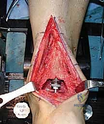
Question 1
A 35-year-old male presents with recurrent clicking and apprehension when pushing up from a chair 6 months after an elbow dislocation. Which ligament is primarily deficient, and what is the typical path of subluxation?
Explanation
Question 2
In a 4-year-old child with developmental dysplasia of the hip, an osteotomy is planned to improve anterolateral acetabular coverage. The osteotomy is described as an incomplete pericapsular cut that hinges on the triradiate cartilage without extending into the sciatic notch. Which osteotomy is being described?
Explanation
Question 3
A 22-year-old rugby player presents with recurrent anterior shoulder instability. A 3D CT scan demonstrates 25% anterior glenoid bone loss and an engaging Hill-Sachs lesion. What is the most appropriate surgical management?
Explanation
Question 4
A 2.5-year-old obese girl presents with bilateral genu varum. Radiographs show a metaphyseal-diaphyseal angle (Drennan's angle) of 18 degrees and medial physeal beaking. What is the most appropriate initial management?
Explanation
Question 5
During surgical reconstruction of a 'terrible triad' injury of the elbow, what is the standard recommended sequence of repair to progressively restore stability?
Explanation
Question 6
A 13-year-old boy presents with severe left hip pain and inability to bear weight following a minor fall. Radiographs confirm a severe, unstable slipped capital femoral epiphysis (SCFE). A modified Dunn procedure is considered. What vascular structure is most at risk and must be meticulously protected during the surgical dislocation and callus debridement?
Explanation
Question 7
What is the primary biomechanical advantage of a reverse total shoulder arthroplasty (RTSA) in the setting of rotator cuff tear arthropathy?
Explanation
Question 8
When performing distraction osteogenesis for a post-traumatic tibial bone defect using an Ilizarov frame, what are the classic optimal latency period and rate of distraction to promote high-quality regenerate bone?
Explanation
Question 9
A 38-year-old female presents with chronic groin pain exacerbated by hip flexion and internal rotation. An AP pelvis radiograph reveals a 'crossover sign.' What underlying pathomorphology does this indicate, and what type of impingement is most likely?
Explanation
Question 10
A 42-year-old female sustains an elbow injury. Radiographs reveal a type IV Bryan and Morrey capitellum fracture. What is the defining characteristic of this fracture pattern?
Explanation
Question 11
A 45-year-old laborer has an irreparable posterosuperior rotator cuff tear with an intact subscapularis, severe external rotation lag, and a positive hornblower's sign. There is no glenohumeral arthritis. Which tendon transfer is most biomechanically appropriate to restore external rotation?
Explanation
Question 12
A 9-year-old boy presents with a cubitus varus deformity 3 years after a supracondylar humerus fracture. He is asymptomatic, but if left uncorrected, what is the most significant potential late functional complication of this deformity?
Explanation
Question 13
A 65-year-old male presents with groin pain and a palpable mass 5 years after receiving a metal-on-polyethylene total hip arthroplasty using a large-diameter cobalt-chromium femoral head. Serum metal ions show significantly elevated cobalt and normal chromium levels. What is the most likely diagnosis?
Explanation
Question 14
During anatomic reconstruction of a chronic type V acromioclavicular (AC) joint separation, the surgeon reconstructs the coracoclavicular (CC) ligaments. To accurately reproduce the biomechanics of the native joint, where should the conoid and trapezoid reconstruction tunnels be placed relative to the distal clavicle?
Explanation
Question 15
A 38-year-old bodybuilder undergoes distal biceps tendon repair using a single anterior incision technique. Postoperatively, he exhibits weakness in extending the wrist and fingers, though wrist drop is incomplete. The affected nerve most likely runs between which two muscle bellies near the level of the radial neck?
Explanation
Question 16
A newborn is noted to have a fully segmented hemivertebra at T8, causing congenital scoliosis. Ultrasound of the renal system and an echocardiogram are both normal. What is the most critical next diagnostic screening study required for this patient prior to any surgical planning?
Explanation
Question 17
A 6-year-old boy is diagnosed with Legg-Calvé-Perthes disease. Which of the following radiographic findings is recognized as one of Catterall's 'head-at-risk' signs, indicating a poorer prognosis?
Explanation
Question 18
In evaluating a proximal humerus fracture for the risk of avascular necrosis (AVN), which of the following is considered the strongest predictor of ischemia to the articular segment according to Hertel's criteria?
Explanation
Question 19
In the pathogenesis of Charcot neuroarthropathy, the neurovascular theory suggests that autonomic neuropathy leads to which of the following physiologic changes?
Explanation
Question 20
A 4-month-old infant with developmental dysplasia of the hip is being treated with a Pavlik harness. During a follow-up visit, the infant is noted to lack active knee extension on the treated side, though there is normal ankle movement. What is the most appropriate next step in management?
Explanation
Question 21
A 72-year-old female undergoes a reverse total shoulder arthroplasty (rTSA) for severe rotator cuff tear arthropathy. Which of the following best describes the fundamental biomechanical alteration achieved by the rTSA implant design compared to native anatomy?
Explanation
Question 22
A 55-year-old male complains of a loud 'squeaking' noise coming from his hip 3 years after a ceramic-on-ceramic total hip arthroplasty. He is otherwise asymptomatic. Radiographs show a well-fixed implant. Which of the following is the most likely primary etiology of this phenomenon?
Explanation
Question 23
A 34-year-old male falls on an outstretched arm and sustains an elbow injury. Examination reveals varus posteromedial rotatory instability (VPMRI). Which of the following injury patterns is most classically associated with this specific physical examination finding?
Explanation
Question 24
According to the principles of deformity correction formulated by Paley, what is the expected outcome if an osteotomy is performed at a level separate from the Center of Rotation of Angulation (CORA), but the correction axis (hinge) is placed exactly on the CORA?
Explanation
Question 25
A 22-year-old rugby player undergoes an open Latarjet procedure for recurrent anterior shoulder instability with 30% glenoid bone loss. Postoperatively, he exhibits profound weakness in elbow flexion and decreased sensation over the lateral forearm. Which of the following structures was most likely injured during the surgical approach?
Explanation
Question 26
In the pathophysiology of Cam-type femoroacetabular impingement (FAI), which of the following best describes the primary mechanism of chondral injury?
Explanation
Question 27
A 45-year-old mechanic presents with insidious onset of weakness in finger and thumb extension, without sensory deficits. Compression of the posterior interosseous nerve (PIN) is suspected. Which of the following represents the most common site of compression for this nerve?
Explanation
Question 28
Which of the following radiographic parameters is considered the most reliable predictor of humeral head ischemia following a proximal humerus fracture, according to Hertel's criteria?
Explanation
Question 29
A 60-year-old male with a metal-on-polyethylene total hip arthroplasty presents with new-onset groin pain and a pseudotumor on MRI. Infection workup is negative. Blood metal ion levels reveal elevated serum cobalt relative to chromium. This clinical picture is most likely driven by mechanically assisted crevice corrosion (MACC) at which interface?
Explanation
Question 30
A 14-year-old elite gymnast presents with lateral elbow pain and catching. Radiographs reveal a radiolucent lesion in the capitellum. The pathogenesis of this condition is most directly related to which of the following?
Explanation
Question 31
A newborn is diagnosed with Congenital Femoral Deficiency (CFD). During the comprehensive orthopedic evaluation, the surgeon must actively screen for commonly associated anomalies. Which of the following conditions is LEAST likely to be associated with CFD?
Explanation
Question 32
During a coracoclavicular (CC) ligament reconstruction for a high-grade acromioclavicular joint separation, the surgeon passes grafts to recreate the native ligaments. Which of the following accurately describes the anatomic relationship of the native CC ligaments?
Explanation
Question 33
A 75-year-old female who underwent revision total hip arthroplasty with a dual mobility construct presents with an acute dislocation. Radiographs reveal the 'bubble sign' with eccentric position of the femoral head within the acetabular shell. What is the precise mechanism of this specific type of intraprosthetic dislocation?
Explanation
Question 34
A 30-year-old female falls on an outstretched hand and sustains a capitellar fracture. On CT scan, the fracture fragment consists of a large, hemispherical piece of articular cartilage with minimal to no attached subchondral bone. This morphology is best classified as which type of capitellar fracture?
Explanation
Question 35
A 4-year-old boy with Proximal Focal Femoral Deficiency (PFFD) is evaluated for a Van Nes rotationplasty. For this procedure to be functionally successful, which of the following anatomic prerequisites is absolutely essential?
Explanation
Question 36
A 28-year-old patient presents with symptomatic scapular winging following a posterior triangle lymph node biopsy. On examination, the scapula rests in a translated lateral and downwardly rotated position. The winging is accentuated when the patient abducts the arm against resistance. Which nerve is most likely injured?
Explanation
Question 37
In the management of Legg-Calve-Perthes disease, the Herring Lateral Pillar Classification is widely used due to its strong prognostic value. Which radiographic feature specifically defines a Herring Group B classification?
Explanation
Question 38
A 65-year-old female with severe rheumatoid arthritis is considering a semiconstrained (linked) Total Elbow Arthroplasty (TEA). When discussing the long-term prognosis and risks, what should the surgeon quote as the most common long-term complication leading to revision?
Explanation
Question 39
A 2-year-old obese child presents with bilateral genu varum. You are distinguishing between physiologic bowing and infantile Blount disease. On standing AP radiographs, measurement of the metaphyseal-diaphyseal angle (MDA) of Drennan is performed. An angle greater than what threshold is highly predictive of progression to infantile Blount disease?
Explanation
Question 40
When using a hexapod external fixator (e.g., Taylor Spatial Frame) for complex lower extremity deformity correction, what represents its primary biomechanical and functional advantage over a traditional Ilizarov circular frame?
Explanation
Question 41
A 45-year-old male sustains a terrible triad injury of the elbow following a fall. Intraoperatively, the surgeon decides on a single lateral approach. What is the recommended sequence of anatomical repair to systematically restore elbow stability?
Explanation
Question 42
A 28-year-old female presents with deep groin pain exacerbated by hip flexion and internal rotation. An AP pelvis radiograph demonstrates a 'cross-over sign'. What type of femoroacetabular impingement and anatomical abnormality does this represent?
Explanation
Question 43
A 72-year-old female undergoes a reverse total shoulder arthroplasty for cuff tear arthropathy. How does the biomechanical design of the prosthesis primarily improve active elevation?
Explanation
Question 44
In deformity correction planning using the principles of Paley, a closing wedge osteotomy is performed on the tibia. If the osteotomy hinge is placed outside the center of rotation of angulation (CORA), what is the expected geometric consequence?
Explanation
Question 45
A 30-year-old male sustains an Essex-Lopresti injury. During surgery, the highly comminuted radial head is excised and not replaced. What is the most likely biomechanical complication of this omission?
Explanation
Question 46
A 6-week-old infant is treated with a Pavlik harness for developmental dysplasia of the hip. At the first follow-up, the parents report the infant is no longer kicking the affected leg. Examination reveals decreased active knee extension but normal foot movements. What is the most appropriate next step?
Explanation
Question 47
A 25-year-old baseball pitcher complains of vague posterior shoulder pain and weakness in external rotation. Examination shows isolated atrophy of the infraspinatus with normal supraspinatus bulk. A paralabral cyst is suspected. Where is the cyst most likely located?
Explanation
Question 48
In the Ponseti method for the treatment of idiopathic clubfoot, what is the correct sequence of deformity correction?
Explanation
Question 49
A 22-year-old collegiate baseball pitcher presents with posterior elbow pain during the deceleration phase of throwing. He lacks 15 degrees of full extension. Radiographs show posteromedial olecranon osteophytes. What is the primary underlying pathomechanical cause?
Explanation
Question 50
A 6-year-old boy is diagnosed with Legg-Calve-Perthes disease. Which of the following radiographic findings represents one of Catterall's 'head-at-risk' signs, indicating a poorer prognosis?
Explanation
Question 51
During an arthroscopic anterior Bankart repair on a right shoulder, the surgeon places a suture anchor at the 5:30 position on the glenoid rim. Which major anatomical structure is primarily tensioned by shifting the labrum superiorly at this position?
Explanation
Question 52
Which of the following is the most common musculoskeletal manifestation requiring surgical intervention in adult patients with achondroplasia?
Explanation
Question 53
A 45-year-old male undergoes a single-incision anterior repair of a distal biceps tendon rupture. Postoperatively, he is unable to actively extend his fingers or thumb, but wrist extension is partially preserved with a radial deviation bias. Which nerve was injured during the approach?
Explanation
Question 54
In reverse total shoulder arthroplasty (RTSA), inferior tilt and inferior overhang of the glenosphere are primarily designed to prevent which of the following complications?
Explanation
Question 55
A 12-year-old obese male undergoes in-situ pinning for a slipped capital femoral epiphysis (SCFE). Intraoperatively, the slip is noted to be unstable as the patient could not bear weight prior to surgery. Compared to a stable SCFE, this patient is at significantly higher risk for which complication?
Explanation
Question 56
During surgical management of a terrible triad injury of the elbow, the radial head is fixed and the lateral ulnar collateral ligament (LUCL) is repaired. The coronoid fracture involves the anteromedial facet. What is the most appropriate management of this coronoid fracture?
Explanation
Question 57
In deformity correction principles, if an opening wedge osteotomy is performed at a level distant to the Center of Rotation of Angulation (CORA) but the mechanical axes are subsequently realigned, what is the expected geometric result at the osteotomy site?
Explanation
Question 58
A 25-year-old male presents with a locked posterior shoulder dislocation. Which of the following mechanisms of injury is most classically associated with a traumatic posterior dislocation?
Explanation
Question 59
A 30-year-old male presents with activity-related groin pain. Pelvic radiographs reveal a crossover sign, a prominent ischial spine sign, and a posterior wall sign. Which anatomic abnormality is primarily responsible for his femoroacetabular impingement?
Explanation
Question 60
A 28-year-old weightlifter undergoes repair of an acute distal biceps rupture via a single-incision anterior approach. Postoperatively, he reports numbness over the radial aspect of his forearm. Which nerve is most likely injured?
Explanation
Question 61
A patient is undergoing deformity correction for a midshaft tibial malunion. According to Paley's rules of osteotomies, if the osteotomy is performed at a site separate from the Center of Rotation of Angulation (CORA) and the proximal and distal mechanical axes are aligned, what is the inevitable geometric result?
Explanation
Question 62
A 40-year-old female presents with posterolateral rotatory instability (PLRI) of the elbow. During the lateral pivot-shift test of the elbow, at what degree of flexion does maximal subluxation of the radial head typically occur?
Explanation
Question 63
A 24-year-old female with developmental dysplasia of the hip undergoes a Bernese periacetabular osteotomy (PAO). Through the standard Smith-Petersen approach, which osteotomy is typically performed first and requires fluoroscopy to ensure it is incomplete and does not enter the joint?
Explanation
Question 64
A 65-year-old male presents with an irreparable subscapularis tear and recurrent anterior shoulder instability. The posterosuperior rotator cuff is intact. Which tendon transfer is most biomechanically appropriate to restore anterior shoulder stability and internal rotation?
Explanation
Question 65
During an open release for severe post-traumatic elbow stiffness via a lateral column approach, the anterior capsule is excised, which improves flexion. However, extension remains severely limited. What is the most appropriate next step in the surgical sequence?
Explanation
Question 66
In an 8-year-old boy undergoing guided growth for genu valgum with a tension band plate (8-plate) on the medial distal femur, how does this implant alter the physis compared to traditional rigid Blount staples?
Explanation
Question 67
A 13-year-old obese boy requires in-situ pinning for a stable slipped capital femoral epiphysis (SCFE). To minimize the risk of joint penetration while maximizing mechanical stability, the single screw should be placed in which zone of the femoral head?
Explanation
Question 68
A 30-year-old male is undergoing reconstruction of a Type V acromioclavicular (AC) joint separation using a free tendon graft. To accurately recreate the native anatomy of the coracoclavicular ligaments, where should the conoid and trapezoid insertions be targeted relative to the distal clavicle?
Explanation
Question 69
During a two-incision distal biceps tendon repair, the surgeon develops the posterior plane between the supinator and the extensor carpi radialis brevis. Which nerve is at greatest risk of injury during this posterior exposure if the forearm is not fully pronated?
Explanation
Question 70
When utilizing a Taylor Spatial Frame to correct a complex multiplanar deformity, the surgeon must input specific parameters into the software. The "mounting parameters" specifically refer to which of the following?
Explanation
Question 71
A 6-year-old boy is diagnosed with Legg-Calve-Perthes disease. Radiographs reveal that exactly 60% of the lateral pillar height is maintained. According to the Herring lateral pillar classification, what is his group and the most appropriate standard initial management?
Explanation
Question 72
A 72-year-old female receives a reverse total shoulder arthroplasty for a 4-part proximal humerus fracture. The surgeon performs a tuberosity repair. Healing of the greater tuberosity is considered most critical for restoring which specific shoulder function?
Explanation
Question 73
In the surgical treatment of a terrible triad injury of the elbow, the coronoid is fixed, the radial head is replaced, and the lateral collateral ligament (LCL) is repaired. However, the elbow remains subluxated and unstable in extension. What is the most appropriate next step?
Explanation
Question 74
A 28-year-old male undergoes arthroscopic osteochondroplasty for femoroacetabular impingement (FAI) due to a large CAM lesion. Over-resection of the anterolateral femoral head-neck junction places the patient at greatest risk for which devastating complication?
Explanation
Question 75
A 24-year-old elite baseball pitcher presents with posterior shoulder pain during the late cocking phase. MRI reveals a partial articular-sided supraspinatus tendon avulsion (PASTA) and a superior labrum anterior-posterior (SLAP) tear. This specific triad of "internal impingement" is most strongly associated with which clinical finding?
Explanation
Question 76
A 3-year-old child presents with bilateral tibia vara. Standing AP radiographs reveal a metaphyseal-diaphyseal angle (Drennan angle) of 18 degrees on both sides. What is the most appropriate initial management for this patient?
Explanation
Question 77
A 15-year-old female gymnast is diagnosed with a capitellar osteochondritis dissecans (OCD) lesion. MRI shows the articular cartilage is intact. Which radiographic view best profiles the capitellum to monitor lesion size and healing during non-operative management?
Explanation
Question 78
During a revision total hip arthroplasty, the surgeon encounters a Paprosky Type IIIB acetabular defect. There is complete loss of the medial wall, superior migration of 3.5 cm, and severe ischial lysis. Which reconstructive option provides the most reliable mechanical fixation for this specific defect pattern?
Explanation
None
