Mastering the Superior Approach for Open Reduction of Chronic Posterior Shoulder Dislocations
Key Takeaway
The superior approach, popularized by Rowe and Zarins, provides unparalleled access for the open reduction of chronic posterior shoulder dislocations. This technique utilizes a superior acromial osteotomy to mobilize the deltoid, allowing comprehensive visualization of the glenohumeral joint. It facilitates safe mobilization of the humeral head, management of reverse Hill-Sachs lesions via McLaughlin or Neer transfers, and precise capsulolabral reconstruction, ensuring optimal joint stability and functional recovery.
INTRODUCTION TO CHRONIC POSTERIOR SHOULDER DISLOCATIONS
Posterior shoulder dislocations are rare, comprising approximately 2% to 5% of all glenohumeral dislocations. Due to their subtle clinical presentation and the frequent misinterpretation of standard anteroposterior (AP) radiographs, up to 50% of posterior dislocations are missed at initial presentation, evolving into chronic, locked posterior dislocations.
When a posterior dislocation becomes chronic, the anterior aspect of the humeral head remains impacted against the posterior glenoid rim, creating an anteromedial impaction fracture known as a reverse Hill-Sachs lesion. The surrounding soft tissues—including the posterior capsule, rotator cuff, and subscapularis—become severely contracted, fibrotic, and adherent to the articular surfaces.
For these complex, chronic presentations, closed reduction is universally contraindicated due to the high risk of iatrogenic humeral head fracture, cartilage shearing, and neurovascular compromise. Open reduction is mandatory. The superior "utility" approach, pioneered and popularized by Rowe and Zarins, remains a gold-standard extensile exposure. It provides unparalleled 360-degree access to the anterior, superior, and posterior aspects of the glenohumeral joint, allowing for safe lysis of adhesions, gentle reduction of the osteopenic humeral head, and concurrent management of the reverse Hill-Sachs defect.
INDICATIONS AND CONTRAINDICATIONS
Indications for the Superior Approach
- Chronic Posterior Dislocations: Dislocations unrecognized for greater than 3 weeks, where soft tissue contracture precludes closed reduction.
- Locked Posterior Dislocations: Acute or subacute dislocations with a significant reverse Hill-Sachs lesion (typically >20% of the articular surface) that is mechanically engaged on the posterior glenoid rim.
- Complex Fracture-Dislocations: Posterior dislocations associated with displaced fractures of the anatomical neck, surgical neck, or tuberosities requiring extensive exposure for open reduction and internal fixation (ORIF).
- Failed Anterior Approaches: Cases where an isolated deltopectoral approach provides insufficient access to the posterior glenoid or posterior rotator cuff.
Contraindications
- Massive Articular Defects (>40-50%): If the reverse Hill-Sachs lesion involves more than 45% of the articular surface, or if the humeral head is severely osteonecrotic, joint-preserving open reduction is likely to fail. Arthroplasty (hemiarthroplasty or total shoulder arthroplasty) is indicated.
- Severe Glenohumeral Osteoarthritis: Pre-existing advanced arthropathy necessitates arthroplasty rather than open reduction.
- Acute, Uncomplicated Dislocations: These should be managed with closed reduction under conscious sedation or general anesthesia.
PREOPERATIVE PLANNING AND IMAGING
Thorough preoperative imaging is the cornerstone of surgical planning for chronic posterior dislocations.
- Standard Radiographs: A true AP (Grashey), scapular Y, and axillary lateral view are mandatory. The axillary view is the most critical radiograph for confirming the posterior direction of the dislocation and identifying the presence of a reverse Hill-Sachs lesion.
- Computed Tomography (CT): A non-contrast CT scan with 3D reconstruction is the gold standard for quantifying the size, depth, and orientation of the anteromedial humeral head defect. It also assesses the posterior glenoid rim for erosion, dysplasia, or posterior Bankart fractures.
- Magnetic Resonance Imaging (MRI): While often degraded by artifact in chronic trauma, MRI can be useful to evaluate the integrity of the rotator cuff, the degree of muscular atrophy/fatty infiltration, and the status of the long head of the biceps (LHB) tendon.
Clinical Pearl: Always calculate the percentage of articular surface involvement of the reverse Hill-Sachs lesion on the axial CT slices. Defects <20% may be stable after reduction; defects 20-40% require a McLaughlin or Neer transfer; defects >40% typically require arthroplasty.
SURGICAL ANATOMY AND BIOMECHANICS
Understanding the superior shoulder anatomy is critical to executing the Rowe and Zarins approach safely.
The Deltoid Muscle and Axillary Nerve
The deltoid arises from the lateral third of the clavicle, the acromion, and the spine of the scapula. The axillary nerve, a terminal branch of the posterior cord of the brachial plexus, exits the quadrangular space and courses transversely along the deep surface of the deltoid.
* The "Safe Zone": The axillary nerve lies approximately 5 to 7 cm distal to the lateral edge of the acromion. When splitting the deltoid distally, the split must never exceed 5 cm from the acromial edge to avoid catastrophic denervation of the anterior and middle deltoid.
The Rotator Cuff Interval and Biceps Tendon
The long head of the biceps (LHB) tendon serves as the primary anatomical landmark in a distorted, chronically dislocated shoulder. It lies within the bicipital groove between the lesser and greater tuberosities and guides the surgeon directly to the rotator cuff interval, separating the subscapularis (anteriorly) from the supraspinatus (superiorly).
SURGICAL TECHNIQUE: STEP-BY-STEP (ROWE AND ZARINS)
1. Patient Positioning and Anesthesia
- Anesthesia: General endotracheal anesthesia combined with an interscalene regional nerve block is recommended for optimal muscle relaxation and postoperative pain control.
- Positioning: Place the patient in the lateral decubitus position on the contralateral side. Secure the patient with a vacuum bean bag.
- Arm Drape: The operative arm must be draped free to allow full manipulation (flexion, extension, internal/external rotation) during the procedure. Use a sterile arm holder or a Mayo stand for support.
2. The "Utility" Skin Incision
- Make a superior "strap" or "saber-cut" incision.
- Begin the incision just anterior to the clavicle, extend it posteriorly over the superior aspect of the acromioclavicular (AC) joint, and carry it down over the spine of the scapula.
- This incision provides excellent cosmetic healing and allows extensile exposure both anteriorly and posteriorly.
3. Acromial Osteotomy and Deltoid Mobilization
Rowe and Zarins emphasized the superiority of an osteotomy over soft-tissue detachment of the deltoid, as bone-to-bone healing is significantly more robust and prevents postoperative deltoid dehiscence.
- Identify the middle third of the deltoid origin on the lateral acromion.
- Using an oscillating saw or a sharp osteotome, perform a sagittal osteotomy to detach a 5-mm-wide piece of the lateral acromial rim, keeping the deltoid origin firmly attached to this bone fragment.
- Sharply dissect and separate the anterior and posterior origins of the deltoid from the clavicle and the spine of the scapula as far as necessary to achieve mobility.
- Deltoid Split: Split the deltoid muscle distally in the direction of its fibers.
> Surgical Warning: Strictly limit this distal split to a maximum of 5 cm from the acromial edge. Place a stay suture at the apex of the split to prevent inadvertent distal propagation and subsequent axillary nerve injury. - Turn the deltoid flap laterally and distally, exposing the underlying subacromial bursa and rotator cuff.
4. Deep Dissection and Joint Exposure
In chronic dislocations, the anatomical landmarks are heavily obscured by dense, fibrotic scar tissue.
- Locate the LHB: The easiest and most reliable landmark is the long head of the biceps tendon. Identify it distally and follow it proximally into the bicipital groove between the lesser and greater tuberosities.
- Rotator Cuff Interval: Incise the rotator cuff interval along the anterior border of the supraspinatus, following the LHB into the joint.
- Capsular Release (Neviaser Technique): Neviaser described a specific sequence for managing the contracted capsule. First, strip the fibrotic capsule directly from the face of the glenoid. Once the glenoid is identified, proceed laterally to lyse the dense adhesions binding the rotator cuff to the anatomical neck of the humerus.
5. Mobilization and Reduction of the Humeral Head
- With the contractures released anteriorly, superiorly, and posteriorly, carefully insert a blunt elevator or a Darrach retractor between the posterior glenoid rim and the anterior aspect of the humeral head.
- Gentle Manipulation: Apply gentle lateral traction and external rotation to the arm while levering the humeral head laterally and anteriorly over the posterior glenoid rim.
> Surgical Warning: In chronic dislocations, the humeral head is profoundly osteopenic due to disuse, and the cartilage is softened. Aggressive levering will cause iatrogenic crush fractures or shear off the remaining articular cartilage. Take time to ensure all soft tissue tethers are released before attempting reduction. - Reduce the head of the humerus into the glenoid cavity. Inspect the joint for stability and assess the size of the reverse Hill-Sachs defect.
MANAGEMENT OF THE ANTERIOR HUMERAL HEAD DEFECT
Once reduced, the shoulder will often remain highly unstable, tending to re-dislocate posteriorly when the arm is internally rotated. This instability is driven by the reverse Hill-Sachs lesion engaging the posterior glenoid rim. Management depends on the size of the defect.
The McLaughlin Procedure (Subscapularis Transfer)
Originally described by McLaughlin in 1952, this technique is indicated for defects comprising 20% to 40% of the articular surface.
* Sharply dissect the subscapularis tendon from its insertion on the lesser tuberosity.
* Debride the reverse Hill-Sachs defect to bleeding cancellous bone to promote healing.
* Transfer the subscapularis tendon directly into the defect.
* Secure the tendon using robust suture anchors placed into the base of the defect.
* Biomechanics: This transfer fills the void, rendering the defect extra-articular, and acts as a mechanical check-rein, preventing the defect from rotating internally and engaging the posterior glenoid rim.
The Neer Modification (Lesser Tuberosity Osteotomy)
Neer modified the McLaughlin procedure to improve the biomechanical strength of the repair by utilizing bone-to-bone healing.
* Instead of detaching the tendon, perform an osteotomy of the lesser tuberosity, keeping the subscapularis tendon attached to the bone fragment.
* Prepare the reverse Hill-Sachs defect as a bleeding bone bed.
* Transfer the lesser tuberosity fragment into the defect.
* Fix the fragment rigidly using two cancellous lag screws with washers.
* Advantage: This provides superior fixation strength and faster incorporation compared to soft-tissue-to-bone healing, allowing for slightly more aggressive early rehabilitation.
CLOSURE AND RECONSTRUCTION
Internal fixation of the glenohumeral joint (e.g., transarticular pinning) is generally not necessary and is discouraged due to the risk of pin breakage and cartilage damage. Stability is achieved through the defect reconstruction and strict postoperative positioning.
1. Rotator Cuff and Capsular Repair
- Thoroughly irrigate the joint to remove all osteochondral debris.
- If the posterior capsule was stretched or torn, perform a posterior capsulorrhaphy or posterior Bankart repair using suture anchors to restore posterior tension.
- Close the rotator cuff interval securely using non-absorbable sutures.
2. Acromial Reattachment
- Bring the deltoid flap back to its anatomical position.
- Reattach the 5-mm bony rim of the acromion to the main body of the acromion.
- Drill three small holes through the main acromion and pass heavy, non-absorbable sutures (e.g., #2 or #5 braided composite sutures) through the drill holes and around the osteotomized fragment.
- Tie the sutures securely, ensuring rigid bone-to-bone compression. This anatomical reattachment ensures the deltoid will heal without displacing distally.
3. Superficial Closure
- Repair the superficial deltoid fascia.
- Close the subcutaneous tissues and skin in a standard layered fashion.
- Apply sterile dressings.
POSTOPERATIVE CARE AND REHABILITATION
The postoperative protocol is highly specialized and differs entirely from the rehabilitation of anterior shoulder dislocations. The primary goal is to prevent the arm from moving anterior to the coronal plane, which would tension the posterior capsule and risk posterior re-dislocation.
Phase I: Immobilization (Weeks 0 to 3)
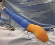
- Positioning: Immediately post-surgery, the arm must be supported in a position posterior to the coronal plane of the body. This is typically achieved using a custom "gunslinger" orthosis or a modified shoulder spica cast that holds the shoulder in slight extension, neutral to slight external rotation, and slight abduction.
- Rationale: If the arm remains posterior to the coronal plane, the anterior structures (including the transferred subscapularis) remain tensioned, and the humeral head is driven anteriorly into the glenoid, preventing posterior subluxation.
- Elbow Mobility: The elbow is left free to allow active flexion and extension, preventing elbow stiffness.
- Shoulder Mobility: The shoulder may be moved actively or passively posteriorly into extension, but strictly restricted from anterior flexion.
Phase II: Early Mobilization (Weeks 3 to 6)
- At 3 weeks, the rigid support is removed.
- Begin gentle, gravity-assisted pendulum exercises.
- Initiate guided isometric exercises for the deltoid and rotator cuff.
- Passive and active-assisted range of motion (ROM) is introduced, gradually allowing the arm to come forward into the coronal plane. Internal rotation is strictly limited to prevent tension on the McLaughlin/Neer transfer and protect the posterior capsule.
Phase III: Strengthening and Progressive Use (Weeks 6 to 12+)
- Progressive use of the arm within the range of comfort is encouraged.
- Begin active strengthening of the dynamic posterior stabilizers (infraspinatus, teres minor, posterior deltoid) and the scapular retractors (rhomboids, middle trapezius).
- Full functional recovery may take 6 to 12 months. Patients must be counseled that while stability and pain relief are highly predictable, a mild to moderate permanent deficit in internal rotation and forward elevation is common and expected.
COMPLICATIONS AND PITFALLS
- Axillary Nerve Injury: The most devastating complication of the superior approach. Strict adherence to the 5-cm rule for the distal deltoid split is mandatory.
- Iatrogenic Humeral Head Fracture: Forcing the reduction against severe soft-tissue contractures in osteopenic bone will result in a crush fracture of the humeral head, necessitating immediate conversion to a shoulder arthroplasty.
- Deltoid Dehiscence: Failure to achieve rigid fixation of the acromial osteotomy can lead to deltoid detachment, resulting in profound, irreversible weakness in shoulder elevation.
- Avascular Necrosis (AVN): Chronic dislocations disrupt the intra-articular blood supply. Even with a successful open reduction, the humeral head may collapse months or years later due to AVN, eventually requiring arthroplasty.
-
Postoperative Stiffness: A degree of stiffness is inevitable and often desirable to maintain stability. Overly aggressive early physical therapy can stretch the posterior repair and lead to recurrent instability.
You Might Also Like