Incision and Drainage of Hand Infections: A Comprehensive Surgical Masterclass
Key Takeaway
Hand infections require prompt surgical intervention to prevent devastating functional loss. This comprehensive guide details the principles of incision and drainage, emphasizing appropriate anesthesia, tourniquet management without exsanguination, and meticulous blunt dissection. It outlines postoperative protocols, including the safe position of splinting and early active motion, alongside a thorough review of targeted antimicrobial, antifungal, and antiviral pharmacotherapy essential for eradicating complex hand infections.
Introduction to Hand Infections
The management of hand infections represents a critical pillar of operative orthopaedics and hand surgery. The intricate anatomy of the hand—comprising tightly compartmentalized fascial spaces, delicate synovial sheaths, and superficial neurovascular bundles—renders it uniquely vulnerable to the rapid spread of pyogenic organisms. Delayed or inadequate surgical intervention can lead to catastrophic outcomes, including tendon necrosis, osteomyelitis, joint destruction, and irreversible stiffness.
The primary objective of an incision and drainage (I&D) procedure in the hand is the complete evacuation of purulence, decompression of fascial spaces, and reduction of the bacterial load, all while meticulously preserving the vital biomechanical structures of the hand. This masterclass delineates the evidence-based principles of surgical drainage, perioperative management, and targeted pharmacotherapy required for the successful eradication of hand infections.
Preoperative Considerations and Anesthesia
The selection of appropriate anesthesia is paramount in the surgical management of hand infections. The physiological environment of an abscess or infected fascial space dictates the anesthetic approach.
🚨 Surgical Warning: The Perils of Local Anesthesia
Never rely on local anesthetic infiltration directly into or adjacent to a septic area in the hand. The acidic environment (low pH) of infected tissue causes local anesthetics to remain in their ionized form, preventing them from crossing nerve cell membranes and rendering them pharmacologically ineffective. Furthermore, injecting fluid into an already swollen, non-compliant compartment exacerbates tissue tension, potentially precipitating microvascular ischemia and mechanically disseminating the infection into adjacent uninfected fascial planes.
Recommended Anesthetic Modalities:
* General Anesthesia: Preferred for extensive infections, uncooperative patients, or when the exact proximal extent of the infection is unknown (e.g., necrotizing fasciitis or severe flexor tenosynovitis extending into the Parona space).
* Distant Regional Blockade: Axillary, supraclavicular, or infraclavicular brachial plexus blocks are excellent alternatives. They provide profound anesthesia and sympathectomy-induced vasodilation without violating the infected surgical field.
Tourniquet Management and Exsanguination Protocols
A bloodless surgical field is absolute necessity in hand surgery to allow for the precise identification of digital nerves, vessels, and tendons. However, the standard protocol for achieving this must be modified in the presence of infection.
🔪 Clinical Pearl: Gravity Elevation over Esmarch Exsanguination
Do not use an Esmarch bandage or elastic wrap to exsanguinate an infected limb. The mechanical compression forces purulent material proximally through lymphatic channels and tendon sheaths, converting a localized infection into a disseminated one.
Step-by-Step Tourniquet Protocol:
1. Apply a well-padded pneumatic tourniquet to the proximal arm.
2. Elevate the operative extremity strictly by gravity for 3 to 6 minutes.
3. Inflate the tourniquet to the standard pressure (typically 250 mm Hg or 100 mm Hg above the patient's systolic blood pressure).
4. Proceed with the operation, ensuring the tourniquet time does not exceed 120 minutes to prevent ischemic neuropraxia.
Surgical Approach and Dissection Principles
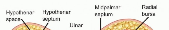
The surgical approach must be tailored to the specific anatomical compartment involved (e.g., midpalmar space, thenar space, flexor tendon sheath, or apical pulp space). Regardless of the specific incision, fundamental principles of hand surgery apply.
Incision Design
- Avoid Flexion Creases: Incisions must never cross flexion creases at a 90-degree angle to prevent postoperative flexion contractures. Use Brunner (zigzag) incisions or mid-axial incisions.
- Adequate Exposure: The incision must be extensile. Inadequate incisions lead to blind dissection, which is the primary cause of iatrogenic neurovascular injury.
Meticulous Dissection
After the skin is incised with a No. 15 scalpel, sharp dissection must cease.
* Utilize blunt dissection exclusively in the deeper subcutaneous tissues.
* Employ a blunt-tipped instrument, such as a mosquito hemostat or tenotomy scissors, spreading the tissues parallel to the neurovascular bundles and tendons.
* Identify and protect the digital nerves and arteries before proceeding into the deep fascial spaces.
Irrigation, Debridement, and Wound Management
The mechanical removal of necrotic debris and bacterial biofilm is the cornerstone of infection eradication.
Copious Irrigation
An incision relieves pressure, but irrigation clears the bacterial load.
* Utilize a pulsatile lavage system or high-volume syringe irrigation.
* Irrigate with several liters of sterile normal saline. The use of antibiotic-impregnated solutions or harsh antiseptics (like undiluted Betadine) in the deep tissues is generally discouraged due to their cytotoxic effects on healthy fibroblasts and tenocytes.
Wound Closure Strategy
🚨 Pitfall: Primary Closure of Infected Wounds
Primary closure of an abscess cavity or heavily contaminated space traps residual bacteria, inevitably leading to recurrence.
- Leave the Wound Open: In the vast majority of cases, the wound should be left open to allow for continuous drainage.
- Delayed Primary Closure: Return the patient to the operating room in 3 to 5 days. If the wound bed exhibits healthy granulation tissue and no further purulence, delayed primary closure can be performed.
- Immediate Coverage of Vital Structures: There is one critical exception to leaving wounds open. If flexor tendons, nerves, or articular cartilage are exposed due to skin necrosis, they must be covered immediately (e.g., via local rotational flaps) to prevent desiccation, necrosis, and catastrophic loss of function.
Postoperative Care and Rehabilitation
The postoperative protocol is as critical as the surgical intervention. Infections involving tendon sheaths and joints inherently result in some degree of functional loss due to adhesive tenosynovitis and capsular fibrosis. Aggressive rehabilitation is mandatory.
The Position of Safe Immobilization (POSI)
Immediately post-surgery, the hand is dressed with bulky, non-adherent gauze to absorb exudate and splinted in the "Position of Function" or "Intrinsic Plus" position. This biomechanical positioning prevents the collateral ligaments from shortening and the volar plates from contracting.
* Wrist: 30 degrees of extension.
* Metacarpophalangeal (MCP) Joints: 60 to 70 degrees of flexion (places the cam-shaped MCP collateral ligaments under maximum tension).
* Interphalangeal (IP) Joints: Full extension (prevents volar plate contracture).
* Thumb: Palmar abducted and opposed.
Rehabilitation and Wound Care
- Elevation: Continuous elevation above the level of the heart is required to minimize interstitial edema.
- Early Active Motion: Supervised active range of motion (AROM) must begin as soon as clinically permissible, often within 24 to 48 hours, to prevent tendon adhesions.
- Whirlpool Therapy: Therapist-supervised dressing changes in a whirlpool bath facilitate mechanical debridement of exudate and encourage early motion in a soothing environment.
- Secondary Coverage: Once the infection is cleared, wounds that cannot be closed primarily may require split-thickness skin grafting or flap coverage.
Antimicrobial Pharmacotherapy in Hand Infections
Surgical drainage must be augmented with appropriate, targeted antimicrobial therapy. Empirical therapy should be initiated immediately after intraoperative cultures are obtained, followed by tailored therapy based on sensitivities.
Antibacterial Agents
Penicillins:
* Natural Penicillins (Penicillin G, V): Effective against Streptococcus viridans and Pasteurella multocida (classic for cat/dog bites). Poor gram-negative coverage.
* Penicillinase-Resistant (Nafcillin, Methicillin, Dicloxacillin): Enhanced coverage for methicillin-susceptible Staphylococcus aureus (MSSA). Nafcillin is highly effective for septic arthritis and osteomyelitis.
* Extended-Spectrum (Ampicillin, Amoxicillin): Better gram-negative coverage.
* With β-lactamase Inhibitors (Amoxicillin/clavulanate [Augmentin], Ampicillin/sulbactam [Unasyn]): The gold standard first-line coverage for human/animal bites and diabetic hand infections due to broad coverage of β-lactamase-producing staphylococci and gram-negative organisms.
Cephalosporins:
* First Generation (Cephalexin, Cefazolin): Excellent against gram-positive organisms. Highly recommended for surgical prophylaxis and uncomplicated cellulitis.
* Second Generation (Cefuroxime, Cefotetan): Increased gram-negative coverage.
* Third Generation (Ceftriaxone, Ceftazidime): Ceftriaxone is the drug of choice for disseminated gonococcal infection (DGI) presenting as tenosynovitis. Ceftazidime covers Pseudomonas.
* Fourth Generation (Cefepime): Broad-spectrum, reserved for severe hospital-acquired infections.
Advanced Gram-Positive Coverage (MRSA):
* Glycopeptides (Vancomycin): Intravenous only. Covers MRSA. Monitor for "red-man syndrome" if infused too rapidly.
* Oxazolidinones (Linezolid): Excellent oral bioavailability and bone/joint penetration. Covers MRSA and Vancomycin-Resistant Enterococci (VRE).
* Streptogramins (Quinupristin-dalfopristin): IV only, good bone penetration for resistant staphylococci and enterococci.
Broad-Spectrum and Gram-Negative Agents:
* Carbapenems (Imipenem, Meropenem): Ultra-broad spectrum. Reserved for severe, limb-threatening polymicrobial infections.
* Aminoglycosides (Gentamicin, Tobramycin): Excellent for Pseudomonas. Requires strict monitoring for nephrotoxicity and ototoxicity.
* Fluoroquinolones (Ciprofloxacin, Levofloxacin): Broad gram-positive and gram-negative coverage. Good oral bioavailability.
* Lincosamides (Clindamycin): Covers staphylococci and anaerobes. High incidence of gastrointestinal side effects (risk of C. difficile).
Specialized Agents:
* Metronidazole: Drug of choice for anaerobes and C. difficile.
* Sulfonamides (Bactrim): Excellent oral option for Community-Acquired MRSA (CA-MRSA).
* Antimycobacterials (Isoniazid, Ethambutol, Rifampin): Used in combination for atypical mycobacterial infections (e.g., Mycobacterium marinum from fish tank exposure). Consult infectious disease specialists.
Antifungal Agents
Fungal infections of the hand, though less common than bacterial, require specific management, particularly in immunocompromised patients or those with chronic paronychia/onychomycosis.
- Polyenes (Amphotericin B, Nystatin): Broad-spectrum. Liposomal Amphotericin B is preferred to reduce severe nephrotoxicity.
- Azoles (Ketoconazole, Fluconazole, Itraconazole): Systemic use, generally well-tolerated for deep fungal infections.
- Allylamines (Terbinafine): The systemic drug of choice for onychomycosis (fungal nail infections). Covers dermatophytes but lacks yeast coverage.
- Nucleoside Analogues (5-Fluorocytosine): Used for Candida and Cryptococcus, though resistance develops rapidly.
Antiviral Agents
Viral infections of the hand, most notably Herpetic Whitlow, must be accurately diagnosed, as surgical incision and drainage of a herpetic lesion is strictly contraindicated and can lead to viral dissemination and bacterial superinfection.
- Nucleoside Analogues (Acyclovir, Valacyclovir): The mainstay of treatment and prophylaxis for Herpetic Whitlow (HSV-1 and HSV-2).
- Topical Fluorouracil: Utilized for the treatment of recalcitrant human papillomavirus (HPV) hand warts.
- Antiretroviral Agents (AZT, Indinavir): Utilized for post-exposure prophylaxis (PEP) following needlestick injuries involving HIV-positive sources.
- Anti-influenza & Interferons: Ribavirin and Interferon-α are utilized for post-exposure prophylaxis against Hepatitis C.
Conclusion
The operative management of hand infections demands a profound respect for the complex anatomy of the upper extremity. Success is predicated on the triad of meticulous surgical decompression, rigid adherence to biomechanically sound postoperative rehabilitation, and the judicious application of targeted antimicrobial therapy. By adhering to these textbook principles, the orthopaedic surgeon can reliably eradicate infection while preserving the intricate function of the human hand.
You Might Also Like