Comprehensive Introduction and Patho-Epidemiology
The evolution of Total Hip Arthroplasty (THA) represents one of the most profoundly successful interventions in the history of modern surgical medicine, fundamentally altering the trajectory of debilitating joint disease. Originally popularized by Sir John Charnley, the paradigm of low-friction arthroplasty utilizing polymethylmethacrylate (PMMA) bone cement and a metal-on-polyethylene articulation set the gold standard for decades. However, as the demographic of patients requiring THA has progressively shifted toward younger, highly active individuals, the limitations of first-generation materials and fixation techniques have become starkly apparent. The primary modes of failure in this high-demand cohort—aseptic loosening secondary to wear-induced osteolysis and mechanical fatigue of the implant-bone interface—have driven an unrelenting quest for the optimal hip construct.

In the pursuit of enhanced longevity, orthopedic surgeons and bioengineers have aggressively explored alternative tribological couplings and advanced fixation methodologies. The resurgence of Metal-on-Metal (MoM) bearings and other hard-on-hard articulations was predicated on the theoretical advantage of elastohydrodynamic lubrication, which drastically reduces volumetric wear compared to conventional metal-on-polyethylene. Concurrently, the philosophy of implant fixation has bifurcated into highly sophisticated cemented and cementless techniques. Although some surgeons advocate a monolithic approach—utilizing either cementless or cemented fixation universally—the prevailing academic consensus demands a rational, patient-specific selection process.

To choose the optimal candidate for a specific femoral stem and bearing couple in THA, the arthroplasty surgeon must synthesize a multitude of variables. These include nuanced cement techniques, modern implant geometries, the historical evolution of osseointegration, patient-specific metabolic factors, and emerging registry data regarding the impact of load transfer on surrounding bone stock. Furthermore, the integration of advanced bearing surfaces, such as MoM, introduces complex considerations regarding metal ion release, hypersensitivity reactions, and trunnion mechanics. This chapter provides an exhaustive, evidence-based roadmap for navigating these choices, ensuring that the orthopedic surgeon can predictably deliver a durable, biomechanically optimized reconstruction.
Detailed Surgical Anatomy and Biomechanics
Proximal Femoral Morphology and the Dorr Classification
A profound understanding of proximal femoral morphology is the cornerstone of successful implant selection and fixation in THA. Bone quality, cortical thickness, and canal geometry dictate the mechanical environment into which the prosthesis is introduced. Dorr et al. developed a seminal classification system that correlates the radiographic appearance of the proximal femur and the cortical index with systemic bone mineral density. This classification remains an indispensable tool for matching the fixation modality to the patient's specific osseous architecture. Strong, dense bone with thick cortices and a narrow diaphyseal canal (Dorr Type A) presents a vastly different biomechanical substrate compared to senile, osteoporotic bone with capacious medullary dimensions (Dorr Type C).
In the Dorr Type A "champagne flute" femur, the diaphysis is narrow, and the metaphyseal-diaphyseal mismatch is pronounced. Attempting to utilize a cemented stem of sufficient size and adequate offset in this morphology often requires aggressive reaming of all cancellous bone, effectively denuding the endosteum. Consequently, the PMMA cement has no trabecular microstructure into which it can interdigitate, severely compromising the shear strength of the bone-cement interface. Therefore, a cementless, proximally coated or tapered stem is biomechanically superior in these patients, relying on initial press-fit stability and subsequent biologic ingrowth.
Conversely, the Dorr Type C "stovepipe" femur is fundamentally ill-suited for standard cementless fixation. The profound osteopenia and thinned cortices fail to provide the necessary hoop stresses required for initial mechanical stability of a press-fit stem. A massive cemented stem is typically required to fill this capacious canal. However, even gentle preparation can strip the fragile endosteum. In these scenarios, advanced cementing techniques—and occasionally primary impaction allografting or compaction of the native cancellous bone—are necessary to facilitate adequate cement intrusion and micro-interlock. Dorr Type B femurs represent a transitional morphology where either cemented or cementless techniques can be successfully deployed, dependent upon the surgeon's expertise and the specific implant design.

Biomechanics of Cemented Femoral Fixation
Within the realm of cemented femoral fixation, two divergent biomechanical philosophies dictate implant design and the mechanism of load transfer at the implant-cement interface. The first is the "tapered slip" philosophy, prototyped by the original flat-back Charnley stem and modernized by the double-tapered EXETER® and triple-tapered C-STEM® designs. These collarless, highly polished stems are engineered to allow controlled, microscopic subsidence through the cement mantle. This dynamic subsidence converts deleterious shear stresses at the implant-cement interface into radial compressive forces, favorably loading the surrounding bone according to Wolff's Law, mitigating stress shielding, and protecting the cement-bone interface from catastrophic loosening. The highly polished surface explicitly prevents chemical or mechanical bonding between the metal and the PMMA.
The second philosophy is the "composite beam" design, which emerged before the long-term benefits of the tapered slip mechanism were fully appreciated. Composite beam stems feature collars, flanges, and roughened or pre-coated surface structures designed to achieve a rigid, unyielding bond with the cement mantle, resisting any subsidence. This rigid construct inevitably generates high shear stresses at both the implant-cement and the cement-bone interfaces. Consequently, the long-term survivorship of composite beam designs is critically dependent upon achieving an absolutely flawless initial bond between the implant and the cement; any debonding leads rapidly to fretting, cement mantle fracture, and aseptic loosening.

The choice between these two cemented philosophies has profound implications for the longevity of the reconstruction, particularly in younger patients. While first-generation cementing techniques yielded suboptimal results in high-demand demographics, the advent of third-generation cementing—incorporating vacuum mixing, distal restrictors, retrograde pulsatile injection, and sustained pressurization—has revolutionized outcomes. Furthermore, the positive bone remodeling observed with collarless polished tapers, combined with the relative ease of a "cement-in-cement" revision should the stem fail, has validated their use even in younger, active cohorts.
Biomechanics of Cementless Osseointegration
Modern cementless femoral components achieve stability through initial rigid mechanical press-fit followed by secondary biologic osseointegration. These implants are broadly categorized into non-modular and modular designs, with surface modifications ranging from extensively porous-coated to proximally coated, utilizing titanium plasma spray, sintered beads, or hydroxyapatite (HA) over grit-blasted substrates. Cylindrical, fully porous-coated stems (e.g., AML®) have demonstrated exceptional survivorship over two decades. However, their biomechanical reliance on diaphyseal fixation often leads to proximal stress shielding—where the unloaded metaphyseal bone resorbs—as well as a higher incidence of anterior thigh pain due to modulus mismatch at the distal tip.
To circumvent the issues of diaphyseal stress shielding, second-generation proximally porous-coated stems were developed. These tapered designs aim to achieve metaphyseal biologic fixation, transferring physiological loads more proximally and preserving calcar bone stock. Mid-term and long-term data have largely validated these biomechanical assumptions, showing reduced incidence of thigh pain and minimized distal osteolysis. The tapered geometry, whether single, double, or flat-wedge, relies on hoop stresses generated in the proximal femur to achieve immediate rotational and axial stability.

More recently, short, neck-preserving stems have been introduced to further optimize proximal load transfer and preserve metaphyseal bone stock. Designs inspired by the Mayo hip (e.g., METHA®, NANOS®) or curved neck-preserving stems (CFP) aim to load the femoral neck and calcar anatomically. These short metaphyseal-engaging implants serve as a biomechanical bridge between hip resurfacing and standard cementless stems. They are particularly advantageous in young, arthritic patients with excellent bone quality, facilitating minimally invasive approaches and simplifying future revision surgeries by preserving the diaphyseal canal.
Exhaustive Indications and Contraindications
Patient Selection for Fixation Modalities
The decision matrix for selecting between cemented and cementless fixation is multifaceted, requiring the surgeon to balance patient age, physiological demand, and intrinsic bone quality. Traditionally, cementless implants were strictly reserved for younger, highly active patients with robust Dorr Type A or B femora, under the premise that biologic ingrowth offers a permanent, self-renewing interface capable of withstanding decades of cyclic loading. Conversely, cemented fixation was the default for elderly, low-demand patients, particularly those with Dorr Type C morphology, where immediate mechanical stability is paramount and the risk of intraoperative periprosthetic fracture during press-fit broaching is unacceptably high.
However, contemporary literature has blurred these rigid boundaries. Improved third-generation cementing techniques have demonstrated excellent survivorship in younger patients, particularly when utilizing polished tapered stems that allow for favorable bone remodeling. Simultaneously, modern highly porous titanium coatings have shown reliable osseointegration even in elderly patients with compromised bone stock, offering the advantage of shorter operative times and eliminating the risk of cement monomer-induced hypotension (bone cement implantation syndrome).
On the acetabular side, the indications heavily favor cementless fixation across almost all demographics. Long-term follow-up of cemented acetabular components has revealed alarming radiologic loosening rates of 24-60% at 10 to 25 years. Unlike the femur, the acetabulum does not benefit significantly from modern cementation techniques due to its complex trabecular anatomy and the difficulty in achieving sustained pressurization. Consequently, cementless hemispherical cups with highly porous coatings are indicated for the vast majority of primary THAs, reserving cemented cups primarily for severe post-radiation osteonecrosis or massive reconstructive cages in revision scenarios.
Metal-on-Metal and Advanced Bearing Considerations
The selection of the tribological bearing is inextricably linked to the patient's functional demands and the chosen fixation strategy. Metal-on-Metal (MoM) articulations were historically indicated for young, highly active patients due to their exceptionally low volumetric wear rates and the ability to utilize large-diameter femoral heads, which dramatically increases the jump distance and reduces the risk of dislocation. These hard-on-hard bearings were viewed as the ultimate solution to the epidemic of polyethylene wear-induced osteolysis that plagued earlier generations of THA.
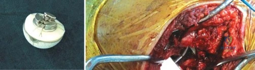
Despite these theoretical advantages, the indications for MoM bearings have been severely restricted in modern practice due to the recognition of Adverse Local Tissue Reactions (ALTR) and Aseptic Lymphocytic Vasculitis-Associated Lesions (ALVAL). Absolute contraindications for MoM articulations now include patients with known metal hypersensitivity, severe renal impairment (due to the inability to excrete cobalt and chromium ions), and women of childbearing age (due to the unknown teratogenic effects of transplacental metal ion transfer).
When a MoM or alternative hard-on-hard bearing (such as ceramic-on-ceramic) is indicated, the surgeon must be acutely aware of the modularity at the head-neck junction. Trunnionosis—mechanically assisted crevice corrosion at the morse taper—has emerged as a critical failure mode in large-head MoM constructs. Therefore, if a surgeon opts for a cementless modular stem to optimize version and offset, they must carefully weigh the risks of introducing an additional modular interface that could serve as a nidus for fretting and corrosion.

| Variable | Cemented Fixation | Cementless Fixation | Metal-on-Metal Bearing |
|---|---|---|---|
| Primary Indication | Dorr Type C bone, elderly, irradiated bone | Dorr Type A/B bone, young/active patients | High dislocation risk, high functional demand (historically) |
| Contraindications | Young patients with Dorr A (relative), monomer allergy | Dorr Type C (relative), prior extensive irradiation | Renal failure, metal allergy, females of childbearing age |
| Biomechanical Goal | Micro-interlock, stress distribution via PMMA | Macro/micro biologic ingrowth, proximal loading | Fluid-film lubrication, massive jump distance |
| Major Risk Factor | Aseptic loosening, cement mantle fracture | Stress shielding, intraoperative fracture | ALVAL, pseudotumor, trunnionosis, systemic ion elevation |

Pre-Operative Planning, Templating, and Patient Positioning
Radiographic Analysis and Digital Templating
Meticulous preoperative planning is the sine qua non of a successful total hip arthroplasty, serving to anticipate anatomical anomalies, restore biomechanical parameters, and select the appropriate implant inventory. The standard radiographic series must include a properly scaled anteroposterior (AP) view of the pelvis with the hips internally rotated 15 degrees to profile the femoral neck, alongside AP and cross-table lateral views of the operative femur. Digital templating software, calibrated with a radiopaque marker, allows the surgeon to accurately assess the center of rotation, femoral offset, and leg length discrepancies.
When templating for a cemented femoral stem, the surgeon must account for a uniform cement mantle of at least 2 to 3 millimeters surrounding the implant. The template should ideally fill the medullary canal while leaving sufficient space for this mantle, particularly in the critical proximal-medial and distal regions. Conversely, when templating for a cementless stem, the goal is to achieve intimate cortical contact. For proximally coated tapered stems, the template should demonstrate tight medial-lateral fill in the metaphysis, while extensively coated stems require a minimum of 4 to 5 centimeters of diaphyseal scratch fit to ensure initial rotational stability.
Acetabular templating involves identifying the radiographic teardrop, the ilioischial line, and the superior acetabular rim to determine the anatomical center of rotation. The cup template is positioned at 40-45 degrees of inclination, ensuring adequate medialization without breaching the quadrilateral plate. For cementless hemispherical cups, the surgeon must plan for 1 to 2 millimeters of under-reaming to achieve a robust press-fit, while also evaluating the availability of superior bone stock should supplemental screw fixation be required.

Patient Positioning and Surgical Exposure
The choice of patient positioning and surgical approach profoundly influences the ease of implant insertion and the accuracy of component orientation. The lateral decubitus position is most commonly utilized for the posterior and direct lateral approaches. Rigid pelvic fixation is critical; any unappreciated pelvic roll during the procedure can lead to catastrophic malpositioning of the acetabular component, predisposing the patient to dislocation, particularly when utilizing large-diameter MoM or hard-on-hard bearings where edge-loading can rapidly accelerate wear and ion release.
The supine position, utilized for the direct anterior approach (DAA), offers the advantage of fluoroscopic guidance and more reliable assessment of leg length and offset. However, femoral elevation and exposure can be challenging, particularly in muscular or obese patients. Regardless of the approach, the surgical exposure must be extensile enough to allow in-line preparation of the femoral canal. Inadequate exposure often leads to varus malalignment of the femoral stem or inadvertent cortical perforation.
Soft tissue tensioning is assessed dynamically throughout the procedure. The surgeon must balance the restoration of femoral offset—which optimizes the moment arm of the abductor musculature—with the restoration of leg length. Failure to restore offset can result in abductor weakness, a persistent Trendelenburg gait, and increased joint reactive forces that accelerate bearing wear. Conversely, excessive lengthening can lead to sciatic or femoral nerve palsy and profound patient dissatisfaction.
Step-by-Step Surgical Approach and Fixation Technique
Cemented Femoral and Acetabular Techniques
Executing a flawless cemented femoral reconstruction requires strict adherence to third-generation cementing principles. Following meticulous broaching of the canal to preserve a rim of cancellous bone, the medullary cavity is aggressively cleansed using pulsatile lavage to remove marrow, fat, and debris. A distal intramedullary plug is deployed to restrict cement flow and allow for subsequent pressurization. The canal is then packed with epinephrine-soaked sponges to achieve absolute hemostasis; a dry cancellous bed is imperative for optimal micro-interlock of the PMMA.
The PMMA is vacuum-mixed to eliminate macroporosities that act as stress risers within the cement mantle. It is delivered via a retrograde injection gun, filling the canal from distal to proximal to avoid air entrapment. A proximal seal is applied, and the cement is sustained under high pressure to force it into the trabecular interstices. The polished tapered stem is then introduced in a controlled, centralized manner, ensuring a circumferential cement mantle. The stem is held rigidly in place until complete polymerization occurs, avoiding any micro-motion that could compromise the nascent implant-cement interface.
Cementing the acetabulum is technically demanding and less forgiving. The sclerotic subchondral bone must be preserved but aggressively prepared with multiple 3-4 millimeter keyholes to provide macro-interlock for the cement. Achieving sustained pressurization in the hemispherical geometry of the acetabulum is notoriously difficult. A flanged, all-polyethylene cup is typically utilized, and pressure is maintained using a specialized pusher until the cement cures. Given the high long-term failure rates, this technique is rarely utilized in primary THA today, superseded by cementless technology.

Cementless Femoral Preparation and Implantation
Preparation for a cementless femoral stem relies on precise broaching to create an envelope that perfectly matches the geometry of the implant. For proximally coated tapered stems, compaction broaching is often preferred over extraction broaching. Compaction broaches preserve the local cancellous bone, compressing it into the endosteal wall to create a dense, biologically active bed that enhances immediate press-fit and subsequent osseointegration. The surgeon incrementally increases broach size until rigid rotational and axial stability is achieved, often confirmed by the distinct acoustic "pitch" or "note" generated by the mallet strikes.
In cases of profound anatomic deformity, such as developmental dysplasia of the hip (DDH) or prior proximal femoral osteotomy, standard non-modular stems may fail to achieve both stability and appropriate version. In these complex scenarios, modular cementless stems (e.g., S-ROM®) are invaluable. These designs allow the surgeon to independently prepare the diaphysis for optimal fit and fill, and subsequently dial in the metaphyseal sleeve to achieve the requisite anteversion. Furthermore, modular neck adapters (e.g., ML KINECTIV®, METHA®) permit fine-tuning of leg length and offset. However, the surgeon must remain cognizant that every modular junction introduces a potential site for fretting corrosion and mechanical failure.


Cementless Acetabular Reaming and Press-Fit
The preparation of the acetabulum for a cementless hemispherical cup involves sequential concentric reaming to expose bleeding subchondral bone while meticulously preserving the structural integrity of the medial wall and peripheral rim. Most modern systems advocate for under-reaming the cavity by
Clinical & Radiographic Imaging Archive


