Orthopedic Ban Review | Dr Hutaif General Orthopedics R -...
20 Jun 2026
55 min read
158 Views
Key Takeaway
This interactive board review contains 100 randomly selected orthopedic surgery questions with clinical images, immediate feedback, and detailed references.
Orthopedic Ban Review | Dr Hutaif General Orthopedics R -...
Comprehensive 100-Question Exam
00:00
Start Quiz
Question 1
A 35-year-old male sustains a high-energy open tibia fracture after a motorcycle collision. The wound is 12 cm long with extensive soft tissue stripping, periosteal damage, and visible bone contamination with soil, but coverage is possible without a free flap. Based on current evidence-based guidelines for preventing infection in this specific injury pattern, which of the following initial intravenous antibiotic regimens is most appropriate?

Explanation
This is a Gustilo-Anderson Type IIIA open fracture (high energy, >10 cm, adequate soft tissue coverage). Standard prophylaxis for Type III open fractures historically includes a first-generation cephalosporin (Cefazolin) and an aminoglycoside (Gentamicin). If organic material (soil/farm injury) is present, adding Penicillin is recommended to cover Clostridium species. Current guidelines typically recommend 72 hours of coverage or 24 hours post-soft tissue coverage.
Question 2
Which of the following processing methods for ultra-high-molecular-weight polyethylene (UHMWPE) in total joint arthroplasty provides the greatest reduction in long-term oxidative degradation while maintaining wear resistance?
Explanation
Irradiation of UHMWPE creates cross-links that improve wear resistance but also creates free radicals that lead to oxidative degradation and embrittlement. Processing the polyethylene in an inert (oxygen-free) environment and subsequently thermally treating it (remelting or annealing) extinguishes the free radicals, significantly reducing long-term oxidative degradation while maintaining the wear properties of highly cross-linked polyethylene.
Question 3
A 28-year-old male presents to the emergency department with severe leg pain out of proportion to his injury following a tibial shaft fracture. Clinical examination raises suspicion for acute compartment syndrome.
The decision to proceed with emergent fasciotomy is most definitively supported by which of the following physiological criteria?
The decision to proceed with emergent fasciotomy is most definitively supported by which of the following physiological criteria?

Explanation
The Delta P (ΔP) is the most reliable threshold for diagnosing acute compartment syndrome. A Delta P (Diastolic Blood Pressure minus the Absolute Compartment Pressure) of less than 30 mmHg is considered an indication for emergent fasciotomy. Relying on absolute pressure alone can lead to unnecessary fasciotomies, especially in hypertensive patients.
Question 4
A 65-year-old female with osteoporosis is started on Denosumab. This medication exerts its primary biological effect by directly interacting with which of the following molecular targets?
Explanation
Denosumab is a human monoclonal antibody that binds to RANKL (Receptor Activator of Nuclear factor Kappa-B Ligand). RANKL is produced by osteoblasts and stromal cells. By binding to RANKL, denosumab prevents RANKL from activating its receptor, RANK, on the surface of osteoclasts and their precursors. This inhibits osteoclast formation, function, and survival, thereby decreasing bone resorption.
Question 5
In evaluating bone graft substitutes for a posterolateral lumbar fusion, a surgeon selects Demineralized Bone Matrix (DBM). Which of the following accurately describes the bone-forming properties of DBM?
Explanation
Demineralized bone matrix (DBM) is an allograft derivative. It provides a scaffold for bone growth (osteoconductive) and contains bone morphogenetic proteins (BMPs) and other growth factors that stimulate mesenchymal stem cells to differentiate into osteoblasts (osteoinductive). However, because it is decellularized during processing, it lacks live bone-forming cells and is therefore NOT osteogenic.
Question 6
A 68-year-old male presents with deep, boring thigh pain and systemic fatigue. Radiographs demonstrate multiple 'punched-out' lytic lesions in the proximal femur.
Laboratory studies reveal hypercalcemia and a normocytic anemia. If a bone marrow biopsy is performed, it would most likely reveal an abnormal proliferation of cells that normally secrete which of the following?
Laboratory studies reveal hypercalcemia and a normocytic anemia. If a bone marrow biopsy is performed, it would most likely reveal an abnormal proliferation of cells that normally secrete which of the following?

Explanation
The clinical presentation (older male, 'punched-out' lytic lesions, hypercalcemia, anemia) is classic for Multiple Myeloma, the most common primary malignancy of bone. Multiple myeloma is a neoplasm of plasma cells. Normal plasma cells secrete immunoglobulins, and malignant plasma cells typically secrete monoclonal immunoglobulins or light chains (Bence Jones proteins), detectable on SPEP/UPEP.
Question 7
A 72-year-old patient requires emergent orthopedic surgery for a displaced femoral neck fracture. The patient is on Warfarin for atrial fibrillation, and the current INR is 3.5. Which of the following is the most rapid and effective method to reverse the coagulopathy prior to surgical intervention?
Explanation
Prothrombin Complex Concentrate (PCC) is the most rapid and effective agent for reversing Warfarin-induced coagulopathy in an emergent setting. It contains concentrated Vitamin K-dependent factors (II, VII, IX, X). Intravenous Vitamin K is given simultaneously to provide sustained reversal once the half-life of the administered PCC factors expires. FFP requires thawing, necessitates large volumes (risk of fluid overload), and reverses INR more slowly than PCC.
Question 8
In the peripheral nerve injury classification described by Sunderland, a third-degree injury (Sunderland III) is characterized by loss of continuity of the axon and which other neural structures?
Explanation
Sunderland classification: Type I is neuropraxia (myelin injury, intact axon). Type II is axonotmesis (axon disrupted, all connective tissues intact). Type III is disruption of the axon and the endoneurium, but the perineurium and epineurium remain intact. Type IV is disruption of the axon, endoneurium, and perineurium (only epineurium intact). Type V is neurotmesis (complete transection).
Question 9
A 62-year-old male with a history of a metal-on-metal total hip arthroplasty presents with a painful, swollen hip. Aspiration yields sterile, thick, cloudy fluid.
The pathogenesis of this adverse local tissue reaction (ALTR) involves a hypersensitivity response mediated primarily by which of the following cell types?
The pathogenesis of this adverse local tissue reaction (ALTR) involves a hypersensitivity response mediated primarily by which of the following cell types?

Explanation
Adverse local tissue reactions (ALTR) or pseudotumors in metal-on-metal hip arthroplasty are typically driven by a delayed, Type IV cell-mediated hypersensitivity reaction. This reaction is primarily mediated by T-lymphocytes (specifically, a lymphocyte-dominated immune response leading to ALVAL - Aseptic Lymphocytic Vasculitis-Associated Lesions) responding to metal ions (cobalt and chromium).
Question 10
During normal human gait, the center of mass reaches its highest vertical displacement at which phase of the gait cycle?
Explanation
During the gait cycle, the body's center of mass undergoes a sinusoidal vertical displacement. It reaches its highest point during mid-stance (when the body passes directly over the supporting limb) and its lowest point during double limb support (which occurs around heel strike of the leading limb and toe-off of the trailing limb).
Question 11
A 55-year-old patient undergoing a total knee arthroplasty is prescribed Enoxaparin for deep vein thrombosis (DVT) prophylaxis. What is the primary molecular mechanism of action of this pharmacological agent?
Explanation
Enoxaparin is a Low Molecular Weight Heparin (LMWH). Unlike unfractionated heparin, which has roughly equal activity against Factor Xa and Factor IIa (thrombin), LMWH primarily binds to Antithrombin III, causing a conformational change that vastly accelerates its inhibition of Factor Xa, with much less effect on Factor IIa. Rivaroxaban and Apixaban are direct Factor Xa inhibitors (do not require ATIII).
Question 12
A 40-year-old male sustains a transverse midshaft humerus fracture, which is treated with open reduction and rigid internal fixation utilizing absolute stability principles (lag screw and compression plate).
Which of the following processes is the primary mechanism of bone healing expected in this scenario?
Which of the following processes is the primary mechanism of bone healing expected in this scenario?
Explanation
Rigid internal fixation with absolute stability (e.g., compression plating) eliminates interfragmentary motion, bypassing the intermediate stage of callus formation. This leads to primary bone healing, characterized by osteoclastic 'cutting cones' directly crossing the fracture line, followed immediately by osteoblast-mediated deposition of new Haversian systems. Endochondral ossification/callus formation happens with relative stability (secondary bone healing).
Question 13
Which of the following cellular markers is most specific for identifying osteoclasts in histological preparations of bone?
Explanation
Tartrate-resistant acid phosphatase (TRAP) is an enzyme highly expressed by osteoclasts and is commonly used as a histological marker to identify them in bone tissue sections. Alkaline phosphatase, Type I collagen, and osteocalcin are markers associated with osteoblasts and bone formation.
Question 14
A 9-year-old boy with sickle cell disease presents with an acute onset of fever, severe left femur pain, and an elevated C-reactive protein. Blood cultures are drawn. Statistically, what is the most common causative organism for hematogenous osteomyelitis in this specific patient population?
Explanation
While Salmonella is classically associated with sickle cell disease and is highly unique to this population (due to hyposplenism and gastrointestinal microinfarctions), Staphylococcus aureus remains the most common overall cause of osteomyelitis in sickle cell patients worldwide in most large epidemiological studies. (Note: Many board questions test Salmonella as the 'classic' association, but careful wording regarding 'most common overall' leads to S. aureus; if asked for the 'classic unique organism', it is Salmonella).
Question 15
In healthy adult articular cartilage, what structural landmark separates the deep uncalcified zone from the calcified cartilage zone?
Explanation
The tidemark is a distinct histological line that separates the uncalcified deep zone of articular cartilage from the calcified cartilage zone. The lamina splendens is the acellular layer at the very surface of the superficial zone. The cement line separates the calcified cartilage from the subchondral bone.
Question 16
During a routine anterior cruciate ligament reconstruction, a pneumatic tourniquet is inflated to 250 mmHg. After 90 minutes of continuous inflation, which of the following systemic metabolic changes is most likely to be observed immediately upon deflation?

Explanation
Tourniquet deflation after a prolonged period of ischemia results in the release of metabolic byproducts into the systemic circulation. This includes a washout of carbon dioxide (leading to an increase in end-tidal CO2), lactic acid (leading to a transient drop in systemic pH/metabolic acidosis), and intracellular contents from ischemic muscle (transient increase in serum potassium). Core temperature usually drops slightly due to reperfusion of the cooled limb.
Question 17
A new diagnostic test for periprosthetic joint infection is evaluated. The study reports that the test has a High Positive Predictive Value (PPV). How is the Positive Predictive Value mathematically defined?
Explanation
Positive Predictive Value (PPV) is the probability that subjects with a positive screening test truly have the disease. It is calculated as True Positives divided by the sum of True Positives and False Positives (all positive tests). Unlike sensitivity and specificity, PPV is dependent on the prevalence of the disease in the studied population.
Question 18
A 32-year-old male sustains a displaced intracapsular femoral neck fracture. To understand the risk of avascular necrosis, an understanding of the adult femoral head blood supply is required.
The predominant blood supply to the weight-bearing dome of the femoral head in an adult arises from which of the following vessels?
The predominant blood supply to the weight-bearing dome of the femoral head in an adult arises from which of the following vessels?

Explanation
In adults, the predominant blood supply to the femoral head, particularly the weight-bearing superior portion, comes from the lateral epiphyseal artery system, which is the terminal branch of the Medial Femoral Circumflex Artery (MFCA). The artery of the ligamentum teres supplies only a small, clinically insignificant volume of the medial head in adults.
Question 19
A 45-year-old male presents with a persistent midshaft clavicle fracture 8 months post-injury. Radiographs reveal a 'horse-shoe' or 'elephant foot' appearance at the fracture site with abundant callus formation but no bridging bone. What is the fundamental biological reason for this specific type of nonunion?
Explanation
An 'elephant foot' or 'horse-shoe' nonunion is a hypertrophic nonunion. The presence of abundant callus indicates that the biological healing potential (blood supply and cellular activity) is adequate. The failure to progress to solid union is fundamentally due to excessive mechanical instability (inadequate immobilization), which prevents the calcification of the fibrocartilaginous callus into woven bone.
Question 20
Which of the following nutritional deficiencies directly impairs the hydroxylation of proline and lysine residues during collagen synthesis, leading to weakened bone matrix and poor wound healing?
Explanation
Vitamin C (ascorbic acid) is an essential cofactor for the enzymes prolyl hydroxylase and lysyl hydroxylase. These enzymes are required for the hydroxylation of proline and lysine residues in procollagen. This hydroxylation is critical for the cross-linking and stabilization of the collagen triple helix. Deficiency leads to Scurvy, characterized by fragile blood vessels, poor wound healing, and weakened bone matrix.
Question 21
A 13-year-old obese male presents with a 2-day history of severe right thigh pain and inability to bear weight after a minor trip. Examination shows obligatory external rotation of the right hip with flexion.
According to the Loder classification, which of the following criteria defines an 'unstable' slipped capital femoral epiphysis (SCFE)?
According to the Loder classification, which of the following criteria defines an 'unstable' slipped capital femoral epiphysis (SCFE)?
Explanation
The Loder classification divides SCFE into stable and unstable based on the patient's ability to bear weight. An unstable SCFE is defined by the inability to ambulate even with crutches, regardless of the duration of symptoms. This classification is clinically highly relevant because unstable SCFE has a significantly higher rate of avascular necrosis (AVN) approaching 50%.
Question 22
During a primary total knee arthroplasty (TKA) using a measured resection technique, trial components are inserted. The surgeon notes that the knee is excessively tight in flexion but symmetrical and perfectly balanced in extension. Which of the following is the most appropriate next intraoperative step to balance the knee?
Explanation
A tight flexion gap with a balanced extension gap requires modifications that exclusively or primarily affect flexion. Increasing the posterior slope of the tibia or releasing the PCL (if retaining) will open the flexion gap without significantly altering the extension gap. Milling more distal femur would loosen the extension gap. Upsizing the femur tightens the flexion gap further. Downsizing the tibial insert would loosen both gaps.
Question 23
A 32-year-old female is involved in a high-speed MVC. An AP Pelvis radiograph demonstrates symphyseal diastasis of 3.5 cm.
Further imaging confirms the posterior sacroiliac (SI) ligaments are intact. According to the Young-Burgess classification, which of the following ligamentous complexes is MOST likely disrupted in this APC-II injury?
Further imaging confirms the posterior sacroiliac (SI) ligaments are intact. According to the Young-Burgess classification, which of the following ligamentous complexes is MOST likely disrupted in this APC-II injury?
Explanation
An Anteroposterior Compression Type II (APC-II) injury involves disruption of the pubic symphysis (>2.5 cm) along with rupture of the sacrotuberous, sacrospinous, and anterior sacroiliac ligaments. The posterior sacroiliac ligaments remain intact, providing vertical stability but resulting in rotational instability (an 'open book' pelvis).
Question 24
In Zone II flexor tendon repairs of the hand, preservation of the vincula is heavily emphasized to optimize intrinsic healing. The vincula brevia to the flexor digitorum profundus (FDP) is located near which of the following anatomical structures?
Explanation
The blood supply to the flexor tendons in the digital sheath relies on both synovial diffusion and vascular perfusion via the vincula. The vincula brevia to the FDP is located near its insertion at the base of the distal phalanx. The vincula longa of the FDP arises near the proximal interphalangeal joint.
Question 25
A 22-year-old football player sustains an axial load injury to a plantarflexed foot. He has severe midfoot pain and bruising on the plantar aspect of the foot. The Lisfranc ligament is crucial for midfoot stability and is anatomically defined as connecting which two structures?
Explanation
The Lisfranc ligament is the strongest interosseous ligament in the midfoot, originating from the lateral aspect of the medial cuneiform and inserting onto the medial aspect of the base of the second metatarsal. There is no direct ligamentous connection between the first and second metatarsal bases, making this articulation vulnerable.
Question 26
A 45-year-old male presents to the emergency department with acute low back pain and bilateral sciatica following heavy lifting. Which of the following is considered the most consistent and sensitive early clinical indicator of cauda equina syndrome?
Explanation
Urinary retention is the most sensitive early sign of cauda equina syndrome (CES), often manifesting with a post-void residual > 100-200 mL. While saddle anesthesia, decreased rectal tone, and fecal incontinence are classic signs, urinary retention generally precedes them, and its absence makes CES highly unlikely.
Question 27
Following an anterior cruciate ligament (ACL) reconstruction using a bone-patellar tendon-bone autograft, the graft undergoes a biological process known as 'ligamentization.' At which post-operative time frame is the graft mechanically at its weakest due to active remodeling and necrosis?
Explanation
The ligamentization process includes phases of necrosis, revascularization, cellular repopulation, and remodeling. The graft is mechanically at its weakest during the revascularization and early remodeling phase, which typically peaks between 6 and 12 weeks post-operatively. This is a critical period where aggressive rehabilitation can stretch or fail the graft.
Question 28
A 15-year-old male presents with localized mid-thigh pain and night sweats. Radiographs reveal a permeative diaphyseal lesion with an 'onion-skin' periosteal reaction.
Biopsy demonstrates small round blue cells. The most common chromosomal translocation associated with this condition results in the fusion of which genes?
Biopsy demonstrates small round blue cells. The most common chromosomal translocation associated with this condition results in the fusion of which genes?
Explanation
The clinical and radiographic presentation is classic for Ewing sarcoma. Over 85% of Ewing sarcomas are characterized by the t(11;22)(q24;q12) chromosomal translocation, which results in the EWS-FLI1 fusion gene. SYT-SSX is associated with synovial sarcoma, and TLS-CHOP is seen in myxoid liposarcoma.
Question 29
Recombinant human Bone Morphogenetic Protein-2 (rhBMP-2) is utilized in spine surgery and fracture repair for its potent osteoinductive properties. Binding of BMP-2 to its cell-surface serine/threonine kinase receptors directly initiates which of the following intracellular signaling cascades?
Explanation
Bone morphogenetic proteins (BMPs) belong to the TGF-beta superfamily. When BMP-2 binds to its cell surface receptors, it causes phosphorylation of the intracellular receptor-regulated Smads, specifically Smad 1, 5, and 8. These then form a complex with Smad 4, translocate to the nucleus, and upregulate osteogenic genes like Runx2.
Question 30
A 3-month-old female is being treated with a Pavlik harness for Developmental Dysplasia of the Hip (DDH). During a follow-up visit, it is noted that the anterior straps are adjusted too tightly, causing excessive hip flexion. This specific positioning error places the child at greatest risk for which of the following complications?
Explanation
In the Pavlik harness, excessive hip flexion (typically > 120 degrees) can impinge the femoral nerve against the inguinal ligament, leading to a femoral nerve palsy (evident by loss of active knee extension/quadriceps function). Conversely, excessive abduction (posterior straps too tight) increases the risk of avascular necrosis (AVN) of the femoral head.
Question 31
A 25-year-old male sustains a high-energy Pauwels type III (vertical) femoral neck fracture. When selecting an internal fixation construct, the surgeon must account for the primary biomechanical force driving failure and varus collapse in this specific fracture pattern. Which of the following forces is most prominent in this scenario?
Explanation
The Pauwels classification is based on the angle of the femoral neck fracture relative to the horizontal plane. A Pauwels type III fracture is steeply oriented (greater than 50 degrees). Because of this vertical orientation, weight-bearing generates extremely high shear forces across the fracture site, predisposing to varus collapse, nonunion, and fixation failure.
Question 32
In total hip arthroplasty (THA), cross-linking of ultra-high-molecular-weight polyethylene (UHMWPE) is performed to dramatically reduce adhesive and abrasive wear. However, irradiation used to create cross-links also produces free radicals. Historically, how are these free radicals most effectively eliminated to prevent long-term oxidative degradation?
Explanation
Irradiation causes cross-linking of UHMWPE but leaves residual free radicals. If exposed to oxygen in vivo, these free radicals cause oxidative degradation, weakening the material. Thermal treatments such as remelting (heating above the melting point) or annealing (heating just below the melting point) quench these free radicals. Modern alternatives include Vitamin E doping, which acts as an antioxidant without the need for thermal treatment.
Question 33
A 55-year-old female with severe, long-standing carpal tunnel syndrome undergoes electromyography (EMG) and nerve conduction studies (NCS). Which of the following specific findings on EMG confirms active axonal loss and ongoing muscle denervation rather than simple demyelination?
Explanation
Nerve conduction studies (NCS) typically detect demyelination (showing increased latencies and decreased conduction velocities). EMG, which involves inserting a needle into the muscle, is used to detect axonal injury. The presence of fibrillation potentials and positive sharp waves on EMG indicates active denervation of the muscle fibers, implying severe nerve compression causing axonal loss.
Question 34
Based on current high-level evidence, when comparing non-operative management featuring early functional rehabilitation to operative repair for acute Achilles tendon ruptures, non-operative management demonstrates:
Explanation
Recent meta-analyses and Level I randomized controlled trials have demonstrated that when acute Achilles tendon ruptures are treated non-operatively with an early weight-bearing and functional rehabilitation protocol, the re-rupture rates are statistically equivalent to operative repair. However, non-operative treatment avoids surgical complications such as wound breakdown, infection, and sural nerve injury.
Question 35
A 68-year-old male complains of worsening fine motor clumsiness, difficulty buttoning his shirt, and gait instability. Exam reveals a positive Hoffmann sign and hyperreflexia. MRI of the cervical spine shows severe stenosis at C5-C6. Which of the following MRI findings is most predictive of a poor neurological recovery following surgical decompression?
Explanation
In Cervical Spondylotic Myelopathy (CSM), signal changes within the spinal cord dictate prognosis. T2 hyperintensity represents edema or gliosis and can be reversible. However, T1 hypointensity ('black holes') in the spinal cord indicates myelomalacia (cystic necrosis/permanent cord damage) and is a strong independent predictor of poor clinical outcome and lack of recovery post-decompression.
Question 36
A 45-year-old male sustains a posterior root tear of the medial meniscus.
Biomechanically, if this injury is left untreated, it alters knee contact pressures and kinematics most similarly to which of the following conditions?
Biomechanically, if this injury is left untreated, it alters knee contact pressures and kinematics most similarly to which of the following conditions?
Explanation
The meniscal roots are essential for anchoring the meniscus and converting axial loads into circumferential 'hoop' stresses. A complete radial tear at the posterior root completely disrupts this ability to contain hoop stresses, causing the meniscus to extrude. Biomechanical studies have shown that the contact pressures in a knee with a medial meniscus root tear are nearly identical to those of a knee that has undergone a complete medial meniscectomy.
Question 37
Within normal human articular cartilage, the extracellular matrix exhibits distinct structural variations by depth. Which zone contains the highest concentration of water and is characterized by collagen fibers oriented strictly parallel to the joint surface?
Explanation
The superficial (tangential) zone of articular cartilage contains the highest water content (approx. 80%), the lowest proteoglycan content, and collagen fibers (primarily Type II) arranged parallel to the articular surface to effectively resist shear forces. The deep zone has the lowest water content and collagen fibers arranged perpendicular to the surface.
Question 38
A 28-year-old male sustains a closed comminuted tibia fracture. Four hours post-admission, he develops severe pain out of proportion to the injury.
Intracompartmental pressure testing is performed. The diagnosis of acute compartment syndrome is most definitively supported by which of the following hemodynamic criteria?
Intracompartmental pressure testing is performed. The diagnosis of acute compartment syndrome is most definitively supported by which of the following hemodynamic criteria?
Explanation
The widely accepted threshold for diagnosing acute compartment syndrome and indicating fasciotomy is a 'delta P' of less than 30 mmHg. Delta P is calculated as the patient's diastolic blood pressure minus the intracompartmental pressure. Relying on an absolute pressure threshold (e.g., >30 mmHg) can lead to unnecessary fasciotomies in hypertensive patients or missed diagnoses in hypotensive patients.
Question 39
The Ponseti method is the gold standard for the conservative management of idiopathic clubfoot, involving sequential manipulation and casting. Based on the Ponseti protocol, which of the following components of the clubfoot deformity is corrected LAST?
Explanation
The Ponseti method addresses clubfoot deformities in a specific sequence described by the acronym CAVE: Cavus (corrected first by elevating the first ray to supinate the forefoot), Adductus, Varus, and finally Equinus. Equinus is corrected last and in the vast majority of cases requires a percutaneous Achilles tenotomy to achieve adequate dorsiflexion.
Question 40
A 35-year-old female presents with a lytic, eccentrically located epiphyseal lesion in the proximal tibia.
Biopsy confirms a Giant Cell Tumor (GCT) of bone. Denosumab, a monoclonal antibody, may be used as neoadjuvant therapy. Denosumab exerts its effect by targeting RANKL. In the pathophysiology of GCT, which cells primarily express RANKL?
Biopsy confirms a Giant Cell Tumor (GCT) of bone. Denosumab, a monoclonal antibody, may be used as neoadjuvant therapy. Denosumab exerts its effect by targeting RANKL. In the pathophysiology of GCT, which cells primarily express RANKL?
Explanation
Giant cell tumor of bone consists of two main cell populations: the reactive, osteoclast-like multinucleated giant cells and the true neoplastic mononuclear stromal cells. The mononuclear cells inappropriately express high levels of Receptor Activator of Nuclear factor Kappa-B Ligand (RANKL), which recruits and activates the multinucleated giant cells to cause massive bone destruction. Denosumab binds to RANKL, preventing this activation.
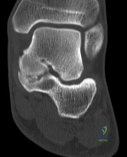
Question 41
According to Perren's strain theory of bone healing, as visually contextualized in the following radiograph
, what specific interfragmentary strain environment is required to promote secondary bone healing with robust callus formation?
, what specific interfragmentary strain environment is required to promote secondary bone healing with robust callus formation?
Explanation
According to Perren's strain theory, an interfragmentary strain of 2% to 10% stimulates secondary bone healing via endochondral ossification, leading to callus formation. A strain less than 2% promotes primary bone healing (cutting cones without callus). Strain greater than 10% results in fibrous tissue formation, and strain over 30% typically leads to a nonunion.
Question 42
Highly cross-linked ultra-high-molecular-weight polyethylene (UHMWPE) is widely used in modern total joint arthroplasty to reduce wear. While cross-linking successfully decreases adhesive and abrasive wear, it significantly diminishes which of the following mechanical properties?
Explanation
The irradiation process used to highly cross-link UHMWPE improves wear resistance but decreases its fatigue resistance, ductility, ultimate tensile strength, and yield strength. To mitigate free radicals generated during this process and prevent oxidation, manufacturers utilize techniques such as remelting, annealing, or adding an antioxidant like Vitamin E.
Question 43
A 15-year-old boy presents with severe, deep bone pain in his leg. Imaging demonstrates a destructive diaphyseal lesion with a classic onion-skin periosteal reaction, as seen in
. A core biopsy reveals a proliferation of small blue round cells. Which of the following chromosomal translocations is most characteristic of this pathology?
. A core biopsy reveals a proliferation of small blue round cells. Which of the following chromosomal translocations is most characteristic of this pathology?
Explanation
The clinical scenario and 'onion-skin' periosteal reaction describe Ewing sarcoma. This malignant bone tumor is characterized by the t(11;22)(q24;q12) translocation in approximately 85% of cases, resulting in the EWS-FLI1 fusion protein. t(9;22) is seen in extraskeletal myxoid chondrosarcoma and CML; t(2;13) in alveolar rhabdomyosarcoma; t(X;18) in synovial sarcoma; and t(12;16) in myxoid liposarcoma.
Question 44
Tranexamic acid (TXA) is extensively utilized in orthopedic surgery to minimize perioperative blood loss. What is the precise molecular mechanism of action for this medication?
Explanation
Tranexamic acid is a synthetic analog of the amino acid lysine. It functions as an antifibrinolytic by reversibly binding to lysine receptor sites on plasminogen. This competitive inhibition prevents plasminogen from binding to fibrin and converting into active plasmin, effectively halting fibrinolysis.
Question 45
Articular cartilage relies on a precise matrix composition for its unique mechanical properties. The remarkable compressive strength of articular cartilage is primarily imparted by which of the following?
Explanation
Aggrecan, the predominant proteoglycan in articular cartilage, possesses highly negatively charged glycosaminoglycan (GAG) side chains. These fixed negative charges draw water into the matrix via osmotic pressure (Donnan swelling effect). The resulting hydrostatic swelling pressure, constrained by the tensile strength of the Type II collagen network, provides cartilage with its ability to withstand immense compressive loads.
Question 46
A patient suffers a traumatic radial nerve palsy after a humerus shaft fracture. After 4 months of observation with no clinical recovery, a surgical exploration is performed. The MRI
guided the decision. Intraoperatively, the nerve is intact in continuity but encased in fibrotic scar tissue disrupting the endoneurial tubes, while the perineurium remains entirely intact. This represents which Sunderland classification of nerve injury?
guided the decision. Intraoperatively, the nerve is intact in continuity but encased in fibrotic scar tissue disrupting the endoneurial tubes, while the perineurium remains entirely intact. This represents which Sunderland classification of nerve injury?
Explanation
Sunderland classified nerve injuries into five degrees. A Third-degree injury involves disruption of the axon and endoneurium, with an intact perineurium and epineurium. Intrafascicular scarring often misdirects regenerating axons or blocks them entirely, meaning spontaneous recovery is variable and often incomplete, frequently necessitating surgical intervention.
Question 47
A specialized physical examination maneuver for diagnosing subtle ligamentous knee injuries has a reported sensitivity of 90% and specificity of 95%. If a referral center begins seeing a higher proportion of acute knee injuries, increasing the overall prevalence of this specific ligament tear in their patient population, which of the following test characteristics will also increase?
Explanation
Positive Predictive Value (PPV) and Negative Predictive Value (NPV) are strongly dependent on the prevalence of the disease in the tested population. As prevalence increases, the PPV increases (a positive test is more likely to be a true positive) and the NPV decreases. Sensitivity, specificity, and likelihood ratios are inherent characteristics of the test and are not affected by disease prevalence.
Question 48
During surgical exploration of the brachial plexus for a traumatic injury, you are evaluating the anatomic structures originating directly from the upper trunk. Which of the following nerves branches exclusively from the upper trunk?
Explanation
The suprascapular nerve and the nerve to the subclavius are the only branches that originate directly from the upper trunk of the brachial plexus (formed by C5 and C6 roots). The dorsal scapular (C5) and long thoracic (C5, C6, C7) nerves branch directly from the roots.
Question 49
Normal bone remodeling relies on a tightly regulated coupling between bone formation and resorption. Which of the following cell types primarily synthesizes and secretes RANKL (Receptor Activator of Nuclear factor Kappa-B Ligand) to stimulate osteoclastogenesis?
Explanation
Osteoblasts (as well as osteocytes and certain stromal cells) secrete RANKL. RANKL binds to the RANK receptor found on the surface of osteoclast precursors and mature osteoclasts, stimulating their differentiation, activation, and survival. Osteoblasts also secrete Osteoprotegerin (OPG), a decoy receptor that binds RANKL to inhibit osteoclast activity.
Question 50
Giant cell tumor (GCT) of bone is a locally aggressive benign tumor. In cases of unresectable or recurrent GCT, Denosumab is often utilized. What is the specific mechanism of action for this therapeutic agent?
Explanation
Denosumab is a fully human monoclonal antibody that specifically binds to and neutralizes RANKL (Receptor Activator of Nuclear factor Kappa-B Ligand). In Giant Cell Tumor of bone, the neoplastic mononuclear stromal cells secrete vast amounts of RANKL, which recruits and activates the reactive, multinucleated osteoclast-like giant cells that cause bone destruction. Denosumab blocks this interaction, leading to rapid tumor consolidation.
Question 51
An intramedullary nail utilized for a tibial shaft nonunion presents with a broken distal locking screw, as conceptualized in
. Microscopic analysis of the retrieved screw shows beach-mark lines on the fracture surface. What is the primary mechanism of failure for this implant?
. Microscopic analysis of the retrieved screw shows beach-mark lines on the fracture surface. What is the primary mechanism of failure for this implant?
Explanation
Orthopedic implants in the setting of delayed union or nonunion typically fail via fatigue failure. Fatigue is the structural failure of a material subjected to repeated cyclic loading at stress levels well below the ultimate tensile strength. Macroscopically, fatigue failure does not present with severe plastic deformation (unlike ductile failure) and is classically identified by 'beach marks' or striations on the failure surface under electron microscopy.
Question 52
Periprosthetic joint infections are notoriously difficult to eradicate due to bacterial biofilm formation. What is the primary chemical constituent of the extracellular polymeric substance (EPS) 'slime layer' formed by mature Staphylococcus epidermidis biofilms?
Explanation
The biofilm matrix (Extracellular Polymeric Substance) in staphylococcal infections is largely composed of polysaccharide intercellular adhesin (PIA). PIA is biochemically identified as poly-N-acetylglucosamine (PNAG). This matrix protects the embedded bacteria from both host immune responses and systemic antibiotics.
Question 53
During a regional block for orthopedic surgery, a patient experiences acute systemic toxicity from an accidental intravascular injection of bupivacaine, leading to severe cardiac dysrhythmias and impending arrest. What is the immediate first-line specific antidote for this local anesthetic systemic toxicity (LAST)?
Explanation
Intravenous lipid emulsion (typically 20% Intralipid) is the rescue treatment of choice for local anesthetic systemic toxicity (LAST), particularly that induced by highly lipophilic amides like bupivacaine. The 'lipid sink' theory postulates that the lipid emulsion creates an expanded intravascular lipid phase that sequesters the lipophilic local anesthetic, removing it from target organs like the heart and brain.
Question 54
Orthopedic surgical implants manufactured from 316L stainless steel perform well in normal host tissues but can be susceptible to localized corrosion. Which specific alloying element is added to 316L stainless steel to enhance its resistance to pitting and crevice corrosion in chloride-rich in vivo environments?
Explanation
In 316L stainless steel, Molybdenum (added at roughly 2-3%) specifically increases resistance to pitting and crevice corrosion in chloride-containing environments (like the human body). Chromium (17-19%) provides the generalized passivating oxide layer. Nickel (13-15%) stabilizes the face-centered cubic austenitic phase, making it non-magnetic. 'L' denotes low Carbon (<0.03%), which prevents chromium carbide precipitation and subsequent intergranular corrosion.
Question 55
A 45-year-old female refugee presents with vague, generalized bone pain and proximal muscle weakness. Plain radiographs reveal bilateral symmetric transverse radiolucencies in the medial femoral necks (Looser zones). Laboratory workup reveals hypocalcemia, hypophosphatemia, highly elevated alkaline phosphatase, and elevated intact PTH. What is the most likely underlying diagnosis?
Explanation
The clinical picture of generalized bone pain, muscle weakness, Looser zones (pseudofractures), and labs showing secondary hyperparathyroidism (low/normal Ca, low P, high PTH, high Alk Phos) points clearly to osteomalacia, most commonly secondary to severe Vitamin D deficiency. Osteomalacia represents defective mineralization of newly formed osteoid in mature bone.
Question 56
The architectural organization of articular cartilage zones reflects their functional biomechanical roles. In which distinct zone of articular cartilage are the collagen fibrils primarily oriented parallel to the joint surface to resist physiological shear stresses?
Explanation
The superficial (tangential) zone comprises the uppermost 10-20% of articular cartilage. Here, chondrocytes are flattened, and the Type II collagen fibrils are aligned parallel to the articular surface. This orientation provides the cartilage with maximal tensile strength to withstand sheer forces during joint articulation. In contrast, the deep (radial) zone features collagen aligned perpendicularly to resist compressive loads.
Question 57
A patient with a well-documented history of advanced breast carcinoma presents with a solitary lytic lesion in the distal femur, confirmed on MRI
. What is the most common physiological route through which this carcinoma metastasized to the appendicular skeleton?
. What is the most common physiological route through which this carcinoma metastasized to the appendicular skeleton?
Explanation
The overwhelming majority of bone metastases from carcinomas (such as breast, prostate, lung, kidney, and thyroid) disseminate via hematogenous spread, specifically through the venous system. Batson's venous plexus is a valveless paraspinal venous network that provides a direct pathway for tumor emboli to reach the axial skeleton, and venous sinuses facilitate spread to the appendicular long bones.
Question 58
Understanding the normal human gait cycle is critical for diagnosing pathological gait patterns. During the normal stance phase of walking on level ground, maximum ankle dorsiflexion occurs precisely during which sub-phase?
Explanation
Maximum ankle dorsiflexion (approximately 10 to 15 degrees) occurs at the very end of terminal stance. During this phase, the heel of the stance foot begins to rise, and the tibia translates forward over the fixed foot. Immediately after terminal stance begins pre-swing, during which the ankle rapidly plantarflexes for toe-off.
Question 59
When stabilizing a highly comminuted diaphyseal fracture using bridge plating techniques, orthopedic surgeons frequently opt for a locked plating construct over conventional non-locked plates. From a biological and biomechanical standpoint, what is the primary advantage of utilizing locked screws in this scenario?
Explanation
Locked plates function as internal fixators. Because the screw heads thread directly into the plate, the construct acts as a single fixed-angle device. This provides stability without the need to aggressively compress the plate against the bone, thereby preserving the periosteal blood supply. In contrast, conventional plates rely on the friction generated by compressing the plate against the bone, which can strangulate local periosteal perfusion.
Question 60
A 65-year-old male develops sudden, severe pain and swelling in his right knee two days following an inguinal hernia repair. Radiographs demonstrate distinct linear calcifications within the menisci
. Aspiration yields inflammatory synovial fluid containing rhomboid-shaped crystals that exhibit weak positive birefringence. What is the precise chemical composition of these crystals?
. Aspiration yields inflammatory synovial fluid containing rhomboid-shaped crystals that exhibit weak positive birefringence. What is the precise chemical composition of these crystals?
Explanation
The clinical presentation (acute monoarthritis often precipitated by stress/surgery), radiologic evidence of chondrocalcinosis, and the characteristic synovial fluid analysis (rhomboid crystals, weakly positively birefringent under polarized light) are pathognomonic for pseudogout. Pseudogout is caused by the intra-articular deposition of Calcium Pyrophosphate Dihydrate (CPPD) crystals. Monosodium urate causes gout (needle-shaped, strongly negatively birefringent).
Question 61
Which of the following biologic grafts possesses osteoconductive, osteoinductive, and osteogenic properties?
Explanation
Fresh autogenous cancellous bone contains a scaffold (osteoconductive), growth factors (osteoinductive), and live cells (osteogenic). Allografts and DBM lack live cells, while synthetic ceramics only provide an osteoconductive scaffold.
Question 62
In internal fixation, the pullout strength of a cortical screw is most significantly increased by altering which of the following parameters?
Explanation
Pullout strength is most significantly influenced by the outer diameter of the screw. Other factors include the thread depth and the shear strength of the bone, but increasing the outer thread diameter yields the greatest biomechanical advantage.
Question 63
Tranexamic acid (TXA) is widely used in orthopedic surgery to reduce perioperative blood loss. Which of the following best describes its mechanism of action?
Explanation
TXA is a synthetic analog of the amino acid lysine. It competitively binds to the lysine-binding sites on plasminogen, preventing its activation to plasmin and thus effectively inhibiting fibrinolysis.
Question 64
A 12-year-old child presents with frequent fractures, blue sclerae, and hearing loss. This condition is primarily caused by a quantitative defect in which of the following structural components?
Explanation
Osteogenesis imperfecta is most commonly caused by an autosomal dominant mutation affecting the COL1A1 or COL1A2 genes. This results in a quantitative or qualitative defect in Type I collagen, which is the major structural protein of bone.
Question 65
During the process of endochondral ossification, the transition of chondrocytes from the proliferative zone to the hypertrophic zone is primarily regulated by which of the following signaling pathways?
Explanation
The Ihh-PTHrP feedback loop is the primary master regulator of chondrocyte maturation. Ihh stimulates the production of PTHrP, which in turn delays the differentiation of proliferating chondrocytes into hypertrophic chondrocytes.
Question 66
A 65-year-old male undergoes a revision total hip arthroplasty. Intraoperatively, extensive black metallic debris and tissue necrosis are found around the modular head-neck junction. Which of the following processes is primarily responsible for this finding?
Explanation
Trunnionosis is the clinical manifestation of fretting and galvanic corrosion at the modular head-neck junction. It is most commonly associated with a cobalt-chromium head on a titanium stem, leading to adverse local tissue reactions (ALTR).
Question 67
Which of the following transcription factors is considered the 'master regulator' of osteoblast differentiation?
Explanation
Runx2 is essential for the commitment of mesenchymal stem cells to the osteoblast lineage. Mice lacking Runx2 exhibit a complete absence of bone formation due to developmental arrest of osteoblasts.
Question 68
A high-energy open tibial shaft fracture with a 6 cm wound and extensive soft tissue stripping is classified as Gustilo-Anderson IIIB. Following debridement and skeletal stabilization, coverage of a middle-third tibial defect is most appropriately achieved with which of the following?
Explanation
The soleus rotational flap is the workhorse for soft tissue coverage of the middle third of the tibia. The gastrocnemius flap is preferred for the proximal third, while free tissue transfer is typically required for the distal third.
Question 69
In the context of fracture healing, low oxygen tension (hypoxia) at the fracture site directly stimulates angiogenesis primarily through the upregulation of which molecule?
Explanation
Hypoxia at the fracture site stabilizes Hypoxia-Inducible Factor-1 alpha (HIF-1 alpha). This leads to the transcriptional upregulation of VEGF, which promotes the angiogenesis necessary for callus formation and subsequent fracture healing.
Question 70
A 45-year-old female presents with a pathological fracture of the proximal femur. Biopsy reveals a primary bone tumor characterized by multinucleated giant cells in a background of mononuclear stromal cells. The neoplastic mononuclear cells primarily express which of the following to recruit the giant cells?
Explanation
In Giant Cell Tumor of bone (GCT), the mononuclear stromal cells are the true neoplastic cells. They express abnormally high levels of RANKL, which recruits and activates the reactive multinucleated giant cells responsible for extreme bone resorption.
Question 71
Sclerostin, a key negative regulator of bone formation targeted by drugs like romosozumab, exerts its effect by binding to LRP5/6 and inhibiting which of the following pathways?
Explanation
Sclerostin is produced by osteocytes and acts as a powerful antagonist to the canonical Wnt/beta-catenin signaling pathway. By binding to the LRP5/6 co-receptors, it effectively inhibits osteoblastogenesis and bone formation.
Question 72
According to the Sunderland classification of nerve injuries, a third-degree injury involves disruption of the axon and which of the following supporting structures?
Explanation
A Sunderland third-degree injury involves disruption of both the axon and the endoneurium, while the perineurium and epineurium remain intact. Internal scarring often limits functional return, making recovery unpredictable compared to second-degree injuries.
Question 73
A patient is prescribed a bisphosphonate for osteoporosis. At the cellular level, nitrogen-containing bisphosphonates (e.g., alendronate) inhibit osteoclast function primarily by targeting which of the following enzymes?
Explanation
Nitrogen-containing bisphosphonates inhibit farnesyl pyrophosphate synthase in the mevalonate pathway. This prevents the prenylation of small GTPases essential for osteoclast survival and cytoskeletal organization, ultimately inducing osteoclast apoptosis.
Question 74
A 25-year-old male sustains a severe crush injury to his lower extremity.
Compartment syndrome is suspected. Which of the following delta pressure measurements definitively indicates the need for emergent fasciotomy?
Compartment syndrome is suspected. Which of the following delta pressure measurements definitively indicates the need for emergent fasciotomy?
Explanation
A delta pressure (Diastolic Blood Pressure minus Compartment Pressure) of less than 30 mmHg is considered the most reliable threshold for diagnosing acute compartment syndrome. Relying purely on absolute pressure readings can be misleading.
Question 75
The primary structural component of articular cartilage is Type II collagen, but its compressive stiffness and load-bearing capacity are primarily provided by the osmotic swelling pressure of which molecule?
Explanation
Aggrecan is the most abundant large aggregating proteoglycan in articular cartilage. Its highly negatively charged glycosaminoglycan chains attract water, creating the swelling pressure that effectively resists compressive joint loads.
Question 76
Which of the following orthopedic implant materials has a modulus of elasticity most similar to that of cortical bone, thereby minimizing stress shielding?
Explanation
Titanium alloy has a modulus of elasticity (approx. 110 GPa) closer to that of cortical bone (approx. 15-20 GPa) compared to stiffer materials like cobalt-chrome (210 GPa) or stainless steel (190 GPa). This closer biomechanical match reduces stress shielding.
Question 77
In the process of bone remodeling, osteoclasts create an acidic microenvironment in the resorption pit by actively pumping protons. Which enzyme is critical for generating these protons within the osteoclast cytoplasm?
Explanation
Carbonic anhydrase II catalyzes the conversion of carbon dioxide and water into carbonic acid, which then dissociates into protons and bicarbonate. The protons are actively pumped across the ruffled border to acidify the resorption pit and dissolve bone mineral.
Question 78
A 45-year-old patient undergoes revision total hip arthroplasty. The surgeon utilizes a titanium alloy femoral stem and a cobalt-chromium femoral head. Over time, the release of metal ions at the modular head-neck junction leads to adverse local tissue reactions. Which of the following electrochemical processes is most primarily responsible for this degradation due to the contact of dissimilar metals?
Explanation
Galvanic corrosion occurs when two dissimilar metals are in direct contact within an electrolytic environment, such as bodily fluids. The less noble metal becomes the anode and corrodes, releasing ions into the surrounding tissue.
Question 79
A 28-year-old male is brought to the trauma bay following a high-speed motor vehicle collision. He is hypotensive and tachycardic. Pelvic radiographs reveal an anteroposterior compression type III (APC-III) pelvic ring injury. A circumferential pelvic binder is requested to reduce pelvic volume. To be most effective, the binder should be centered directly over which of the following anatomic landmarks?
Explanation
To effectively reduce pelvic volume and stabilize the pelvic ring, a pelvic binder must be centered over the greater trochanters. Placement higher over the iliac crests can actually paradoxically open the pelvic ring further or fail to reduce it.
Question 80
According to Perren's strain theory of bone healing, different types of tissues can tolerate varying amounts of strain before failing. Which of the following represents the maximum tissue strain tolerated by lamellar bone?
Explanation
According to Perren's strain theory, lamellar bone can only tolerate up to 2% strain before failing. Woven bone can tolerate up to 10%, cartilage up to 30%, and granulation tissue up to 100%.
Question 81
A 24-year-old male sustains a closed bilateral femoral shaft fracture. On post-injury day two, he develops confusion, tachypnea, and a petechial rash over his axillae and conjunctivae. Based on Gurd's criteria for Fat Embolism Syndrome (FES), which of the following is considered a major criterion?
Explanation
Gurd's major criteria for Fat Embolism Syndrome include respiratory insufficiency, cerebral involvement (altered mental status), and a petechial rash. Tachycardia, fever, retinal changes, and anemia are considered minor criteria.
Question 82
A 68-year-old female presents with severe back pain and generalized fatigue. Laboratory workup reveals hypercalcemia and a monoclonal spike on serum protein electrophoresis. Radiographs demonstrate multiple punched-out lytic lesions in her skull and vertebral bodies. Which of the following urinary findings is most characteristic of her underlying diagnosis?
Explanation
The patient's presentation is classic for multiple myeloma, the most common primary malignancy of bone in adults. Bence Jones proteins (free light chains) are typically found in the urine of these patients.
Question 83
A 13-year-old obese male presents with left groin pain and an obligatory external rotation of the hip during active flexion. An AP pelvis radiograph is obtained.
What is the most appropriate management for a stable presentation of this condition?
What is the most appropriate management for a stable presentation of this condition?
Explanation
The presentation is classic for a Slipped Capital Femoral Epiphysis (SCFE). The gold standard treatment for a stable SCFE is in situ fixation across the physis with a single cannulated screw.
Question 84
A 30-year-old male sustains an isolated open fracture of the tibial shaft (Gustilo-Anderson Type II) without severe soft tissue stripping or gross contamination. According to current evidence-based guidelines, which of the following is true regarding the timing of initial surgical debridement?
Explanation
Recent high-level evidence demonstrates that the historical '6-hour rule' is not strictly required. As long as early intravenous antibiotics are administered, surgical debridement within 24 hours yields comparable infection rates for Type I and II open fractures.
Question 85
A 45-year-old male suffers a burst fracture of T12 with complete spinal cord injury. On arrival at the emergency department, he is warm, flushed, and has a blood pressure of 80/40 mmHg with a heart rate of 52 beats per minute. Which of the following best describes the pathophysiology of his hemodynamic state?
Explanation
The patient is experiencing neurogenic shock, characterized by hypotension and bradycardia due to the loss of sympathetic vasomotor tone and unopposed vagal (parasympathetic) activity. This must be distinguished from spinal shock, which refers to the transient loss of spinal reflexes.
Question 86
When utilizing a cementless titanium component in total joint arthroplasty, optimizing the surface topography is critical for long-term biological fixation. What is the optimal pore size range required to promote maximal bone ingrowth?
Explanation
Extensive biomechanical and histological studies show that the optimal pore size for biological bone ingrowth into porous-coated implants is between 50 and 300 micrometers. Pores smaller than this do not allow adequate vascularization, while larger pores lead to fibrous tissue formation.
Question 87
A 22-year-old carpenter lacerates his index finger at the level of the proximal phalanx, resulting in both flexor digitorum superficialis (FDS) and profundus (FDP) transection (Zone II injury). Primary repair is planned. Blood supply to the flexor tendons within this specific zone is primarily provided by which of the following structures?
Explanation
In flexor tendon Zone II (from the A1 pulley to the FDS insertion), the tendons are enclosed in a synovial sheath. The primary blood supply to the tendons in this relatively avascular zone comes dorsally through the vincula brevis and longa.
Question 88
During clinical evaluation of a patient with a suspected anterior cruciate ligament (ACL) tear, the examiner performs a pivot shift test. The palpable clunk that occurs during knee flexion from an extended position represents the reduction of the anteriorly subluxated tibia. Which structure is mechanically responsible for providing the force that reduces the tibia during this maneuver?
Explanation
The pivot shift test relies on the iliotibial (IT) band. As the knee flexes past approximately 20-30 degrees, the IT band transitions from an extensor to a flexor, creating a posterior force vector that reduces the anteriorly subluxated lateral tibial plateau.
Question 89
A 35-year-old male presents 8 months after a tibial shaft fracture treated with cast immobilization. Radiographs reveal a hypertrophic nonunion characterized by abundant callus formation but a persistent radiolucent line at the fracture site. What is the most appropriate management strategy for this condition?
Explanation
Hypertrophic nonunions have adequate biology and blood supply (evidenced by callus formation) but lack sufficient mechanical stability. The optimal treatment is to provide rigid stabilization, typically with internal fixation (plating or intramedullary nailing), which alone will allow the fracture to heal.
Question 90
Articular cartilage relies on its unique extracellular matrix composition to provide a frictionless surface and withstand compressive forces. Which of the following collagen types represents the predominant structural protein within normal adult hyaline articular cartilage?
Explanation
Type II collagen constitutes approximately 90-95% of the collagen found in normal hyaline articular cartilage. Type I collagen is predominant in bone, tendon, and fibrocartilage.
Question 91
A 19-year-old male sustains a low-velocity civilian handgun injury to the right thigh. Radiographs demonstrate a minimally displaced midshaft femur fracture with retained bullet fragments in the soft tissues. Distal pulses are intact, and the neurologic exam is normal. Which of the following is the most appropriate initial management?
Explanation
Low-velocity gunshot wounds resulting in fractures are generally treated as Gustilo-Anderson Type I open fractures. They do not routinely require formal tract debridement or bullet removal unless inside a joint; they are managed with local wound care, antibiotics, and appropriate stabilization (such as IM nailing).
Question 92
A 15-year-old female undergoes neoadjuvant chemotherapy followed by limb-salvage surgery for osteosarcoma of the distal femur. Pathological evaluation of the resected tumor is performed. Which of the following is the single most important prognostic factor for long-term survival in this patient?
Explanation
The degree of tumor necrosis after neoadjuvant chemotherapy (specifically >90% necrosis) is the single most important prognostic indicator for overall survival in patients with high-grade osteosarcoma.
Question 93
A 6-year-old boy falls from monkey bars and sustains a completely displaced, extension-type supracondylar humerus fracture (Gartland Type III). Radiographs show the distal fragment is displaced posteromedially. Which of the following peripheral nerves is at highest risk of injury with this specific displacement pattern?
Explanation
In a posteromedially displaced supracondylar fracture, the proximal fragment displaces anterolaterally, commonly piercing the brachialis muscle and tenting or injuring the radial nerve. Posterolateral displacement endangers the anterior interosseous nerve (AIN).
Question 94
Bone Morphogenetic Protein-2 (BMP-2) is utilized clinically to enhance spinal fusion and fracture healing. Upon binding to its cell surface receptor, BMP-2 primarily initiates intracellular osteogenic signaling through which of the following pathways?
Explanation
BMP-2 binds to serine/threonine kinase receptors, which leads to the phosphorylation and activation of the Smad 1/5/8 complex. This complex then translocates to the nucleus to upregulate osteogenic gene expression.
Question 95
A 32-year-old male sustains a high-energy trauma resulting in a vertically oriented (Pauwels type III) femoral neck fracture.
Which of the following fixation constructs provides the greatest biomechanical stability for this specific fracture pattern?
Which of the following fixation constructs provides the greatest biomechanical stability for this specific fracture pattern?
Explanation
Pauwels type III femoral neck fractures are highly vertical and experience significant shear forces. A fixed-angle sliding hip screw provides superior biomechanical stability compared to parallel cancellous screws, reducing the risk of varus collapse and nonunion.
Question 96
A 40-year-old recreational tennis player suffers an acute complete tear of the Achilles tendon. During shared decision-making regarding operative versus non-operative management, the patient asks about the primary advantage of surgical repair. According to classical meta-analyses, which of the following is the most significant statistical advantage of operative treatment?
Explanation
Historically, the main advantage of operative repair of an acute Achilles tendon rupture has been a significantly lower re-rupture rate compared to traditional non-operative casting. However, modern functional rehabilitation protocols have narrowed this gap.
Question 97
Following total knee arthroplasty, a patient is prescribed rivaroxaban for chemical thromboprophylaxis. What is the specific pharmacological mechanism of action of this medication?
Explanation
Rivaroxaban is a direct oral anticoagulant (DOAC) that works by directly inhibiting Factor Xa, preventing the conversion of prothrombin to thrombin. Dabigatran, in contrast, is a direct thrombin inhibitor.
None
Previous ChapterOrthopedic A Review | Dr Hutaif General Orthopedics Rev -...
Next Chapter Approaches Orthopedic B Review | Dr Hutaif General Orth -...

Medically Verified Content by
Prof. Dr. Mohammed Hutaif Clinic
Consultant Orthopedic & Spine Surgeon