Operative Management of the Thumb: Trapeziometacarpal Synovectomy and Interphalangeal Soft Tissue Reconstruction
Key Takeaway
Thumb trapeziometacarpal synovectomy and interphalangeal soft tissue reconstruction are critical procedures for managing inflammatory arthritis and extension contractures. This guide details the dorsal surgical approaches, meticulous soft tissue handling, and precise capsular releases required to restore function. Proper management of the superficial radial nerve, extensor tenolysis, and postoperative Kirschner wire fixation are emphasized to optimize outcomes and mitigate complications such as extensor lag or recurrent deformity.
INTRODUCTION TO THUMB JOINT PRESERVATION AND RECONSTRUCTION
The thumb is the cornerstone of hand biomechanics, contributing to approximately 40% to 50% of overall hand function. Pathologic conditions affecting the thumb—ranging from inflammatory arthropathies such as rheumatoid arthritis to severe post-traumatic contractures—can profoundly diminish a patient's quality of life, grip strength, and fine motor dexterity.
This comprehensive academic treatise details two critical joint-preserving and function-restoring procedures: Thumb Trapeziometacarpal (TMC) Joint Synovectomy and Interphalangeal (IP) Soft Tissue Reconstruction. These procedures demand a profound understanding of the intricate capsuloligamentous anatomy, precise surgical execution, and rigorous postoperative rehabilitation protocols. The techniques described herein are designed for the postgraduate orthopedic surgeon, fellow, and practicing consultant seeking to optimize functional outcomes in complex thumb deformities.
THUMB TRAPEZIOMETACARPAL JOINT SYNOVECTOMY
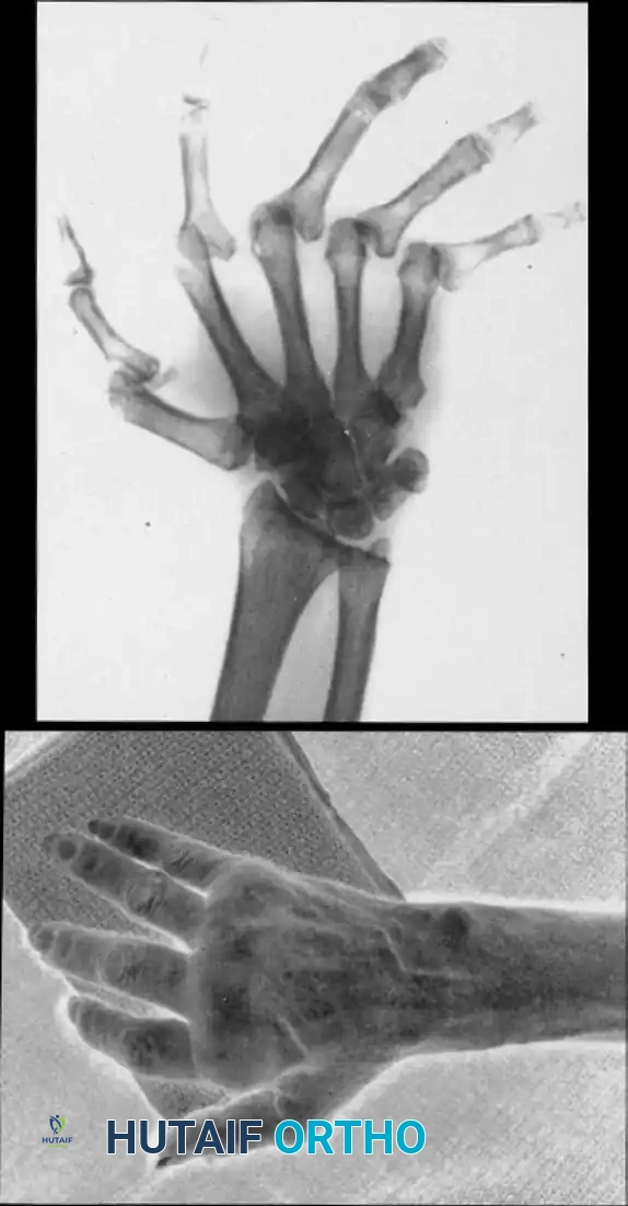
Synovial hypertrophy at the trapeziometacarpal (basal) joint is a hallmark of early-stage inflammatory arthritis and, occasionally, early primary osteoarthritis. If left untreated, aggressive synovitis leads to attenuation of the critical stabilizing ligaments—most notably the anterior oblique (beak) ligament and the dorsoradial ligament—culminating in dorsal subluxation of the metacarpal base, articular cartilage destruction, and the classic adduction contracture of the thumb web space.
Synovectomy is indicated in patients with refractory pain and swelling who have failed conservative management (e.g., splinting, NSAIDs, intra-articular corticosteroid injections) but still possess well-preserved articular cartilage and a passively correctable joint.
Surgical Anatomy and Biomechanics
The TMC joint is a highly mobile, biconcave saddle joint. Its stability relies heavily on its capsuloligamentous envelope rather than its bony architecture.
* Dorsoradial Ligament (DRL): The primary restraint to dorsal subluxation of the metacarpal base.
* Anterior Oblique Ligament (AOL): Often referred to as the "beak ligament," it acts as a critical volar stabilizer during pinch mechanisms.
* Superficial Radial Nerve (SRN): The terminal sensory branches of the SRN cross directly over the operative field. Injury to these branches can result in debilitating neuromas (Wartenberg’s syndrome), which often overshadow the benefits of the primary surgery.
Preoperative Preparation and Positioning
- Anesthesia: Regional anesthesia (brachial plexus block) or general anesthesia is utilized based on patient comorbidities and surgeon preference.
- Positioning: The patient is positioned supine with the operative extremity extended on a radiolucent hand table.
- Tourniquet: A well-padded pneumatic upper arm tourniquet is applied and inflated to 250 mm Hg (or 100 mm Hg above systolic blood pressure) following exsanguination with an Esmarch bandage.
- Equipment: Loupe magnification (minimum 2.5x to 3.5x) is mandatory for the identification and protection of the SRN branches. Fine rongeurs, sharp curettes, and bipolar electrocautery must be available.
Surgical Approach
The approach to the TMC joint must provide adequate exposure for complete synovectomy while minimizing the risk of iatrogenic destabilization.
- Incision: Make a straight dorsal incision, approximately 3 to 4 cm in length, centered over the TMC joint. As the incision proceeds distally, gently curve it toward the palmar aspect of the thumb metacarpal. This modified Wagner or dorsal approach allows for excellent visualization of the dorsal capsule and the abductor pollicis longus (APL) insertion.
- Superficial Dissection: Carefully incise the skin and subcutaneous tissues.
🔪 Surgical Warning: Superficial Radial Nerve
The sensory branches of the superficial radial nerve are highly variable and exquisitely sensitive to traction. Employ blunt dissection with tenotomy scissors spread parallel to the nerve fibers. Retract the skin and nerve branches gently using Ragnell or Sen retractor, strictly avoiding excessive tension.
Capsulotomy and Synovectomy Technique
- Capsular Exposure: Identify the interval between the extensor pollicis brevis (EPB) and the abductor pollicis longus (APL).
- Capsulotomy: Open the joint capsule on the dorsal side of the APL tendon. A longitudinal or slightly L-shaped capsulotomy is preferred to facilitate robust closure. Elevate the capsule as a continuous full-thickness flap to preserve its vascularity and structural integrity.
- Joint Inspection: Distract the thumb longitudinally to open the joint space. Inspect the articular surfaces of the trapezium and the first metacarpal base to confirm the absence of advanced eburnation (which would necessitate an arthroplasty or arthrodesis).
- Synovectomy:
- Utilize a fine pituitary rongeur and a small, sharp curet to systematically excise the hypertrophic synovial tissue.
- Pay particular attention to the volar and ulnar recesses of the joint, where synovitis often hides and erodes the AOL.
- Irrigate the joint copiously with sterile saline to flush out loose synovial debris and cartilage fragments.
Assessment of Ligamentous Laxity
Following the synovectomy, the stability of the TMC joint must be rigorously assessed.
- Apply a dorsal-to-volar shear stress to the metacarpal base.
- If extensive ligamentous laxity is noted (subluxation greater than one-third of the articular surface), a simple capsulorrhaphy will be insufficient.
- Contingency Plan: In cases of profound instability, reconstruction of the trapeziometacarpal capsuloligamentous structures is required. This is typically achieved via a ligament reconstruction using a slip of the flexor carpi radialis (FCR) or APL tendon (e.g., Eaton-Littler reconstruction) to recreate the volar beak ligament and stabilize the metacarpal base.
Closure and Postoperative Protocol
- Capsular Closure: Repair the dorsal capsule meticulously using non-absorbable or slowly absorbable braided sutures (e.g., 3-0 or 4-0 Ethibond or PDS). The capsule should be imbricated slightly to eliminate redundant tissue and reinforce dorsal stability.
- Skin Closure: Deflate the tourniquet, achieve meticulous hemostasis with bipolar cautery, and close the skin with interrupted non-absorbable sutures (e.g., 4-0 or 5-0 Nylon).
- Immobilization: Apply a bulky, sterile compressive dressing and a well-molded thumb spica splint. The thumb must be immobilized in extension and palmar abduction to maintain the joint in its most stable, congruent position and to prevent adduction contracture.
Postoperative Care:
* The initial splint remains in place for 10 to 14 days.
* Sutures are removed at the 2-week mark.
* A custom thermoplastic thumb spica splint is fabricated, and the patient begins a supervised hand therapy program focusing on active range of motion (ROM) while avoiding forceful pinch activities for 6 weeks.
INTERPHALANGEAL SOFT TISSUE RECONSTRUCTION
Severe extension contractures of the thumb interphalangeal (IP) joint present a formidable reconstructive challenge. These contractures frequently arise secondary to prolonged immobilization, trauma, burns, or inflammatory conditions. The pathoanatomy typically involves a combination of dorsal capsular contracture, shortening of the collateral ligaments, and dense adhesions of the extensor pollicis longus (EPL) tendon.
Indications and Patient Selection
Surgical release (Surgical Technique 73-18) is indicated when:
1. The IP joint is passively correctable (or has the potential to be correctable once soft tissues are released).
2. There are no significant radiographic changes (e.g., advanced joint space narrowing, osteophytes, or articular destruction).
3. The extension contracture severely limits the patient's ability to perform opposition and grasp.
If the joint exhibits severe arthritic changes or is rigidly fixed due to bony ankylosis, an IP joint arthrodesis is the procedure of choice rather than soft tissue reconstruction.
Surgical Approach and Skin Management
- Incision: Expose the IP joint via a dorsal longitudinal or gently curved incision.
- Managing Severe Contractures: In cases of severe, long-standing extension contracture, the dorsal skin will have contracted significantly. A simple linear incision will not allow for closure once the joint is flexed.
- Technique: Plan to convert the linear incision into a Z-plasty during wound closure. This involves creating triangular flaps with angles of approximately 60 degrees to recruit lateral skin laxity and lengthen the dorsal skin envelope.
💡 Clinical Pearl: Z-Plasty Planning
Always design the Z-plasty flaps before making the initial incision. Ensure the central limb of the Z-plasty aligns with the axis of maximum tension (the longitudinal axis of the thumb) to maximize the lengthening effect upon transposition.
Extensor Tenolysis and Capsular Release
The core of the procedure involves a systematic, step-wise release of the dorsal tethering structures.
- Extensor Tenolysis:
- Identify the EPL tendon proximal to the IP joint.
- Perform a meticulous tenolysis, freeing the tendon from surrounding scar tissue and adhesions along the proximal phalanx.
- Use a Freer elevator or tenotomy scissors to glide circumferentially around the tendon.
- Dorsal Capsulotomy:
- Retract the mobilized extensor tendon laterally (usually to the ulnar side) to expose the underlying dorsal capsule.
- Incise the dorsal part of the IP joint capsule transversely.
- Collateral Ligament Release:
- The dorsal portions of the collateral ligaments are often contracted and contribute to the extension deformity.
- Carefully incise the dorsal fibers of the radial and ulnar collateral ligaments.
- Caution: Do not completely transect the collateral ligaments, as this will result in gross mediolateral instability. Release only the dorsal fibers until the joint can be passively flexed.
Joint Stabilization and Fixation
Once the soft tissue release is complete, the joint must be stabilized in a functional position to allow the soft tissues to heal at their new, lengthened resting state.
- Positioning: Flex the IP joint to 20 to 30 degrees. This is the optimal functional position for the thumb IP joint, balancing the needs of pinch strength and clearance during grasp.
- Kirschner Wire Fixation:
- Drive a smooth Kirschner wire (typically 0.045-inch or 1.14 mm) across the IP joint to hold it rigidly in the 20 to 30-degree flexed position.
- The wire can be driven retrograde from the joint surface out the tip of the distal phalanx, and then antegrade across the joint into the proximal phalanx.
- Cut the wire outside the skin and bend it to prevent migration and facilitate easy removal in the clinic.
Wound Closure Strategies
Closure over a newly flexed joint that was previously contracted in extension is notoriously difficult due to skin shortage.
- Z-Plasty Execution: If a Z-plasty was planned, transpose the flaps and suture them using 5-0 or 6-0 non-absorbable monofilament. This will immediately relieve dorsal skin tension.
- Secondary Intention: If there is still a small portion of the wound, particularly distally near the eponychial fold, that cannot be closed without excessive tension, leave it open.
- Attempting to force closure under tension will lead to skin necrosis, wound breakdown, and potential infection of the underlying joint or tendon.
- These small, open distal wounds typically heal remarkably well by secondary intention within 10 to 14 days with simple non-adherent dressings.
🚨 Pitfall: Forcing Skin Closure
Never compromise the flexion achieved during the release just to close the skin. The primary goal is functional joint position. Skin can heal by secondary intention or be grafted later; a recurrent contracture due to compromised positioning is a failure of the procedure.
Postoperative Care and Rehabilitation (Surgical Technique 73-19)
The postoperative protocol is a delicate balance between protecting the surgical release and preventing recurrent stiffness.
- Initial Immobilization: Apply a protective volar splint to support the thumb, leaving the K-wire exposed but protected.
- Day 10 - K-Wire Removal: The splint and the Kirschner wire are removed at 10 days postoperatively in the clinic.
- Rehabilitation: Gentle, active, and active-assisted range of motion exercises are initiated immediately following pin removal.
- Day 14 - Suture Removal: Sutures are removed at approximately 14 days, assuming adequate wound healing.
- Continued Splinting: The thumb is splinted in a functional position for another 2 to 3 weeks. The splint is worn continuously, removed only for the prescribed periods of exercise.
- Managing Extensor Lag: If an incomplete extension (extensor lag) develops during the rehabilitation phase—a common occurrence due to the relative lengthening of the extensor mechanism—splinting is continued for an additional 2 to 3 weeks, often incorporating a dynamic extension component.
Prognosis and Patient Counseling:
It is imperative to manage patient expectations preoperatively. While this reconstructive procedure is highly effective at restoring a functional arc of motion and improving grasp, normal motion is rarely regained. The goal is a functional, stable thumb, not a return to pre-pathologic kinematics.
CONCLUSION
Surgical intervention for thumb trapeziometacarpal synovitis and interphalangeal extension contractures requires a masterful command of hand anatomy and biomechanics. The dorsal approaches described provide excellent exposure but demand meticulous handling of the superficial radial nerve and the delicate dorsal skin envelope.
Whether performing a meticulous synovectomy to halt the progression of basal joint arthritis or executing a complex tenolysis and capsulotomy to salvage a stiff IP joint, the surgeon must adhere strictly to the principles of tissue preservation, precise stabilization (via K-wire fixation or ligamentous reconstruction), and rigorous postoperative therapy. By following these textbook-level protocols, orthopedic surgeons can reliably restore critical thumb function and significantly enhance patient outcomes.
You Might Also Like