Operative Management of Painful Paraarticular Calcifications and Snapping Scapula Syndrome
Key Takeaway
Painful paraarticular calcifications arise from focal tendon degeneration, leading to acute chemical inflammation and severe pain during the resorptive phase. While conservative measures like corticosteroid injections or extracorporeal shock wave therapy often suffice, refractory cases necessitate surgical excision. Concurrently, periscapular pain may manifest as snapping scapula syndrome, requiring precise surgical resection of the superomedial scapular angle to alleviate mechanical impingement and restore scapulothoracic kinematics.
INTRODUCTION TO PERIARTICULAR SOFT-TISSUE LESIONS
Painful paraarticular calcifications and periscapular friction syndromes represent a spectrum of debilitating soft-tissue and osteotendinous pathologies encountered frequently in orthopedic practice. While calcific tendinitis is most classically associated with the rotator cuff of the shoulder, analogous calcific deposits frequently develop around the wrist, elbow, hip, knee, foot, and hand. These lesions typically arise within a tendon or the adjacent soft tissues near the osteotendinous junction.
Occasionally, localized periscapular pain is not driven by intrinsic tendinous calcification but rather by mechanical friction and bursal inflammation at the scapulothoracic articulation—a condition known as snapping scapula syndrome. This comprehensive masterclass delineates the pathophysiology, clinical evaluation, and evidence-based operative management of both painful paraarticular calcifications and snapping scapula syndrome, providing a rigorous framework for orthopedic residents, fellows, and practicing consultants.
PAINFUL PARAARTICULAR CALCIFICATIONS
Pathophysiology and Histology
The etiology of paraarticular calcifications is fundamentally linked to focal tissue hypoxia, microtrauma, and subsequent fibrocartilaginous metaplasia. As in the rotator cuff of the shoulder, the calcification is most probably located in an area of focal necrosis or degeneration. The natural history of calcific tendinitis follows a distinct, cell-mediated cycle (Uhthoff’s cycle) comprising three phases:
1. Precalcific Phase: Fibrocartilaginous metaplasia occurs within the tendon substance due to localized ischemia.
2. Calcific Phase: Subdivided into formative, resting, and resorptive stages.
3. Postcalcific Phase: Fibroblasts remodel the defect with type I collagen, restoring tendon integrity.
Clinical Pearl: The resorptive phase is the most acutely painful stage. During this phase, the calcium deposit assumes a "toothpaste-like" consistency and incites a profound chemical inflammatory reaction characterized by neovascularization and macrophage infiltration.
Gärtner and Simons evaluated acute and chronic calcific deposits using infrared spectroscopy, revealing variable water, carbon dioxide, and phosphate contents in all samples. Notably, no significant differences in these proportions were seen between acute and chronic deposits. Furthermore, scanning and electron microscopy have demonstrated similar crystal morphologies across both phases, confirming that the acute pain is driven by the inflammatory response rather than a sudden shift in crystal composition.
Regional Anatomic Manifestations
While the shoulder remains the most common site, paraarticular calcifications manifest in several distinct anatomic locations, often mimicking other common orthopedic pathologies:
- The Wrist: Calcifications are predominantly found in the flexor carpi ulnaris (FCU) tendon near its insertion into the pisiform, or in the flexor carpi radialis (FCR) tendon near its insertion into the base of the second metacarpal.
- The Elbow: Calcific deposits frequently localize to the common extensor tendon origin at the lateral epicondyle. The signs and symptoms closely resemble those of acute lateral epicondylitis (tennis elbow), though the onset of pain in calcific tendinitis is typically much more abrupt and severe.
- The Knee (Pellegrini-Stieda Disease): The presence of calcification in the medial collateral ligament (MCL) is known as Pellegrini-Stieda disease. Unlike spontaneous calcific tendinitis, this is usually directly related to prior trauma, such as a severe sprain or partial tear of the MCL.
- The Ankle and Distal Tibiofibular Syndesmosis: Calcifications between the distal tibia and fibula are similarly trauma-induced, often following syndesmotic sprains or tears of the interosseous membrane.
Clinical Evaluation and Diagnostic Imaging
Patients presenting with an acute lesion in the resorptive phase exhibit signs of a severe chemical inflammatory reaction. Clinical examination reveals exquisite point tenderness, localized swelling, erythema, and increased local heat. The pain is often severe enough to cause pseudoparalysis of the affected joint.
Radiographic evaluation is the cornerstone of diagnosis. Standard anteroposterior (AP) and lateral radiographs may miss smaller deposits.
* Supplementary Oblique Views: Often necessary to project the calcific deposit away from the underlying bone.
* Soft-Tissue Density Films: Highly valuable if the deposit is small, faint, and not readily visible on standard exposures.
* Ultrasonography: Highly sensitive for identifying calcifications, assessing their consistency (hard vs. soft/resorptive), and guiding therapeutic injections.
Nonoperative Management
The natural history of paraarticular calcification is generally self-limiting, and spontaneous recovery may occur without treatment as the deposit is resorbed.
- Pharmacologic Therapy: Nonsteroidal anti-inflammatory drugs (NSAIDs) are the first line of defense to manage the acute chemical inflammation.
- Corticosteroid Injections: Infiltration with a local anesthetic agent, supplemented by the injection of 40 mg of methylprednisolone (Depo-Medrol) or its equivalent, produces immediate relief and can be curative. The injection should be directed into the peritendinous bursa rather than directly into the tendon substance to avoid tendon rupture.
- Advanced Modalities: Ultrasound-guided barbotage (needling and lavage) and Extracorporeal Shock Wave Therapy (ESWT) have demonstrated significant efficacy in fragmenting the deposits and stimulating neovascularization, particularly around the shoulder.
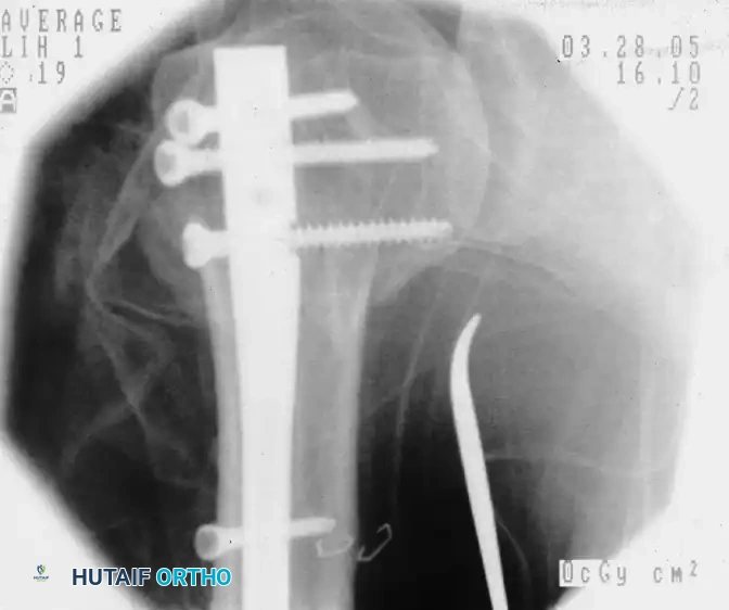
Operative Management: Arthroscopic Excision
Surgical intervention is indicated when the response to nonoperative measures is unsatisfactory, typically after 3 to 6 months of conservative care, or in cases of chronic, unremitting pain.
For chronic symptomatic calcification of the rotator cuff, arthroscopic excision is the gold standard.
1. Localization: The lesion is localized intraoperatively using a spinal needle. A hyperemic "strawberry lesion" on the bursal surface of the rotator cuff often overlies the deposit.
2. Bursectomy: For lesions on the superior surface, a thorough subacromial bursectomy is generally required to achieve adequate visualization.
3. Excision: Once localized, the tendon is incised longitudinally in line with its fibers. The calcium deposit is manually released and evacuated using a small curet or a motorized shaver.
4. Subacromial Decompression: Ellman recommended performing a subacromial decompression (acromioplasty) after the release of the calcific deposit to prevent secondary impingement of the inflamed, thickened rotator cuff.
Evidence-Based Outcome: Ark et al. reported 91% good or excellent results in 23 patients with chronic calcific rotator cuff tendinitis treated by arthroscopic excision of the calcium deposits and subacromial bursectomy. This protocol remains highly reliable in contemporary practice.
SNAPPING SCAPULA SYNDROME AND SCAPULOTHORACIC BURSITIS
While paraarticular calcifications cause pain via chemical inflammation, periscapular pain often arises from mechanical friction. Snapping scapula syndrome (washboarding syndrome) is characterized by painful crepitus during scapulothoracic motion.
Pathomechanics and Indications for Surgery
The scapulothoracic articulation lacks a true synovial joint capsule; instead, movement is facilitated by the serratus anterior and subscapularis muscles gliding over the thoracic rib cage, cushioned by the supraserratus and infraserratus bursae.
Snapping scapula can be caused by:
* Osseous Lesions: Osteochondromas (most common benign tumor of the scapula), healed rib fractures, or Luschka’s tubercle (an anatomic variant where the superomedial angle is abnormally prominent).
* Soft-Tissue Lesions: Elastofibroma dorsi, fibromas, or chronic fibrotic bursitis.
When conservative management (postural rehabilitation, periscapular strengthening, and corticosteroid injections into the scapulothoracic bursa) fails, surgical resection of the superomedial angle of the scapula is indicated.
Surgical Technique: Open Resection of the Superomedial Scapular Angle
The following technique expands upon the foundational principles of scapular resection, ensuring meticulous soft-tissue handling and protection of critical neurovascular structures.
1. Patient Positioning and Anesthesia
- Administer general endotracheal anesthesia.
- Position the patient prone on a radiolucent Jackson table with chest rolls to allow free excursion of the thorax.
- The operative arm is draped free. Place the arm in the "chicken-wing" position (internal rotation with the dorsum of the hand resting on the lumbar spine). This maneuver wings the scapula, lifting the medial border away from the thoracic wall and making the superomedial angle highly prominent.
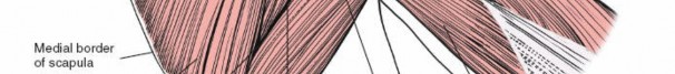
2. Surgical Approach and Superficial Dissection
- Incision: Make a 7 to 10 cm longitudinal incision parallel to and approximately 2 cm lateral to the medial border of the scapula, centered over the intended area of resection (the superomedial angle).
- Flap Mobilization: Develop full-thickness fasciocutaneous flaps medially and laterally to expose the trapezius muscle.
- Trapezius Split: Split the trapezius muscle bluntly in line with its fibers directly over the superomedial angle of the scapula.
🚨 SURGICAL WARNING: Do not extend the trapezius split too far medially. Aggressive medial dissection endangers the spinal accessory nerve (Cranial Nerve XI) and the transverse cervical artery, which course along the deep surface of the trapezius.
3. Deep Dissection and Muscle Detachment
- Rhomboid Release: Identify the insertion of the rhomboid minor and the superior portion of the rhomboid major on the medial border of the scapula. Carefully free the rhomboid muscles from the osseous edge using electrocautery, leaving a small cuff of tissue for later repair.
- Subperiosteal Elevation:
- Anteriorly: Subperiosteally dissect the serratus anterior and subscapularis muscles from the inferior (ventral) surface of the scapula.
- Posteriorly: Subperiosteally elevate the supraspinatus muscle from the posterior (dorsal) surface of the scapula, taking care not to violate the suprascapular notch laterally, which houses the suprascapular nerve and vessels.
4. Scapular Resection (Richards and McKee Technique)
- After exposing the medial 3 to 4 cm of the scapula, inspect the ventral surface to determine the cause of the snapping. Look for an osteochondroma, an osseous exostosis, or an abnormally hooked superomedial angle.
- If no discrete mass is identified, the prominent superomedial angle itself is the mechanical culprit and must be resected.
- Protection: As suggested by Richards and McKee, place a sterile folded towel or a broad malleable retractor deep to the scapula to protect the underlying chest wall, pleura, and neurovascular structures.
- Osteotomy: Use an oscillating saw to perform the osteotomy. Cut a triangle with 4- to 5-cm borders from the superomedial angle of the scapula. The cut should run obliquely from the superior border (medial to the suprascapular notch) to the medial border.
- Contouring: Use a high-speed burr or a bone rasp to meticulously contour and smooth the osteotomy edges. Any sharp osseous spicules left behind will cause recurrent bursitis and crepitus.
5. Closure
- Thoroughly irrigate the surgical bed to remove all bone debris.
- Achieve meticulous hemostasis.
- Muscle Approximation: Securely reattach the rhomboid muscles and the serratus anterior fascia to the remaining medial border of the scapula. Drill holes may be placed in the scapular edge to facilitate a robust transosseous repair using heavy nonabsorbable sutures (e.g., #2 FiberWire).
- Close the trapezius split with interrupted absorbable sutures.
- Insert a closed-suction drain deep to the muscle layer to prevent postoperative hematoma formation.
- Close the subcutaneous tissue and skin in the usual layered manner.
Postoperative Rehabilitation Protocol
Successful outcomes following superomedial angle resection depend heavily on structured postoperative rehabilitation to prevent arthrofibrosis and restore scapulothoracic kinematics.
- Phase I (Weeks 0-3): The patient is placed in a broad arm sling for comfort. Immediate postoperative management focuses on pain control and wound healing. Pendulum exercises and passive range of motion (ROM) of the glenohumeral joint are initiated on postoperative day 1. Active scapular retraction is strictly avoided to protect the rhomboid repair.
- Phase II (Weeks 4-6): The sling is discontinued. Active-assisted and active ROM exercises are commenced. Gentle periscapular isometric exercises are introduced.
- Phase III (Weeks 7-12): Progressive resistance training is initiated, focusing on the serratus anterior, rhomboids, and lower trapezius to restore dynamic scapular stability. Return to heavy lifting or overhead sports is typically permitted after 3 to 4 months, contingent upon the recovery of full, pain-free periscapular strength.
You Might Also Like