Arthroscopic Suprascapular Nerve Release: Surgical Technique
Key Takeaway
Suprascapular nerve entrapment is an increasingly recognized etiology of posterior shoulder pain, often associated with massive rotator cuff tears, paralabral ganglia, or transverse scapular ligament hypertrophy. Arthroscopic suprascapular nerve release provides excellent visualization and decompression of the suprascapular notch. This comprehensive guide details the indications, pathoanatomy, and step-by-step arthroscopic technique—including portal placement, ligamentous landmarks, and neurovascular preservation—to ensure safe and effective surgical outcomes for refractory cases.
SUPRASCAPULAR NERVE ENTRAPMENT: PATHOPHYSIOLOGY AND CLINICAL EVALUATION
Suprascapular nerve entrapment has drawn significant attention in recent orthopedic literature as a primary or concomitant cause of persistent parascapular and posterior shoulder pain. Historically underdiagnosed, this compressive neuropathy is frequently associated with massive, retracted rotator cuff tears. The medial retraction of the supraspinatus and infraspinatus tendons places pathological traction on the suprascapular nerve, which is anatomically tethered at the suprascapular notch.
Beyond traction neuropathy secondary to rotator cuff pathology, other established etiologies of suprascapular nerve entrapment include space-occupying lesions—most notably paralabral ganglia extending into or around the suprascapular notch—and altered intrinsic anatomy of the notch itself. This altered anatomy often manifests as hypertrophy or ossification of the superior transverse scapular ligament (STSL) and constricting bony morphology of the notch (e.g., a V-shaped notch).
Indications for Surgical Intervention
Lafosse et al. established that chronic, debilitating posterior shoulder pain coupled with electrodiagnostic evidence (EMG/NCS) of suprascapular nerve compression that remains refractory to conservative management (e.g., physical therapy, NSAIDs, image-guided perineural injections) constitutes a definitive indication for surgical nerve release.
Clinical Pearl:
When evaluating a patient with a massive rotator cuff tear, profound atrophy of the supraspinatus and infraspinatus fossae out of proportion to the tear size, combined with a dull, aching posterior shoulder pain, should raise high clinical suspicion for secondary suprascapular nerve entrapment.
SURGICAL ANATOMY AND BIOMECHANICS
A profound understanding of the neurovascular anatomy is paramount for safe arthroscopic decompression. The suprascapular nerve arises from the upper trunk of the brachial plexus (C5, C6) and courses posteriorly beneath the trapezius and omohyoid muscles to reach the suprascapular notch.
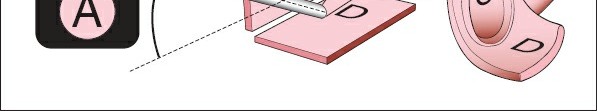
At the notch, the anatomical relationship is classically described by the mnemonic "Army over, Navy under": the suprascapular Artery passes superior to the transverse scapular ligament, while the suprascapular Nerve passes inferior to the ligament, directly through the bony notch.
Warner et al. provided critical topographic mapping for arthroscopic orientation, demonstrating that the suprascapular notch is located approximately 4.5 cm medial to the posterolateral corner of the acromion. The nerve is highly vulnerable to iatrogenic injury during blind dissection or aggressive medial mobilization of retracted rotator cuff tendons.
ARTHROSCOPIC SUPRASCAPULAR NERVE RELEASE (LAFOSSE, TOMASI, AND CORBETT TECHNIQUE)
The arthroscopic approach to the suprascapular nerve, popularized by Lafosse, provides unparalleled illumination and magnification of the suprascapular notch without the morbidity of extensive open muscle detachment.
Patient Positioning and Setup
- Positioning: Place the patient in a standard "beach chair" position. Ensure the head and neck are secured in a neutral position to avoid excessive traction on the contralateral brachial plexus.
- Arm Suspension: The operative arm is held in slight flexion and abduction with approximately 3 kg of longitudinal traction to open the subacromial and glenohumeral spaces.
- Anesthesia: General anesthesia is typically employed, often supplemented with an interscalene block or a supraclavicular catheter for postoperative pain management.
Portal Placement Strategy
Precise portal placement is the linchpin of a successful arthroscopic nerve release.
1. Standard Posterior Portal: Used for initial intra-articular diagnostic arthroscopy.
2. Standard Lateral Portal: Used for subacromial viewing and bursectomy.
3. Anterolateral Portal: Created at the anterolateral corner of the acromion. This is the optimal working portal for completing the dissection of the transverse scapular notch using shavers and radiofrequency (RF) devices.
4. Suprascapular Nerve Portal: Positioned between the clavicle and the scapular spine, approximately 7 cm medial to the lateral border of the acromion. This portal is approximately 2 cm medial to the classic Neviaser portal.
🚨 CRITICAL WARNING:
The spinal accessory nerve traverses near the medial border of the scapula. To avoid catastrophic iatrogenic injury to the spinal accessory nerve, ensure the suprascapular nerve portal remains strictly lateral to the medial third of the scapular spine. The portal should be more than 5 cm lateral to the spinal accessory nerve's expected course.
Step-by-Step Surgical Technique
1. Diagnostic Arthroscopy and Subacromial Preparation
After a thorough inspection of the glenohumeral joint through the posterior portal, introduce the arthroscope into the subacromial space through the lateral portal.
- Utilize a motorized shaver and a radiofrequency ablation device through the posterior portal to perform a meticulous anteromedial bursectomy.
- Clearing the bursa is critical to provide unobstructed access to the coracoid base and the suprascapular notch.
Surgical Pitfall:
Fluid extravasation and soft tissue swelling during the procedure add significantly to the difficulty of gaining adequate exposure to the transverse scapular ligament. Therefore, if a distal clavicular resection or subacromial decompression (SAD) is planned, it must be deferred until after the suprascapular nerve decompression is complete.
2. Identification of Coracoclavicular Landmarks
Once the anteromedial bursectomy is complete, switch the arthroscope to the lateral portal for optimal viewing. Establish the anterolateral working portal.
- Coracoacromial (CA) Ligament: First, identify the CA ligament and follow its course medially and inferiorly to its insertion at the base of the coracoid process.
- Coracoclavicular (CC) Ligaments: Carry the dissection posteriorly and medially from the coracoid base to identify the conoid and trapezoid ligaments.
- The Transverse Scapular Ligament (TSL): The medial border of the CC ligaments at the base of the coracoid defines the lateral insertion of the superior transverse scapular ligament. The TSL is reliably identified as the medial continuity of the conoid ligament, bridging the suprascapular notch.
3. Establishing the Suprascapular Nerve Portal
This portal is created under direct arthroscopic visualization using an outside-in technique.
- Once visualization of the TSL is adequate, use an 18-gauge spinal needle to guide the placement of the new suprascapular nerve portal.
- Insert the needle through the trapezius at an angle orthogonal to the suprascapular fossa, directing it slightly anteriorly toward the transverse scapular notch.
- Verification: If the spinal needle is oriented correctly, the tip of the needle should be visualized immediately anterior to the anterior border of the supraspinatus muscle belly.
4. Dissection and Decompression of the Notch
- Once the spinal needle is appropriately positioned, use a #11 scalpel to incise the skin.
- Introduce a blunt trocar through the trapezius and surrounding soft tissues, directing it toward the suprascapular nerve.
- Use the blunt trocar to gently dissect the fatty areolar tissues surrounding the suprascapular nerve within the notch and to further clarify the anterior and posterior borders of the TSL.
- Neurovascular Identification: The suprascapular artery is easily visualized pulsating superior to the ligament. The suprascapular nerve is identified as a distinct white band traveling underneath the ligament.
- If necessary, use radiofrequency or shaver devices to enhance the dissection. However, ensure that all motorized and thermal instruments remain strictly superior and lateral to the conoid ligament insertion at the base of the coracoid to avoid catastrophic injury to the suprascapular artery.
5. Ligament Release
- Once the ligament and nerve are definitively identified, position the blunt tip of the trocar or a specialized arthroscopic nerve elevator lateral to the suprascapular nerve within the notch.
- Protect the nerve inferiorly while using arthroscopic scissors or a specialized RF probe to transect the transverse scapular ligament from lateral to medial.
- Confirm complete release by visualizing the nerve floating freely within the notch without tethering.
MANAGEMENT OF CONCOMITANT PATHOLOGY: ADHESIONS AND CAPSULAR CONTRACTURE
In many cases of chronic suprascapular nerve entrapment, particularly those associated with massive, retracted rotator cuff tears or prior failed surgical interventions, patients present with profound secondary shoulder stiffness. Significant adhesion contractures of the humeroscapular motion interface are frequently encountered.
These dense adhesions connect the deep surface of the deltoid to the rotator cuff and the proximal humerus. Because they obliterate the normal bursal gliding planes, they severely limit both active and passive shoulder range of motion.
Arthroscopic and Open Lysis of Adhesions
- Arthroscopic Release: Following nerve decompression, a thorough resection of the capsular margins and an intra-articular capsular release may be necessary to prevent early scar formation and restore capsular volume.
- Open Deltopectoral Approach (For Severe Revision Cases): In extreme cases of revision surgery where arthroscopic mobilization is insufficient, an axillary deltopectoral approach may be required. Through this approach, the surgeon must perform a complete, circumferential lysis of adhesions between the bursal surface around the proximal humerus and the surfaces of the entire rotator cuff, coracoacromial arch, coracoid base, and conjoined tendon.
POSTOPERATIVE CARE AND REHABILITATION PROTOCOL
The success of a suprascapular nerve release, particularly when combined with extensive lysis of adhesions or rotator cuff repair, relies heavily on a rigorous and immediate postoperative rehabilitation protocol.
Pain Management
- A supraclavicular or interscalene perineural catheter is ideally placed before surgery under ultrasound guidance by the anesthesia team.
- This catheter is left in place to provide continuous regional analgesia for the first 48 to 72 hours postoperatively, which is critical for tolerating immediate physical therapy.
Early Mobilization Phase (0-3 Weeks)
- Immediate Motion: After surgery, the patient and their family members are instructed on immediate active-assisted range-of-motion (AAROM) exercises. These must be repeated continuously throughout the day to prevent the recurrence of adhesions.
- Formal Physical Therapy: Formal physical therapy for passive range of motion (PROM) and AAROM is initiated within 24 hours of surgery.
- Stretching Protocol: Stretching exercises must be performed aggressively in all four quadrants (forward elevation, external rotation at the side, internal rotation, and cross-body adduction). Patients are instructed to perform these stretches five times a day, with five repetitions of each stretching maneuver per session.
Clinical Follow-Up
- We recommend a mandatory reexamination within 3 weeks postoperatively to assess the restoration of motion, monitor wound healing, and evaluate early signs of neurological recovery.
- While pain relief is often rapid following nerve decompression, patients must be counseled that the recovery of muscle trophism and motor strength in the supraspinatus and infraspinatus may take 6 to 12 months, depending on the chronicity of the preoperative entrapment and the degree of Wallerian degeneration.
You Might Also Like