Trapeziometacarpal Arthrodesis: Comprehensive Surgical Principles and the Doyle Technique
Key Takeaway
Trapeziometacarpal arthrodesis is a highly effective surgical intervention for advanced basal joint arthritis of the thumb, particularly in young, high-demand patients. This procedure provides a stable, pain-free thumb pillar while preserving grip strength. The Doyle technique utilizes a dorsal sliding corticocancellous graft from the thumb metacarpal, combined with precise joint positioning and Kirschner wire fixation, to achieve reliable osseous union and optimal biomechanical function.
INTRODUCTION TO TRAPEZIOMETACARPAL ARTHRODESIS
Trapeziometacarpal (TMC) arthrodesis is a time-honored, highly durable surgical intervention for the management of advanced osteoarthritis, post-traumatic arthritis, and inflammatory arthropathies affecting the basal joint of the thumb. While suspensionplasty and interpositional arthroplasty techniques (such as the ligament reconstruction and tendon interposition [LRTI]) are frequently utilized in older, lower-demand populations, TMC arthrodesis remains the gold standard for young, active patients, manual laborers, and individuals requiring maximal grip and key pinch strength.
By fusing the saddle joint of the thumb, the surgeon effectively eliminates pain while providing a rigid, stable pillar against which the index and middle fingers can forcefully oppose. The primary historical challenge associated with TMC arthrodesis has been the risk of nonunion, given the complex multi-planar forces acting across the joint and the relatively small surface area available for osseous integration. To mitigate this, various techniques have been developed, including the use of plates, screws, memory staples, and tension band wiring.
The technique popularized by Doyle, detailed extensively in this masterclass, employs a meticulously contoured joint preparation combined with a dorsal sliding corticocancellous graft from the thumb metacarpal. This autologous graft acts as both an osteogenic bridge and a biologic tension band, significantly enhancing union rates while avoiding the complications associated with prominent metallic hardware.
INDICATIONS AND CONTRAINDICATIONS
Primary Indications
- High-Demand Patients: Manual laborers, heavy lifters, and young patients (< 55 years) who require absolute preservation of grip and pinch strength.
- Isolated TMC Joint Arthritis: Eaton-Littler Stage II or III osteoarthritis where the scaphotrapezial-trapezoid (STT) joint is radiographically and clinically spared.
- Post-Traumatic Arthritis: Sequelae of intra-articular Bennett or Rolando fractures resulting in painful articular incongruity.
- Paralytic Deformities: Stabilization of the thumb ray in patients with peripheral nerve injuries (e.g., median or ulnar nerve palsy) to facilitate tendon transfers.
- Failed Conservative Management: Persistent, debilitating pain despite exhaustive non-operative measures, including splinting, NSAIDs, and intra-articular corticosteroid injections.
Contraindications
- Pantrapezial Arthritis: Degenerative changes extending into the STT joint. Fusing the TMC joint in the presence of STT arthritis will exponentially increase stress on the diseased STT joint, leading to rapid clinical deterioration.
- Elderly, Low-Demand Patients: Individuals who prioritize mobility over absolute strength are generally better served by trapeziectomy with or without LRTI.
- Severe Metacarpal Bone Loss: Inadequate bone stock to support a sliding graft or rigid fixation.
- Active Infection: Absolute contraindication to any elective arthrodesis.
Clinical Pearl: Always obtain a dedicated Robert's view (true AP of the thumb) and a stress view to accurately stage basal joint arthritis and definitively rule out STT joint involvement prior to committing to a TMC arthrodesis.
BIOMECHANICS AND JOINT POSITIONING
The most critical determinant of a successful clinical outcome in TMC arthrodesis—assuming osseous union is achieved—is the precise spatial positioning of the fused joint. The thumb must be positioned to allow for forceful opposition to the index and middle fingers (key pinch and chuck pinch) while simultaneously permitting the hand to open wide enough to grasp large objects (e.g., a large cylinder or a doorknob) and lay flat on a surface.
The "Clenched Fist" Position
The optimal position for fusion is often described as the "clenched fist" or "holding a tennis ball" position. Biomechanically, this translates to:
* Palmar Abduction: 30 to 40 degrees.
* Radial Abduction: 30 to 35 degrees.
* Pronation: 10 to 15 degrees (to allow the pulp of the thumb to face the pulp of the index finger).
Surgical Warning: Excessive palmar abduction or extension will severely limit the patient's ability to cup the hand or perform fine motor tasks. Conversely, excessive adduction will prevent the patient from grasping large objects and may lead to a debilitating adduction contracture of the first web space. Intraoperative clinical assessment of the thumb's relationship to the index finger is paramount before final fixation.
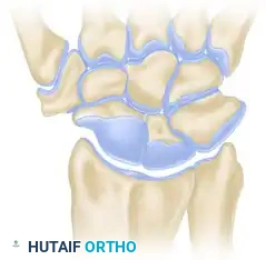
SURGICAL ANATOMY
A profound understanding of the radial-sided anatomy of the wrist and hand is essential to execute this approach safely.
* Superficial Radial Nerve (SRN): The terminal branches of the SRN arborize extensively over the first dorsal compartment and the anatomic snuffbox. Iatrogenic injury or aggressive retraction can lead to a devastating, painful neuroma.
* Extensor Pollicis Brevis (EPB) and Abductor Pollicis Longus (APL): These tendons define the volar border of the anatomic snuffbox and cross directly over the TMC joint. The APL frequently has multiple slips inserting onto the base of the first metacarpal and the trapezium.
* Radial Artery: Courses dorsally deep to the APL and EPB tendons, resting on the scaphoid and trapezium before diving between the two heads of the first dorsal interosseous muscle. It must be protected during dorsal capsular dissection and graft harvesting.
SURGICAL TECHNIQUE: THE DOYLE METHOD
1. Anesthesia, Positioning, and Preparation
- Anesthesia: The procedure is typically performed under regional anesthesia (supraclavicular or axillary brachial plexus block) supplemented with intravenous sedation or general anesthesia.
- Positioning: Place the patient in the supine position. The operative arm is extended onto a radiolucent hand table. The hand is fully supinated to expose the palmar-radial aspect of the thumb.
- Tourniquet: Exsanguinate the limb with an Esmarch bandage and inflate a well-padded pneumatic upper arm tourniquet to 250 mm Hg (or 100 mm Hg above systolic blood pressure) to ensure a bloodless surgical field.
2. Incision and Superficial Dissection
- Make a palmar curved incision (often referred to as a modified Wagner approach) centered over the TMC joint. The incision should expose the proximal two-thirds of the thumb metacarpal and allow access to both the dorsal and palmar aspects of the trapeziometacarpal joint.
- Carefully dissect through the subcutaneous tissues using blunt dissection (e.g., with tenotomy scissors) to identify and meticulously protect the terminal sensory branches of the superficial radial nerve. Retract these branches gently using vessel loops; avoid rigid retractors that may cause traction neuropraxia.
- Identify the extensor pollicis brevis (EPB) tendon and retract it dorsally.
3. Deep Dissection and Joint Exposure
- Identify the multiple slips of the abductor pollicis longus (APL) tendon. Detach the APL tendons along with a robust portion of the underlying joint capsule. Tag this capsulotendinous flap with a non-absorbable suture for later reattachment and robust closure.
- Reflect the thenar musculature (abductor pollicis brevis and opponens pollicis) distally off the base of the first metacarpal to expose the palmar capsule.
- Incise the dorsal and palmar capsule longitudinally to fully expose the articular surfaces of the trapezium and the base of the first metacarpal. Use a periosteal elevator to clear the joint margins, ensuring excellent visualization of the saddle joint anatomy.
4. Joint Preparation and Contouring
- Cartilage Removal: Utilize a high-speed, air-driven burr under continuous cool saline irrigation to meticulously remove all articular cartilage and sclerotic subchondral bone down to healthy, bleeding cancellous bone. Continuous irrigation is critical to prevent thermal necrosis of the osteocytes, which would severely compromise arthrodesis.
- Joint Shaping: Slightly round the base of the thumb metacarpal to create a convex surface. Conversely, use the burr to create a matching, shallow concavity (a "cup") in the distal articular surface of the trapezium.
- Preservation of Length: It is imperative to preserve the overall shape and length of the joint. Over-resection of bone will lead to undue shortening of the thumb ray, altering the biomechanics of the extrinsic tendons and potentially weakening pinch strength.
- Feathering the Bone: Use a small, sharp osteotome to make multiple small, shallow cuts (often termed "fish-scaling" or "feathering") into the prepared cancellous bone surfaces. This technique significantly increases the surface area available for osteogenesis and releases osteoprogenitor cells from the marrow space.
Surgical Pearl: The "cup and cone" geometry achieved by rounding the metacarpal and cupping the trapezium provides intrinsic stability and allows for fine-tuning of the thumb's position in all planes prior to definitive fixation.
5. Positioning and Provisional Fixation
- Manually reduce the joint and place the thumb into the optimal "clenched fist" position: 30 to 40 degrees of palmar abduction, 30 to 35 degrees of radial abduction, and slight pronation.
- Verify the position clinically by ensuring the thumb pulp can comfortably oppose the index and middle fingers, and that the hand can be laid flat on the hand table.
- Once the ideal position is achieved, insert two crossed Kirschner wires (typically 0.045 or 0.062 inches) across the joint. Drive the wires from the metacarpal base into the trapezium. Ensure the wires cross outside the fusion interface to maximize rotational stability.
- Confirm joint position, hardware placement, and absence of STT joint penetration using intraoperative fluoroscopy (mini C-arm).
6. The Sliding Corticocancellous Graft
This step is the hallmark of the Doyle technique, providing a biologic tension band and a rich source of autologous bone graft.
* Marking the Graft: On the exposed dorsum of the thumb metacarpal, outline a rectangular corticocancellous graft. The graft should be approximately 1.5 to 2.0 cm in length and 0.5 to 0.8 cm in width.
* Drilling: Use a small drill point or a fine Kirschner wire to make multiple contiguous drill holes along the outlined rectangle. This prevents stress risers and inadvertent fracturing of the metacarpal shaft during graft harvest.
* Mobilization: Connect the drill holes using a small, sharp osteotome to carefully mobilize the corticocancellous "sliding graft."
* Trapezial Recess: Create a corresponding rectangular recess in the dorsum of the trapezium, perfectly sized to receive the proximal end of the sliding graft.
* Graft Placement: Slide the mobilized graft proximally across the arthrodesis site so that it bridges the joint and seats firmly into the trapezial recess. Impact the graft gently to ensure a flush, press-fit interface.
* Graft Stabilization: While the press-fit is often stable, if there is any micromotion, stabilize the graft with an additional small Kirschner wire or a low-profile mini-fragment screw to absolutely prevent graft displacement during the postoperative period.
7. Closure
- Thoroughly irrigate the wound to remove any bone debris.
- Release the tourniquet and achieve meticulous hemostasis using bipolar electrocautery. Hematoma formation can increase the risk of infection and fibrosis.
- Reattach the previously tagged abductor pollicis longus (APL) tendon and the capsular flap to the periosteum and remaining capsule using strong, absorbable sutures (e.g., 2-0 or 3-0 Vicryl). This restores the dynamic stabilizing forces across the base of the thumb.
- Close the subcutaneous tissue and skin using standard techniques (e.g., 4-0 Monocryl deep, 5-0 nylon for skin).
POSTOPERATIVE CARE AND REHABILITATION
The success of a TMC arthrodesis relies heavily on strict adherence to postoperative immobilization protocols to ensure undisturbed osseous union.
Phase 1: Immediate Postoperative (Days 0 to 10)
- Apply a nonadhering dressing (e.g., Xeroform or Adaptic) over the incision.
- Apply generous cast padding and a rigid, well-molded thumb spica splint in the operating room. The splint must immobilize the wrist and the thumb up to the interphalangeal (IP) joint, leaving the IP joint free to prevent stiffness.
- Instruct the patient to keep the hand strictly elevated above heart level to minimize edema and throbbing pain.
- Encourage immediate active range of motion of the fingers and the thumb IP joint to promote venous return and prevent tendon adhesions.
Phase 2: Subacute Immobilization (10 Days to 8 Weeks)
- At 10 to 14 days postoperatively, the patient is seen in the clinic. The splint and skin sutures are removed, provided wound healing is satisfactory.
- A short-arm thumb spica cast (fiberglass) is applied. The cast must be meticulously molded around the base of the thumb to provide absolute rigid immobilization of the arthrodesis site.
- The patient remains in this cast for an additional 6 weeks (totaling 8 weeks of immobilization).
Phase 3: Hardware Removal and Rehabilitation (8 Weeks and Beyond)
- At 8 weeks postoperatively, the cast is removed.
- Serial radiographs (AP, lateral, and Robert's views) are obtained to assess for bridging trabecular bone and obliteration of the joint space, indicating radiographic union.
- If clinical and radiographic union is confirmed, the percutaneous or buried Kirschner wires are removed in the clinic or minor procedure room.
- Therapy Initiation: The patient is transitioned to a custom-molded, removable thermoplastic thumb spica splint, to be worn during heavy activities and at night.
- A formal hand therapy program is initiated, focusing on:
- Gentle active and active-assisted range of motion of the wrist and thumb.
- Desensitization techniques for the surgical scar and superficial radial nerve distribution.
- Progressive strengthening exercises (putty, grip strengtheners) beginning at 10 to 12 weeks postoperatively.
- Unrestricted heavy manual labor is typically permitted between 4 to 6 months postoperatively, depending on the robustness of the radiographic union and the patient's clinical strength recovery.
COMPLICATIONS AND MANAGEMENT
While highly successful, TMC arthrodesis is a technically demanding procedure with several potential pitfalls.
1. Nonunion
Historically, nonunion has been the most frequently cited complication of TMC arthrodesis, with older literature reporting rates as high as 15% to 20%. However, with the advent of meticulous joint preparation (feathering) and the Doyle sliding corticocancellous graft technique, nonunion rates have plummeted to less than 5%.
* Management: Asymptomatic nonunions (fibrous unions) often require no intervention if the patient is pain-free and stable. Symptomatic nonunions require revision surgery, typically involving takedown of the pseudarthrosis, rigid internal fixation (e.g., locking plate), and structural iliac crest bone grafting.
2. Malunion
Fusing the thumb in excessive adduction or extension is a severe biomechanical failure.
* Management: Prevention is paramount. Intraoperative clinical assessment of the "clenched fist" position is mandatory. Established, symptomatic malunions require corrective osteotomy and revision arthrodesis.
3. Superficial Radial Nerve Neuritis
Injury or traction to the SRN branches can result in debilitating neuropathic pain or neuroma formation, which can be more disabling than the original arthritis.
* Management: Meticulous blunt dissection and gentle retraction are critical. Postoperative desensitization therapy is the first line of treatment. Refractory neuromas may require surgical excision and burying of the nerve stump into muscle or bone.
4. Adjacent Segment Disease
Fusing the TMC joint alters the kinematics of the thumb ray, theoretically increasing stress on the adjacent STT and metacarpophalangeal (MCP) joints.
* Management: Ensure the STT joint is completely free of arthritis preoperatively. If STT arthritis develops years later, it can be managed with an STT arthrodesis or trapeziectomy.
CONCLUSION
Trapeziometacarpal arthrodesis utilizing the Doyle sliding graft technique is a masterful surgical solution for advanced basal joint arthritis in the high-demand patient. By combining meticulous articular preparation, precise biomechanical positioning, and the biologic advantage of a local corticocancellous graft, the orthopedic surgeon can reliably achieve a solid, pain-free fusion. This restores the critical pillar of the thumb, allowing patients to return to heavy manual labor and forceful gripping activities with excellent long-term durability.
You Might Also Like