Principles of Spinal Arthrodesis: Biomechanics, Bone Grafting, and Surgical Techniques
Key Takeaway
Spinal arthrodesis remains a cornerstone of operative orthopaedics for treating instability, deformity, and degenerative disc disease. This comprehensive guide explores the biological principles of bone grafting, including autograft, allograft, and bone morphogenetic proteins (BMPs). It details evidence-based surgical approaches across the cervical, thoracic, and lumbar spine, emphasizing biomechanics, complication avoidance, and postoperative protocols to optimize fusion rates and clinical outcomes in complex spinal pathology.
Introduction to Spinal Arthrodesis
The evolution of spinal arthrodesis represents one of the most profound advancements in orthopaedic surgery. Since the pioneering works of Hibbs and Albee in the early 20th century for the treatment of spinal tuberculosis and deformity, the indications for spinal fusion have expanded exponentially. Today, spinal arthrodesis is the definitive surgical intervention for a myriad of pathologies, including degenerative disc disease, spondylolisthesis, spinal trauma, neoplastic reconstruction, and complex scoliotic deformities.
Achieving a successful arthrodesis requires a meticulous understanding of spinal biomechanics, the biological cascade of bone healing, and precise surgical execution. The surgeon must navigate the delicate balance between rigid mechanical stabilization and the optimization of the biological environment to facilitate solid osseous union.
Biological Principles of Bone Grafting
The success of any spinal fusion is fundamentally dependent on the biological environment. The ideal bone graft or substitute must possess three critical properties: osteogenesis, osteoinduction, and osteoconduction.
The Triad of Bone Healing
- Osteogenesis: The presence of surviving, viable osteoprogenitor cells and osteoblasts within the graft material that directly produce new bone.
- Osteoinduction: The ability of the graft to recruit mesenchymal stem cells (MSCs) from the host bed and stimulate their differentiation into bone-forming osteoblasts. This process is primarily mediated by bone morphogenetic proteins (BMPs) and other growth factors.
- Osteoconduction: The provision of a three-dimensional structural scaffold that facilitates the ingrowth of neovasculature and the migration of host osteogenic cells.
Autogenous Bone Graft (Autograft)
Iliac crest bone graft (ICBG) remains the "gold standard" for spinal arthrodesis, as it is the only material that inherently possesses all three properties of the bone healing triad.
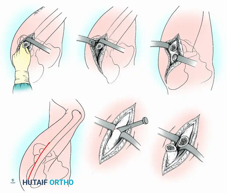
- Harvesting Technique: The anterior or posterior iliac crest can be utilized depending on patient positioning. The posterior superior iliac spine (PSIS) provides a significantly larger volume of cancellous bone. Meticulous subperiosteal dissection is required to avoid injury to the superior cluneal nerves.
- Complications: Despite its biological superiority, ICBG harvest is associated with significant donor-site morbidity, including chronic pain, hematoma, pelvic fracture, and infection.
Clinical Pearl: When harvesting posterior iliac crest bone graft, limit the harvest to lateral to the sacroiliac (SI) joint to prevent iatrogenic SI joint instability, and avoid violating the inner table to protect the superior gluteal artery and sciatic nerve.
Allograft Bone
Allograft bone, typically harvested from cadaveric donors, provides an excellent osteoconductive scaffold and is available in structural (cortical) or non-structural (cancellous) forms.
* Processing: Allografts are processed via freeze-drying or fresh-freezing to reduce immunogenicity. However, this processing destroys viable cells (eliminating osteogenesis) and significantly diminishes osteoinductive proteins.
* Indications: Structural allografts (e.g., fibular struts, femoral rings) are highly effective for anterior column support in the cervical and lumbar spine, particularly when placed under compressive loads.
Osteoinductive Biologics: Bone Morphogenetic Proteins (BMPs)
The discovery and synthesis of recombinant human bone morphogenetic proteins (rhBMP-2 and rhBMP-7/OP-1) have revolutionized spinal arthrodesis. These potent osteoinductive agents eliminate the need for autograft harvest in many cases.
* Mechanism: BMPs bind to specific serine/threonine kinase receptors on the surface of MSCs, activating the intracellular SMAD signaling pathway, which upregulates osteogenic gene expression.
* Application: rhBMP-2 is typically applied via an absorbable collagen sponge (ACS) carrier. It has demonstrated fusion rates equal to or greater than ICBG in anterior lumbar interbody fusion (ALIF) and posterolateral fusion (PLF).
Surgical Warning: The use of rhBMP-2 in the anterior cervical spine is associated with severe, potentially life-threatening prevertebral soft tissue swelling and dysphagia. Its use in this anatomical region must be approached with extreme caution and is generally considered off-label.
Surgical Approaches and Techniques
Anterior Cervical Discectomy and Fusion (ACDF)
First described by Smith and Robinson, and later modified by Cloward, the ACDF is the workhorse procedure for cervical radiculopathy and myelopathy caused by anterior compression.
Positioning and Setup:
The patient is positioned supine with the neck slightly extended using a radiolucent bump beneath the interscapular region. Gardner-Wells tongs or a halo may be used for in-line traction.
Surgical Approach:
1. A transverse incision is made within a natural skin crease, typically on the left side to reduce the risk of injury to the recurrent laryngeal nerve (RLN), which has a more predictable, vertical course on the left.
2. The platysma is divided in line with the incision.
3. Blunt dissection develops the avascular plane between the visceral fascia (containing the trachea, esophagus, and thyroid medially) and the carotid sheath (containing the carotid artery, internal jugular vein, and vagus nerve laterally).
4. The prevertebral fascia is incised to expose the longus colli muscles, which are elevated subperiosteally to allow placement of self-retaining retractors.
Decompression and Grafting:
Following complete discectomy, removal of the posterior longitudinal ligament (PLL), and resection of posterior osteophytes, the cartilaginous endplates are meticulously prepared using a high-speed burr and curettes. Bleeding subchondral bone must be exposed without compromising the structural integrity of the endplate, which could lead to graft subsidence. A structural graft (allograft or synthetic cage packed with autograft/biologics) is impacted into the interspace, followed by the application of an anterior cervical plate to provide immediate biomechanical stability.
Pitfall: Over-distraction of the intervertebral space can lead to severe postoperative axial neck pain and facet joint arthropathy. Distraction should only be sufficient to restore normal foraminal height and cervical lordosis.
Posterolateral Lumbar Fusion (PLF)
Posterolateral fusion remains a highly effective technique for the treatment of lumbar instability, particularly when combined with pedicle screw instrumentation.
Positioning and Setup:
The patient is positioned prone on a specialized radiolucent frame (e.g., Jackson table) that allows the abdomen to hang free. This reduces intra-abdominal pressure, thereby decreasing epidural venous engorgement and intraoperative blood loss.
Surgical Approach and Decortication:
1. A standard midline or paramedian (Wiltse) muscle-sparing approach is utilized to expose the posterior elements.
2. Exposure must extend laterally to the tips of the transverse processes and the pars interarticularis.
3. Decortication: This is the most critical step in a PLF. Using a high-speed burr or gouge, the cortex of the transverse processes, lateral aspect of the superior articular facets, and the pars interarticularis are aggressively decorticated until bleeding cancellous bone is exposed.
4. Bone graft (autograft, DBM, or BMP mixed with local bone) is meticulously packed into the lateral gutters, bridging the decorticated transverse processes.
Anterior Lumbar Interbody Fusion (ALIF)
ALIF provides direct access to the anterior column, allowing for maximal disc removal, insertion of a large structural graft, and excellent restoration of lumbar lordosis.
Indications:
ALIF is ideal for L4-L5 and L5-S1 discogenic pain, low-grade spondylolisthesis, and sagittal plane deformity correction.
Surgical Approach:
A retroperitoneal approach is typically utilized, often with the assistance of an access surgeon (vascular or general surgeon).
* At L5-S1, the approach is usually directly between the bifurcation of the aorta and inferior vena cava (IVC).
* At L4-L5, the great vessels must be mobilized from left to right. The iliolumbar vein must be identified and ligated to allow mobilization of the left common iliac vein and prevent catastrophic hemorrhage.
Biomechanics of ALIF:
By placing a large structural graft in the anterior column, ALIF places the graft under compression (following Wolff's Law), which biologically favors osteogenesis. Furthermore, it maximizes the surface area for fusion compared to posterior interbody techniques.
Complications and Risk Factors in Spinal Arthrodesis
Pseudarthrosis (Nonunion)
Pseudarthrosis is the failure of the spine to achieve a solid osseous union, typically defined as a lack of bridging bone at 1 year postoperatively.
- Diagnosis: Patients often present with recurrent or persistent axial pain. Dynamic (flexion-extension) radiographs may demonstrate motion (>3 degrees or >2mm translation). High-resolution computed tomography (CT) with multiplanar reconstructions is the definitive imaging modality for assessing fusion mass integrity.
- Management: Symptomatic pseudarthrosis requires revision surgery. Principles of revision include rigid internal fixation (often extending the construct), aggressive re-decortication, and the use of potent osteoinductive biologics (e.g., rhBMP-2) combined with autograft.
The Impact of Smoking
Nicotine is a potent vasoconstrictor that significantly diminishes tissue oxygenation and microvascular blood flow to the healing fusion mass. Furthermore, carbon monoxide in cigarette smoke displaces oxygen from hemoglobin, and cellular toxins directly inhibit osteoblast proliferation.
* Clinical studies consistently demonstrate that smoking increases the rate of pseudarthrosis by up to 300%, particularly in multilevel fusions and posterolateral constructs.
* Protocol: Strict smoking cessation is mandatory for a minimum of 4 to 6 weeks preoperatively and 3 to 6 months postoperatively.
Adjacent Segment Disease (ASD)
Following a successful rigid arthrodesis, biomechanical stress is transferred to the adjacent unfused mobile segments. Over time, this increased mechanical load can accelerate disc degeneration, facet arthropathy, and ligamentous hypertrophy at the adjacent levels.
* Prevention: Meticulous surgical technique is required to avoid violating the adjacent facet joint capsules during pedicle screw insertion. Preserving the sagittal balance and natural lordosis of the spine also minimizes abnormal stress distribution to adjacent segments.
Postoperative Protocols and Rehabilitation
The postoperative management of the spinal fusion patient is tailored to the surgical approach, the quality of the patient's bone, and the rigidity of the internal fixation.
- Mobilization: Early mobilization is universally advocated to prevent deep vein thrombosis (DVT), pulmonary atelectasis, and generalized deconditioning. Patients are typically mobilized on postoperative day one.
- Orthoses (Bracing): The use of postoperative bracing (e.g., rigid cervical collars, Thoracolumbosacral orthoses [TLSO]) remains controversial. With the advent of modern, rigid pedicle screw and rod constructs, the biomechanical necessity of external bracing has diminished. However, bracing may still be indicated in patients with severe osteoporosis, multilevel deformity corrections, or those with questionable compliance.
- Pharmacology: Non-steroidal anti-inflammatory drugs (NSAIDs) should be strictly avoided for a minimum of 3 to 6 months postoperatively. NSAIDs inhibit cyclooxygenase (COX) enzymes, thereby suppressing prostaglandin synthesis, which is a critical early mediator in the inflammatory phase of bone healing and osteoinduction.
Conclusion
Spinal arthrodesis is a complex, highly technical procedure that demands a profound understanding of both biomechanics and biology. The modern orthopaedic surgeon has an extensive armamentarium of surgical approaches, instrumentation systems, and biological graft substitutes. By adhering to strict evidence-based indications, executing meticulous surgical technique, and optimizing the host's biological environment, surgeons can maximize fusion rates and achieve excellent, durable clinical outcomes for patients suffering from debilitating spinal pathology.
📚 Medical References
- spinal arthrodesis, Spine 25:82, 2000.
- Violas P, Chapuis M, Bracq H: Local autograft bone in the surgical management of adolescent idiopathic scoliosis, Spine 29:189, 2004.
- Waisman M, Saute M: Thoracoscopy spine release before posterior instrumentation and scoliosis, Clin Orthop Relat Res 336:130, 1997.
- Wall EJ, Bylski-Austrow DI, Shelton FS, et al: Endoscopic discectomy increases thoracic spine fl exibility as effectively as open discectomy: a mechanical study in a porcine model, Spine 23:9, 1998.
- Weatherly CR, Draycott V, O’Brien JF, et al: The rib deformity in adolescent idiopathic scoliosis: a prospective study to evaluate changes after Harrington distraction and posterior fusion, J Bone Joint Surg 69B:179, 1987.
- Weinstein JN, Rydevik BL, Rauschning W, et al: Anatomic and technical considerations of pedicle screw fi xation, Clin Orthop Relat Res 284:34, 1992.
- Weinstein JN, Spratt KF, Spengler D, et al: Spinal pedicle fi xation: reliability and validity of roentgenogram-based assessment and surgical factors on successful screw placement, Spine 13:1012, 1988.
- Weis JC, Betz RR, Clements DH: The prevalence of perioperative complications following anterior spinal fusion in patients with idiopathic scoliosis. Paper presented at the 28th annual meeting of the Scoliosis Research Society, Dublin, Sept 1993.
- Wenger DR, Mubarak SJ, Leach J: Managing complications of posterior spinal instrumentation and fusion, Clin Orthop Relat Res 284:24, 1992.
- Westfall SH, Akbarnia BA, Merenda JT, et al: Exposure of the anterior spine: technique, complications, and results in 85 patients, Am J Surg 154:700, 1987.
- Willers U, Hedlund R, Aaro S, et al: Long-term results of Harrington instrumentation in idiopathic scoliosis, Spine 18: 713, 1993.
- Winter RB: Posterior spinal fusion in scoliosis: indications, techniques, and results, Orthop Clin North Am 10:787, 1979.
- Winter RB: The idiopathic double thoracic curve pattern: its recognition and surgical management, Spine 14:1287, 1989.
- Winter RB, Lovell WW, Moe JH: Excessive thoracic lordosis and loss of pulmonary function in patients with idiopathic scoliosis, J Bone Joint Surg 57A:972, 1974.
- Wojcik AS, Webb JK, Burwell RG: An analysis of the effect of the Zielke operation on S-shaped curves in idiopathic scoliosis, Spine 14:625, 1989.
- Wojcik AS, Webb JK, Burwell RG: An analysis of the effect of the Zielke operation on S-shaped curves in idiopathic scoliosis: a follow-up study revealing some skeletal and soft tissue factors involved in curve progression, Spine 15:816, 1990.
- Wojcik AS, Webb JK, Burwell RG: Harrington-Luque and Cotrel-Dubousset instrumentation for idiopathic scoliosis: a postoperative comparison using segmental radiologic analysis, Spine 15:424, 1990.
- Wood KB, Dekutoski MB, Schendel MJ: Rotational changes of the vertebral-pelvic axis following Isola sublaminar instrumentation. Paper presented at the 28th annual meeting of the Scoliosis Research Society, Dublin, Sept 1993.
- Wood KB, Olsewski JM, Schendel MJ et al: Rotational changes of vertebral pelvic access after sublaminar instrumentation in adolescent idiopathic scoliosis, Spine 22:51, 1997.
- Wood KB, Transfeldt EE, Ogilvie JW, et al: Rotational changes of the vertebral-pelvic axis following Cotrel-Dubousset instrumentation, Spine 16:S404, 1991.
- Woodson ST, Marsh JS, Tanner JB: Transfusion of previously deposited autologous blood for patients undergoing hipreplacement surgery, J Bone Joint Surg 69A:325, 1987.
- Wu Z: Posterior vertebral instrumentation for correction of scoliosis, Clin Orthop Relat Res 215:40, 1987.
- York DH, Cabot RJ, Gaines RW: Response variability of somatosensory-evoked potentials during scoliosis surgery, Spine 12:864, 1987.
- Yoslow W, Becker MH, Bartels J, et al: Orthopaedic defects in familial dysautonomia: a review of 65 cases, J Bone Joint Surg 53A:1541, 1971.
- Zagra A, Lamartina C, Pace A, et al: Posterior spinal fusion in scoliosis: computer-assisted tomography and biomechanics of the fusion mass, Spine 13:155, 1988.
- Zielke K: Derotation and fusion-anterior spinal instrumentation, Orthop Trans 2:270, 1978.
- Zielke K, Berthet A: Ventral derotation spondylodesis: preliminary report on 58 cases, Beitr Orthop Traumatol 25:85, 1978.
- Zindrick MR: Clinical pedicle anatomy, Spine: State of the Art Reviews 6:11, 1992.
- Zindrick MR, Wiltse LL, Doornik A, et al: Analysis of the morphometric characteristics of the thoracic and lumbar pedicles, Spine 12:160, 1987.
You Might Also Like