Hip Arthroscopy Portals: Anatomy, Technique, and Safe Zones
Key Takeaway
Establishing safe and precise portals is the most critical step in hip arthroscopy. The supine position utilizes three standard portals: anterolateral, anterior, and posterolateral. The anterolateral portal is established first under fluoroscopic guidance, serving as the primary viewing portal. Understanding the complex regional anatomy, particularly the lateral femoral cutaneous and sciatic nerves, is paramount to avoiding iatrogenic injury while maximizing intra-articular access and surgical maneuverability.
Introduction to Hip Arthroscopy Portals
Hip arthroscopy is a technically demanding procedure characterized by a steep learning curve. Unlike the knee or shoulder, the hip is a highly constrained, deep ball-and-socket joint enveloped by a thick, robust capsule and a massive muscular envelope. Consequently, the establishment of safe, precise, and reproducible hip arthroscopy portals is the most critical foundational step of the operation.
Improper portal placement can lead to catastrophic iatrogenic injuries, including scuffing of the femoral head articular cartilage, laceration of the acetabular labrum, or permanent damage to major neurovascular structures. This comprehensive guide details the anatomical landmarks, biomechanical positioning, and step-by-step surgical techniques required to safely establish the standard and accessory portals in the supine position.
Preoperative Positioning and Biomechanics
While hip arthroscopy can be performed in the lateral decubitus position, the supine position is the most widely utilized globally due to its superior ease of patient setup, excellent fluoroscopic visualization, and straightforward anatomical orientation.
The Distraction Vector
To safely access the central compartment of the hip, the femoral head must be distracted from the acetabulum. A minimum of 10 mm of joint distraction is required to safely introduce instruments without causing iatrogenic chondral injury.
- Positioning: The patient is placed supine on a specialized hip distraction table.
- The Perineal Post: A well-padded, oversized perineal post is positioned slightly lateralized against the medial thigh of the operative leg. It must not rest directly in the perineum, as this drastically increases the risk of pudendal nerve neuropraxia.
- Operative Leg: Positioned in slight flexion (10° to 15°), neutral abduction, and approximately 15° of internal rotation. Internal rotation is critical as it horizontalizes the femoral neck, maximizing the safe space for anterior portal placement and moving the greater trochanter anteriorly.
- Traction Application: Gross traction is applied first, followed by fine traction under live fluoroscopy until the vacuum seal of the hip joint is broken (often visualized as a radiolucent crescent sign in the joint space).
Surgical Warning: Traction time should be strictly monitored. To minimize the risk of perineal necrosis and sciatic or pudendal neuropraxia, continuous traction should ideally not exceed 90 to 120 minutes.
Anatomic Landmarks and The "Safe Zone"
Before skin preparation and draping, meticulous palpation and marking of bony landmarks are mandatory. The primary landmarks include:
1. Anterior Superior Iliac Spine (ASIS)
2. Greater Trochanter (GT) (Anterior, posterior, and superior borders)
The concept of the arthroscopic "Safe Zone" is dictated by the surrounding neurovascular anatomy. The safe zone is generally located laterally and anteriorly, bounded by the lateral femoral cutaneous nerve (LFCN) anteriorly and the sciatic nerve posteriorly.
The Three Standard Portals
Supine position arthroscopy traditionally utilizes three standard portals: the anterolateral, anterior, and posterolateral.
1. The Anterolateral (AL) Portal
The anterolateral portal is the "workhorse" of hip arthroscopy. It is almost universally established first under fluoroscopic guidance and serves as the primary viewing portal for the central compartment.
- Location: Approximately 1 cm superior and 1 cm anterior to the anterior edge of the greater trochanter.
- Anatomic Path: The trocar pierces the skin, subcutaneous tissue, the gluteus medius muscle, and finally the lateral hip capsule.
- Neurovascular Risks: The nearest neurovascular structures are the superior gluteal nerve and the sciatic nerve. However, in a properly placed AL portal, the superior gluteal nerve lies approximately 4 to 5 cm proximal to the portal trajectory, making it highly safe. The sciatic nerve is protected by its posterior anatomical position.
2. The Anterior (AP) Portal
The anterior portal is traditionally the primary working portal, used for labral repair, osteochondroplasty, and anchor insertion.
- Location: Determined by the intersection of a horizontal line drawn from the tip of the greater trochanter and a vertical line extending inferiorly from the ASIS.
- Anatomic Path: Passes through the sartorius and the rectus femoris muscles, piercing the anterior hip capsule.
- Neurovascular Risks: This portal carries the highest risk of neurovascular injury. It passes dangerously close to the Lateral Femoral Cutaneous Nerve (LFCN) and the ascending branch of the lateral femoral circumflex artery.
Clinical Pearl: To minimize injury to the LFCN, skin incisions for the anterior portal should be strictly superficial. Blunt dissection with a mosquito hemostat down to the fascial layer is recommended before introducing the cannula.
3. The Posterolateral (PL) Portal
The posterolateral portal is frequently used as an outflow portal or for viewing the posterior aspect of the joint and posterior labrum.
- Location: 1 cm posterior and 1 cm superior to the tip of the greater trochanter.
- Anatomic Path: Passes through the gluteus medius and gluteus minimus muscles.
- Neurovascular Risks: The closest major structure is the sciatic nerve.
Surgical Warning: The distance from the PL portal to the sciatic nerve is approximately 2.5 to 3 cm. Placing the hip in 15° of internal rotation during setup moves the greater trochanter anteriorly, thereby increasing the distance between the PL portal and the sciatic nerve, enhancing safety.
Accessory Portals
As hip arthroscopy techniques have evolved, particularly for the management of Femoroacetabular Impingement (FAI) and complex labral reconstructions, numerous accessory portals have been developed to optimize the angle of approach.
- Mid-Anterior Portal (MAP): Located approximately 2 to 3 cm distal and slightly lateral to the standard anterior portal. The MAP has largely replaced the classic anterior portal for many high-volume surgeons because it provides a superior trajectory parallel to the acetabular rim for anchor placement and is further away from the main trunk of the LFCN.
- Proximal Mid-Anterior Portal (PMAP): Located proximal to the MAP, useful for superior labral pathology.
- Proximal Accessory Lateral (PALA): Located proximal to the AL portal, providing an excellent trajectory for viewing the lateral cam lesion during peripheral compartment work.
- Posterosuperior Portal (PSP): Used for specific posterior pathology, though rarely required for standard FAI surgery.
Step-by-Step Surgical Technique: Portal Establishment
The establishment of portals must be methodical, relying on tactile feedback, fluoroscopic confirmation, and precise angulation.
Step 1: Establishing the Anterolateral (AL) Portal
- Verify adequate joint distraction (minimum 10 mm) via fluoroscopy.
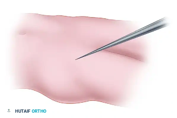
- Make a superficial 5 mm stab incision at the marked AL portal site.
- Introduce a 17-gauge, 6-inch spinal needle.
- Trajectory: Direct the needle approximately 45° cephalad and 30° posterior, aiming for the lateral aspect of the femoral head-neck junction.

- Under live fluoroscopy, advance the needle until it rests on the capsule.
- Pop through the capsule. A distinct tactile "give" is felt. If the joint vacuum is intact, a drop of saline placed in the hub of the needle will be sucked into the joint (the "drop sign").
- Inject 10 to 20 cc of normal saline to distend the capsule, pushing it away from the labrum.
- Pass a flexible Nitinol guidewire through the needle.
- Remove the needle and pass a cannulated dilator and arthroscopic sheath over the wire. Introduce the 70° arthroscope.
Step 2: Establishing the Anterior (AP) or Mid-Anterior (MAP) Portal
The second portal is established under direct intra-articular visualization combined with fluoroscopy.
- With the arthroscope in the AL portal, visualize the anterior triangle (bounded by the anterior labrum, femoral head, and anterior capsule).
- Introduce a spinal needle at the marked AP or MAP site.
- Trajectory: Direct the needle 45° cephalad and 30° toward the midline.
- Watch the needle pierce the capsule on the monitor. Ensure it enters the joint safely away from the labrum and the femoral head articular cartilage.
- Once the trajectory is perfected, pass a Nitinol wire, followed by a cannulated working sheath.
Step 3: The Interportal Capsulotomy
To maneuver instruments freely between the AL and anterior portals, the capsule between them must be incised.
1. Introduce an arthroscopic scalpel (e.g., Beaver blade) through the anterior working portal.
2. Under direct vision from the AL portal, incise the capsule connecting the two portals, running parallel to the acetabular rim, approximately 5 to 8 mm away from the labrum.
3. This capsulotomy allows for the introduction of larger instruments, shavers, and burrs without capsular restriction.
Complications and Avoidance Strategies
1. Neurovascular Injury
- LFCN Neuropraxia: The most common complication in hip arthroscopy. It can occur from direct trauma during anterior portal placement or from prolonged traction. Avoidance: Use the MAP instead of the classic AP, utilize superficial skin incisions, and limit traction time.
- Sciatic Nerve Injury: Rare, but devastating. Avoidance: Ensure internal rotation of the leg during setup. Avoid plunging when establishing the PL portal.
- Pudendal Nerve Neuropraxia: Exclusively a traction-related complication. Avoidance: Use a well-padded, lateralized perineal post. Release traction immediately after central compartment work is completed.
2. Iatrogenic Cartilage and Labral Damage
Forcing a trocar into a tightly constrained joint will inevitably scuff the femoral head or puncture the labrum.
* Avoidance: Never advance a rigid trocar without a guidewire. Ensure a minimum of 10 mm of fluoroscopic distraction before attempting to breach the capsule. Always distend the joint with fluid via the spinal needle before passing the guidewire.
3. Fluid Extravasation
The hip joint requires high pump pressures (often 50-70 mmHg) to maintain visualization due to the thick muscular envelope. Prolonged surgery can lead to massive fluid extravasation into the thigh, or rarely, into the retroperitoneal space, causing abdominal compartment syndrome.
* Avoidance: Monitor core body temperature (fluid can cause hypothermia). Palpate the abdomen periodically during prolonged cases. Ensure outflow is adequate.
Postoperative Protocol and Portal Care
Proper management of the portal sites postoperatively is essential for optimal healing and minimizing infection risks.
- Closure: Following the procedure, the capsule may be repaired depending on the surgeon's preference and the patient's risk for microinstability. The portal skin incisions are typically closed with simple interrupted non-absorbable sutures (e.g., Nylon) or buried absorbable sutures (e.g., Monocryl) combined with sterile adhesive strips.
- Dressings: A bulky, absorbent compressive dressing is applied to manage the expected serosanguinous drainage from the portal sites, which is common in the first 24 to 48 hours due to the high-pressure fluid used during the case.
- Rehabilitation: Weight-bearing status is dictated by the intra-articular procedures performed (e.g., 20 lbs flat-foot weight-bearing for 2-4 weeks following labral repair or microfracture). Early passive range of motion, particularly circumduction, is initiated immediately to prevent intra-articular adhesions and capsular scarring at the portal entry sites.
Mastery of hip arthroscopy portals requires a profound understanding of three-dimensional anatomy, meticulous preoperative setup, and disciplined surgical technique. By adhering to these evidence-based principles, the orthopedic surgeon can safely navigate the "safe zones" of the hip, minimizing complications while achieving optimal therapeutic outcomes.
You Might Also Like