Mastering Hip Arthroscopy: Advanced Surgical Techniques, Complication Management, and Rehabilitation
Key Takeaway
Hip arthroscopy is a highly effective intervention for femoroacetabular impingement (FAI), yet it carries unique risks. While overall complication rates remain low at approximately 1.4%, surgeons must meticulously manage traction times to prevent neurapraxia and carefully execute bony resections to avoid iatrogenic fractures or instability. This guide details advanced surgical techniques, comprehensive complication avoidance strategies, and evidence-based postoperative rehabilitation protocols essential for optimizing patient outcomes.
INTRODUCTION TO HIP ARTHROSCOPY AND FEMOROACETABULAR IMPINGEMENT
Hip arthroscopy has evolved from a diagnostic modality into a highly sophisticated therapeutic intervention, primarily driven by the recognition and surgical management of femoroacetabular impingement (FAI) and associated labral pathology. As indications have expanded, so too has the complexity of the procedures performed, necessitating a profound understanding of hip biomechanics, intricate regional anatomy, and the precise execution of bony and soft-tissue work.
While hip arthroscopy is generally considered safe, the deep, highly constrained nature of the hip joint requires specialized distraction techniques and instrumentation. Consequently, the procedure carries a unique profile of potential complications. Reported complication rates for hip arthroscopy generally remain low—approximately 1.4% in large, consecutive cohorts (e.g., 1,054 consecutive hip arthroscopies). However, avoiding these complications requires meticulous preoperative planning, flawless intraoperative execution, and strict adherence to evidence-based postoperative rehabilitation protocols.
This comprehensive guide delineates the surgical techniques for addressing FAI, provides an exhaustive analysis of potential complications, and outlines the standard of care for postoperative management.
SURGICAL TECHNIQUE: ARTHROSCOPIC MANAGEMENT OF FAI
The surgical correction of FAI involves the precise reshaping of the proximal femur (cam decompression) and/or the acetabular rim (pincer resection), alongside the repair or reconstruction of the acetabular labrum.
Patient Positioning and Joint Distraction
Proper positioning is the foundation of a safe and successful hip arthroscopy. The procedure is typically performed with the patient in either the supine or lateral decubitus position on a specialized traction table.
- Traction Application: Adequate joint distraction (typically 10 to 15 mm) is required to safely access the central compartment without causing iatrogenic scuffing of the articular cartilage.
- The Perineal Post: A well-padded, oversized perineal post is utilized to provide counter-traction. The post must be positioned laterally against the medial thigh rather than directly in the perineum to minimize pressure on the pudendal nerve and local soft tissues.
Surgical Warning: Traction time should be strictly monitored. The general consensus dictates that continuous traction should not exceed 120 minutes, and traction force should be limited to 50 lbs (or less, depending on patient size and ligamentous laxity) to mitigate the risk of traction neurapraxia.
Portal Placement and Capsulotomy
Safe portal establishment is critical to avoiding neurovascular injury, particularly to the lateral femoral cutaneous nerve (LFCN) and the femoral neurovascular bundle.
- Anterolateral (AL) Portal: Established first under fluoroscopic guidance, serving as the primary viewing portal. It is located 1 cm proximal and 1 cm anterior to the tip of the greater trochanter.
- Mid-Anterior Portal (MAP): Established under direct arthroscopic visualization. It is typically placed 5 to 7 cm distal and slightly medial to the AL portal.
- Capsulotomy: To achieve adequate visualization of the peripheral compartment and the cam lesion, a capsulotomy is essential. Surgeons typically begin with an interportal cut parallel to the labrum. To fully expose the femoral neck, a T-shaped capsulotomy is performed by extending a vertical limb distally down the femoral neck, parallel to the axis of the neck.
Clinical Pearl: The lateral femoral cutaneous nerve (LFCN) is at significant risk during the establishment of anterior portals. If the anterior portal is placed too far medially, the branches of the LFCN may be transected or stretched, leading to painful neuromas or lateral thigh numbness.
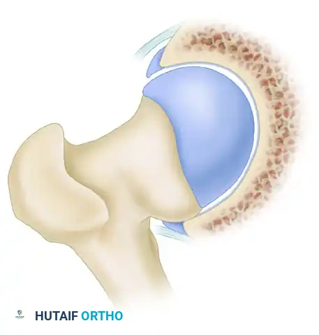
Osteochondroplasty (Cam Lesion Resection)
The goal of cam resection is to re-create a spherical femoral head-neck junction, restoring normal offset and eliminating impingement during terminal hip flexion and internal rotation.
- Exposure and Dynamic Positioning: Once the T-shaped capsulotomy is complete, the cam lesion is inspected. The hip must be dynamically manipulated to bring different aspects of the femoral neck into the arthroscopic field of view:
- Flexion and External Rotation: This maneuver helps expose inferior and medial cam lesions.
- Extension and Internal Rotation: This maneuver brings the superolateral aspect of the femoral neck into view.
- Resection Technique: Introduce a high-speed arthroscopic burr to resect the cam deformity. The resection must be meticulously contoured to blend smoothly with the native articular cartilage proximally and the normal femoral neck distally.
- Fluoroscopic Confirmation: Intraoperative fluoroscopy (utilizing AP, Dunn, and cross-table lateral views) is mandatory to assist in determining the depth and extent of the resection and to confirm that a spherical femoral head has been re-created.
Surgical Warning: Take extreme care to avoid the retinacular vessels when treating lesions on the superolateral neck. These terminal branches of the medial circumflex femoral artery (MCFA) perforate the capsule near the base of the femoral neck and travel proximally to supply the femoral head. Disruption can lead to catastrophic osteonecrosis.
Dynamic Assessment and Capsular Closure
Following bony resection, a dynamic assessment of the hip is performed. The traction is released, and the hip is taken through a full range of motion—specifically maximum flexion, internal rotation, and external rotation—under direct arthroscopic visualization to ensure there is no residual impingement between the femoral neck and the acetabular rim.
Finally, the capsule must be managed. The limb of the T-shaped capsulotomy that extends down the femoral neck should be repaired in a side-to-side fashion using high-strength sutures. Capsular closure restores the biomechanical integrity of the iliofemoral ligament, preventing postoperative microinstability and macroscopic dislocation.
COMPLICATIONS OF HIP ARTHROSCOPY
While the reported complication rate of 1.4% is reassuringly low, the complications that do occur can be profoundly debilitating. A master surgeon must anticipate, recognize, and manage these adverse events.
Traction Neurapraxia
Traction neurapraxia is the most commonly reported complication of hip arthroscopy. It can affect the pudendal, sciatic, femoral, or lateral femoral cutaneous nerves.
- Pathophysiology: Neurapraxia is usually caused by the length of time the leg is placed in traction (stretch injury) or excessive pressure from the perineal post (compression injury).
- Clinical Presentation: Patients may present with perineal numbness, erectile dysfunction (pudendal nerve), foot drop or posterior leg numbness (sciatic nerve), or anterior thigh weakness (femoral nerve).
- Prognosis: Fortunately, these nerve palsies are almost exclusively neurapraxic in nature and typically resolve spontaneously within days to weeks.
- Prevention: Limit traction time to under 2 hours, use a well-padded perineal post, position the post laterally against the medial thigh, and release traction immediately when central compartment work is completed.
Iatrogenic Chondral and Labral Injury
Scuffing of the articular surfaces (femoral head or acetabulum) or iatrogenic puncture of the labrum may occur during initial portal placement and instrument insertion. This complication is likely underreported in the literature. It is mitigated by ensuring adequate joint distraction (minimum 10 mm) prior to introducing the sharp trocar and by using fluoroscopy to guide the initial needle trajectory precisely into the joint space.
Fluid Extravasation and Abdominal Compartment Syndrome
Hip arthroscopy requires high fluid pump pressures (often 40 to 60 mmHg) to overcome the ambient tissue pressure of the thick muscular envelope surrounding the hip. This creates a risk for massive fluid extravasation.
- Soft Tissue Swelling: Extravasation into the thigh is common but usually benign, resolving rapidly postoperatively.
- Abdominal Compartment Syndrome (ACS): This is a rare but potentially fatal complication. Fluid can track into the retroperitoneal space or the peritoneal cavity.
- Risk Factors: In one documented case, ACS was believed to be related to fluid tracking to the retroperitoneal space following an arthroscopic psoas tenotomy. In another catastrophic case, fatal abdominal compartment syndrome occurred after the arthroscopic removal of a loose body in a patient with a prior acetabular fracture, which provided a direct conduit for fluid into the pelvis.
- Management: Anesthesiologists must monitor core temperature and peak airway pressures. A sudden drop in temperature or a spike in airway pressure suggests intra-abdominal fluid accumulation, necessitating immediate cessation of the procedure and potential surgical decompression.
Osteonecrosis of the Femoral Head
Osteonecrosis (avascular necrosis) of the femoral head has been reported after hip arthroscopy. This devastating complication is primarily iatrogenic, resulting from the direct transection or thermal ablation of the retinacular vessels (branches of the MCFA) during superolateral cam resection or aggressive capsulotomy. Surgeons must maintain a safe distance from the posterosuperior retinacular fold and avoid excessive use of radiofrequency ablation in this zone.
Complications of Bony Resection
Arthroscopic treatment of FAI requires a delicate balance; both over-resection and under-resection carry significant consequences.
- Under-resection: Incomplete removal of pincer (acetabular) or cam (femoral) deformities is the leading cause of revision hip arthroscopy. Under-resection leads to persistent impingement, ongoing pain, and continued degradation of the chondrolabral junction.
- Over-resection of the Acetabulum: Resecting too much of the acetabular rim (pincer resection) reduces the center-edge angle and compromises the suction seal of the hip, leading to iatrogenic hip instability and accelerated osteoarthritis.
- Over-resection of the Femoral Neck: Aggressive resection of a femoral neck cam lesion places the femoral neck at severe risk for a postoperative stress fracture.
Clinical Pearl: As a biomechanical rule of thumb, the depth of the cam resection should never exceed 30% of the diameter of the femoral neck. Resections exceeding this threshold exponentially increase the risk of catastrophic femoral neck fracture.
Heterotopic Ossification (HO)
As with open hip surgery, there is a well-documented risk of heterotopic ossification after hip arthroscopy, particularly following extensive bony resection (osteochondroplasty).
- Incidence: A landmark report of 300 cases demonstrated a 1.6% rate of heterotopic ossification. Notably, all instances of HO occurred in a control group of 15 patients who did not receive any pharmacological prophylaxis.
- Prophylaxis: In the same study, none of the 285 patients who received nonsteroidal anti-inflammatory drugs (NSAIDs) developed heterotopic ossification.
- Standard of Care: It is now standard practice to prescribe an NSAID (such as Indomethacin 75 mg sustained-release daily, or Naproxen 500 mg twice daily) for 3 weeks postoperatively to prevent HO, provided there are no gastrointestinal or renal contraindications.
POSTOPERATIVE CARE AND REHABILITATION
The success of hip arthroscopy is inextricably linked to a structured, phased postoperative rehabilitation program. The protocol must balance the need to protect healing tissues (capsular repair, labral repair, and osteochondroplasty sites) with the need to prevent adhesions and restore normal kinematics.
Phase I: Protection and Early Motion (Weeks 0 to 2)
- Weight Bearing: Patients are strictly limited to touchdown weight bearing (TTWB), typically defined as 20 lbs of pressure, for the first 2 weeks. This protects the femoral neck from stress fractures following cam resection and protects the labral repair from excessive compressive loads.
- Range of Motion (ROM): Physical therapy and passive range of motion are initiated in the first 24 to 48 hours. Early motion is critical to prevent intra-articular adhesions.
- Restrictions: Extremes of motion are avoided for several weeks. Specifically, combined flexion and internal rotation are restricted to protect the capsular closure.
- Conditioning: A stationary bike (with high seat placement to avoid deep flexion) can be used immediately to promote joint nutrition and mobility.
Phase II: Intermediate Rehabilitation (Weeks 2 to 6)
- Progression: Crutches are gradually weaned between weeks 2 and 4, provided the patient demonstrates a non-antalgic gait and adequate gluteal control.
- Strengthening: Focus shifts to isometric and gentle isotonic strengthening of the core, gluteus medius, and periarticular musculature.
- Precautions: Active straight leg raises are avoided to prevent hip flexor tendinitis and excessive stress on the anterior capsule.
Phase III: Advanced Strengthening and Return to Play (Months 2 to 6)
- Impact Activities: Impact activities (running, jumping) are strictly not recommended for the first 2 to 3 months to allow for complete bony remodeling of the osteochondroplasty site.
- Sport-Specific Training: At 3 months, patients may begin a gradual return to running program, followed by agility and sport-specific drills.
- Return to Sports: Full, unrestricted return to competitive sports typically takes 4 to 6 months, contingent upon the patient achieving symmetrical strength, full pain-free ROM, and successful completion of functional testing.
CONCLUSION
Hip arthroscopy is a technically demanding but highly rewarding procedure for the treatment of femoroacetabular impingement and labral pathology. By adhering to meticulous surgical techniques—including precise portal placement, dynamic assessment of bony resections, and robust capsular closure—surgeons can optimize clinical outcomes. Furthermore, a deep understanding of potential complications, ranging from traction neurapraxia to catastrophic fluid extravasation, combined with strict adherence to postoperative rehabilitation protocols, ensures that the risks of this advanced procedure are minimized, allowing patients to safely return to their highest level of function.
You Might Also Like