Arthroscopic Management of Acetabular Labral Tears: A Comprehensive Surgical Guide
Key Takeaway
The acetabular labrum is a critical fibrocartilaginous structure providing a fluid seal and biomechanical stability to the hip joint. Arthroscopic management of labral tears focuses on restoring this suction seal through precise repair or selective debridement. This guide details the vascular anatomy, pathomechanics, clinical evaluation—including the classic "C" sign—and step-by-step surgical techniques required to optimize patient outcomes and delay degenerative joint disease.
INTRODUCTION TO ACETABULAR LABRAL PATHOLOGY
The advent and rapid evolution of hip arthroscopy have fundamentally transformed our understanding of hip joint preservation. Central to this paradigm shift is the management of the acetabular labrum. Once considered a vestigial structure and routinely debrided when torn, the labrum is now recognized as a critical biomechanical stabilizer of the hip. Arthroscopic management of labral tears has evolved from simple excision to complex, anatomically precise repair and reconstruction, aimed at restoring the joint's native kinematics and delaying the onset of osteoarthritis.
This comprehensive guide details the anatomy, biomechanics, classification, clinical evaluation, and advanced arthroscopic management of acetabular labral tears, providing a rigorous framework for orthopaedic residents, fellows, and practicing hip preservation surgeons.
ANATOMY AND VASCULARITY OF THE LABRUM
The acetabular labrum is a continuous, ring-like fibrocartilaginous structure that surrounds the periphery of the acetabulum. It is widest anteriorly and thickest superiorly, bridging the acetabular notch inferiorly by merging with the transverse acetabular ligament (TAL).
Microstructure and Zonal Anatomy
Histologically, the labrum is composed of dense type I collagen bundles oriented circumferentially, which allows it to resist hoop stresses generated during axial loading. The labrum transitions into the articular hyaline cartilage of the acetabulum through a narrow transition zone (the chondrolabral junction), which is highly susceptible to shear forces.
Vascular Supply
Understanding the vascularity of the labrum is paramount for surgical decision-making regarding repair versus debridement.
* Arterial Sources: Blood supply to the acetabulum and labrum is derived primarily from the obturator artery, the superior gluteal artery, and the inferior gluteal artery.
* Capsular Penetration: These vessels form a periacetabular anastomotic ring. Small penetrating branches enter the labrum from the capsular side.
* Healing Potential: The periphery of the labrum (the capsular side) is significantly more vascularized than the articular region. The inner one-third (articular side) is largely avascular, relying on synovial fluid diffusion for nutrition.
Clinical Pearl: Similar to the meniscus of the knee, tears occurring at the peripheral, capsular junction (the "red-red" or "red-white" zones) possess a robust healing response when anatomically repaired and biologically stimulated.
BIOMECHANICS AND PATHOPHYSIOLOGY
The labrum serves multiple indispensable functions in the preservation of the hip joint. Its primary roles include enhancing joint stability, distributing contact pressures, and maintaining the intra-articular fluid seal.
The "Suction Seal" Effect
The labrum physically deepens the acetabulum by approximately 21% and increases the articular surface area by 28%. More importantly, it creates a highly competent "suction seal" around the femoral head. This seal prevents the escape of synovial fluid from the central compartment to the peripheral compartment, maintaining a layer of pressurized fluid that supports the femoral head.
Consequences of Labral Deficiency
In the presence of a labral tear, the fluid seal is compromised. The loss of hydrostatic pressurization leads to direct cartilage-on-cartilage contact.
* Increased Contact Pressure: Studies demonstrate that labral tears increase peak contact pressures within the joint by up to 92%.
* Chondral Degradation: This elevated pressure is a primary catalyst for degenerative joint disease. In a landmark study of 436 patients, 73% of those with labral tears or fraying exhibited concomitant articular cartilage damage.
* Spatial Correlation: The majority of chondral damage is located in the exact same geographic zone as the labral damage (most commonly the anterosuperior quadrant). Furthermore, the severity of chondral damage is directly proportional to the severity of the labral tear.
CLASSIFICATION OF LABRAL TEARS
Accurate classification of labral tears aids in standardizing research and guiding surgical intervention. Several classification systems exist, categorizing tears by morphology, etiology, and location.
The Seldes Classification
Seldes et al. described two primary types of labral injuries based on histological and morphological evaluation:
* Type 1 (Detachment): A separation of the labrum from its articular cartilage attachment at the chondrolabral junction. This is the most common type of tear and is highly amenable to arthroscopic repair.
* Type 2 (Intrasubstance): Tears occurring in various planes within the substance of the labrum itself. These are often degenerative and may require selective debridement or reconstruction if the tissue is non-viable.
The Lage Etiological Classification
Lage et al. proposed an arthroscopic classification based on the underlying etiology of the tear:
1. Traumatic: Often resulting from high-energy impact, hip dislocation, or repetitive microtrauma in athletes.
2. Degenerative: Associated with early osteoarthritis and chronic wear.
3. Idiopathic: Tears with no clear inciting event or morphological abnormality.
4. Congenital: Associated with underlying developmental dysplasia of the hip (DDH).
Morphological Classification
During diagnostic arthroscopy, tears are further described by their morphological appearance:
* Radial Flap Tears: Discrete flaps of tissue that can catch within the joint.
* Radial Fibrillated Tears: Fraying of the free edge, often seen in early degenerative states.
* Longitudinal Peripheral Tears: Tears running parallel to the acetabular rim.
* Unstable Tears: Grossly hypermobile labral tissue that displaces into the joint under probing.
Modern Etiological Drivers
Contemporary hip preservation recognizes that isolated labral tears are rare. The vast majority are secondary to underlying pathomorphology, including:
* Femoroacetabular Impingement (FAI): Cam (femoral) and Pincer (acetabular) lesions cause repetitive shear and abutment against the labrum.
* Laxity/Hypermobility: Connective tissue disorders or microinstability leading to labral overload.
* Dysplasia: Under-coverage of the femoral head places excessive shear stress on the hypertrophied labrum.
CLINICAL EVALUATION AND DIAGNOSIS
Patient History
Patients typically present with insidious onset anterior groin pain, which may radiate to the lateral hip, anterior thigh, or buttocks. Pain is exacerbated by prolonged sitting, pivoting, or deep flexion activities. Mechanical symptoms such as clicking, catching, or a sensation of "giving way" are highly indicative of an unstable labral tear.
Physical Examination
A meticulous physical examination is critical. The hallmark sign of intra-articular hip pathology, including labral tears, is the "C" sign.
When asked to localize their pain, the patient will cup their hand over the greater trochanter, with the thumb positioned posteriorly and the fingers wrapping deep into the anterior groin.
Provocative Testing:
* FADIR Test (Flexion, Adduction, Internal Rotation): Highly sensitive for anterior labral tears and anterior FAI. Elicits sharp groin pain.
* FABER Test (Flexion, Abduction, External Rotation): May elicit anterior pain (indicating labral pathology) or posterior pain (indicating sacroiliac joint pathology).
* Dial Test: Evaluates for capsular laxity and microinstability.
Imaging Modalities
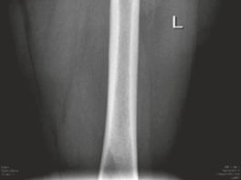
- Radiographs: An AP pelvis, false-profile, and Dunn lateral view are mandatory to assess for FAI (alpha angle, crossover sign) and dysplasia (lateral center edge angle).
- Magnetic Resonance Arthrography (MRA): The gold standard for diagnosing labral tears. The intra-articular gadolinium distends the capsule, allowing contrast to interpose between the torn labrum and the acetabular rim.
SURGICAL TECHNIQUE: ARTHROSCOPIC MANAGEMENT
The surgical management of labral tears has shifted decisively from debridement to preservation. The goal is to restore the suction seal and address the underlying bony pathomorphology.
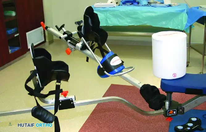
1. Patient Positioning and Setup
Hip arthroscopy can be performed in either the supine or lateral decubitus position, based on surgeon preference.
* Supine Position: The patient is placed on a specialized traction table. A well-padded, oversized perineal post is utilized to lateralize the vector of traction and minimize the risk of pudendal nerve neuropraxia.
* Traction: Gentle, controlled traction is applied to achieve a minimum of 10 mm of joint distraction, confirmed via fluoroscopy, to allow safe instrument passage into the central compartment.
Surgical Warning: Traction time should be strictly limited to less than 2 hours (ideally under 90 minutes) to prevent devastating complications such as pudendal nerve palsy or perineal skin necrosis.
2. Portal Placement
Precise portal placement is critical to avoid iatrogenic injury to the labrum, articular cartilage, and neurovascular structures.
* Anterolateral (AL) Portal: Established first under fluoroscopic guidance. Located 1 cm anterior and 1 cm superior to the tip of the greater trochanter. This is the primary viewing portal.
* Mid-Anterior (MAP) Portal: Established under direct arthroscopic visualization. Located approximately 5-7 cm distal and slightly anterior to the AL portal.
* Proximal Mid-Anterior (PMA) Portal: Often used as an accessory working portal for anchor placement.
3. Capsulotomy and Diagnostic Arthroscopy
An interportal capsulotomy is performed connecting the AL and MAP portals. This allows for adequate maneuverability of instruments and visualization of the peripheral compartment.
A systematic diagnostic sweep of the central compartment is performed. The labrum is probed meticulously to assess for detachment, intrasubstance tearing, and chondrolabral junction integrity. The "wave sign" (delamination of the adjacent articular cartilage) is frequently observed in conjunction with labral tears.
4. Labral Debridement (Selective)
While repair is preferred, debridement is indicated for severely macerated, degenerative, or ossified labral tissue that cannot hold a suture or contribute to the fluid seal.
* Technique: An arthroscopic shaver or radiofrequency (RF) wand is used to resect the unstable, frayed edges.
* Goal: To leave a stable, contoured rim of labral tissue without resecting into the capsular reflection, which would destabilize the hip.
5. Labral Repair Technique
For Type 1 detachments and viable labral tissue, anatomical repair is the gold standard.
Step A: Rim Preparation (Pincer Resection)
If a pincer lesion is present, or if the bone bed is sclerotic, the acetabular rim is decorticated using a 4.0 mm or 5.5 mm burr.
* This serves two purposes: it corrects the bony impingement (over-coverage) and creates a bleeding, cancellous bone bed to stimulate a biological healing response for the repaired labrum.
* Care must be taken to preserve the underlying articular cartilage.
Step B: Anchor Placement
Suture anchors (typically 1.5 mm to 2.9 mm, biocomposite or all-suture constructs) are placed along the acetabular rim.
* Trajectory: Anchors must be placed at the articular margin but angled 10 to 15 degrees away from the joint surface to prevent devastating iatrogenic articular penetration.
* Spacing: Anchors are typically placed 12 to 15 mm apart along the length of the tear.
Pitfall: Penetration of the articular cartilage with a drill bit or anchor is a catastrophic complication. Always confirm drill trajectory with fluoroscopy if the arthroscopic view is compromised, and maintain spatial awareness of the subchondral bone contour.
Step C: Suture Passage and Knot Tying
Sutures are passed through or around the labrum using specialized arthroscopic suture passing devices.
* Simple Loop (Circumferential): The suture is passed around the labrum, capturing the capsular side and pulling the labrum down to the rim. This is excellent for restoring the suction seal.
* Base Stitch (Mattress): The suture is passed through the substance of the labrum near its base. This prevents eversion of the labral edge and is preferred for larger, thicker labra.
* Fixation: Knotted or knotless anchors can be utilized. Knotless constructs are increasingly popular as they eliminate the risk of knot impingement on the articular cartilage.
6. Management of Concomitant Pathology
Repairing the labrum without addressing the underlying cause guarantees failure.
* Cam Decompression: Once the central compartment work is complete, traction is released, and the peripheral compartment is accessed. A cam lesion (loss of femoral head-neck offset) is resected using a burr under dynamic fluoroscopic and arthroscopic visualization to restore a spherical femoral head-neck junction.
* Capsular Closure: Following the procedure, the capsulotomy should be meticulously closed or plicated, particularly in patients with borderline dysplasia or generalized laxity, to prevent postoperative microinstability and protect the labral repair.
POSTOPERATIVE REHABILITATION
Rehabilitation following labral repair is highly structured and requires strict patient compliance to protect the healing tissue while preventing intra-articular adhesions.
Phase 1: Protection (Weeks 0-4)
- Weight Bearing: Flat-foot touch weight-bearing (approx. 20 lbs) using crutches for 2 to 4 weeks to protect the repair and the rim decortication site.
- Range of Motion (ROM): Continuous Passive Motion (CPM) machines or stationary biking (no resistance) are initiated immediately to prevent adhesions.
- Restrictions: Flexion is limited to 90 degrees. Extension and external rotation are strictly limited to protect the anterior capsule and the anterior labral repair.
Phase 2: ROM and Early Strengthening (Weeks 4-8)
- Wean off crutches to full weight-bearing.
- Progressive restoration of full, pain-free ROM.
- Initiate isometric and closed-chain kinetic exercises focusing on the gluteus medius, core, and pelvic stabilizers.
Phase 3: Advanced Strengthening (Weeks 8-12)
- Open-chain exercises and progressive resistance training.
- Focus on neuromuscular control and proprioception.
Phase 4: Return to Sport (Months 4-6)
- Sport-specific drills, plyometrics, and cutting maneuvers.
- Clearance for return to play is typically granted between 4 to 6 months postoperatively, contingent upon symmetrical strength, lack of pain, and successful completion of functional testing.
COMPLICATIONS
While hip arthroscopy is generally safe, the learning curve is steep, and complications can occur:
1. Nerve Injury: Pudendal nerve neuropraxia (from the perineal post) and Lateral Femoral Cutaneous Nerve (LFCN) neuropraxia (from portal placement) are the most common, though usually transient.
2. Iatrogenic Cartilage Injury: Scuffing of the femoral head during instrument insertion.
3. Heterotopic Ossification (HO): Can occur in the capsular tissues postoperatively. Prophylaxis with NSAIDs (e.g., Naproxen or Indomethacin) for 2-3 weeks is routinely recommended.
4. Failure of Repair: Often due to under-resection of FAI, failure to close the capsule, or poor tissue quality.
CONCLUSION
The arthroscopic management of acetabular labral tears represents a pinnacle of modern joint preservation surgery. By understanding the critical biomechanical role of the labral suction seal, accurately diagnosing the underlying pathomorphology, and executing precise, anatomically sound repairs, orthopaedic surgeons can significantly alleviate pain, restore function, and alter the natural history of degenerative hip disease. Mastery of these techniques requires a profound respect for the complex three-dimensional anatomy of the hip and a commitment to rigorous, evidence-based rehabilitation protocols.
You Might Also Like