Mastering Congenital Hand Reconstruction: Rotational Osteotomy of the First Metacarpal and Syndactyly Release
Key Takeaway
The Broudy and Smith rotational osteotomy of the first metacarpal is a definitive procedure for correcting congenital supination deformities of the thumb. By utilizing a specialized racquet-shaped incision and dorsal V-flap, surgeons can achieve optimal pronation and restore opposition. Concurrently, understanding the embryological failure of differentiation is critical for managing syndactyly, the most common congenital hand anomaly, requiring meticulous surgical release and reconstruction.
CONGENITAL HAND ANOMALIES AND RECONSTRUCTIVE PRINCIPLES
The human hand is a marvel of biomechanical engineering, relying on the precise spatial orientation of its digits to achieve both power grasp and fine pinch. Congenital anomalies of the hand present unique reconstructive challenges that demand a profound understanding of embryology, functional anatomy, and spatial geometry. Two of the most critical areas of congenital hand surgery involve the restoration of thumb opposition through rotational osteotomy and the surgical separation of fused digits in syndactyly.
This comprehensive masterclass details the evidence-based protocols, biomechanical principles, and step-by-step surgical techniques for the Broudy and Smith rotational osteotomy of the first metacarpal, followed by an in-depth analysis of the failure of differentiation leading to syndactyly.
ROTATIONAL OSTEOTOMY OF THE FIRST METACARPAL (BROUDY AND SMITH TECHNIQUE)
Indications and Biomechanical Rationale
The thumb contributes approximately 40% to 50% of overall hand function. Its unique ability to oppose the lesser digits is predicated on the specialized saddle geometry of the trapeziometacarpal (carpometacarpal) joint and the precise rotational alignment of the first metacarpal.
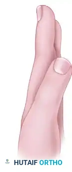
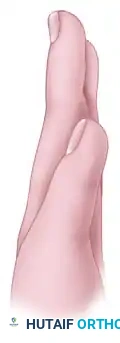
Congenital anomalies, such as thumb hypoplasia (Blauth classification), congenital clasped thumb, or severe supination deformities, often leave the thumb lying in the plane of the palm. A supinated thumb cannot effectively oppose the index and middle fingers, severely compromising tip-to-tip and tripod pinch kinematics.
The Broudy and Smith rotational osteotomy is a highly specialized, soft-tissue-accommodating procedure designed to derotate the first metacarpal into functional pronation. Unlike simple transverse osteotomies, which often suffer from severe soft-tissue tethering and skin tension upon rotation, the Broudy and Smith technique utilizes a mathematically designed local skin flap to recruit tissue, allowing for tension-free pronation of the thumb ray.
Clinical Pearl: The primary goal of the rotational osteotomy is to position the thumb pulp so that it directly faces the pulp of the index and long fingers. A failure to address the soft-tissue envelope during this bony rotation will inevitably lead to vascular compromise or gradual recurrence of the deformity.
Preoperative Planning and Patient Positioning
- Clinical Evaluation: Assess the degree of passive mobility at the carpometacarpal (CMC) joint. The joint must have adequate mobility to allow for opposition once the bone is rotated. Evaluate the intrinsic thenar musculature; if severely hypoplastic, an opponensplasty (e.g., Huber transfer or FDS transfer) may be required concomitantly or as a staged procedure.
- Radiographic Evaluation: Standard posteroanterior (PA), lateral, and Robert's views of the thumb are mandatory to assess the basilar joint architecture and the medullary canal diameter for planned Kirschner wire (K-wire) fixation.
- Anesthesia and Positioning: The procedure is performed under general anesthesia, often supplemented with a regional brachial plexus block for postoperative analgesia. The patient is positioned supine with the operative arm extended on a radiolucent hand table.
- Tourniquet: A well-padded pneumatic upper arm tourniquet is applied and inflated to 250 mm Hg (or 100 mm Hg above systolic pressure in pediatric patients) following exsanguination with an Esmarch bandage.
Surgical Technique: Step-by-Step
1. Incision Design and Flap Elevation
The brilliance of the Broudy and Smith technique lies in its geometric incision, which transposes skin to accommodate the new rotational axis of the thumb.
- The Primary Incision: Make a transverse, racquet-shaped skin incision on the volar aspect of the thumb. Extend this incision dorsally to form a V-shaped tongue at the middorsum of the first metacarpal.
- Apex Positioning: It is critical that the apex of the "V" lies exactly at the level of the first metacarpal base. This precise placement allows adequate exposure for the subsequent metaphyseal osteotomy while preserving the vascularity of the flap.
- The Secondary Incision: Make a proximal longitudinal incision on the radiovolar side of the first metacarpal. This incision must be oriented exactly 120 degrees from the apex of the dorsal "V". This 120-degree angle is mathematically calculated to match the desired degree of pronation required to restore opposition.
2. Exposure and Osteotomy
- Carefully elevate the full-thickness skin flaps, protecting the superficial radial nerve branches dorsally and the neurovascular bundles volarly.
- Expose the base of the first metacarpal subperiosteally. Retract the extensor pollicis brevis (EPB) and abductor pollicis longus (APL) tendons.
- The Osteotomy: Perform a transverse osteotomy at the metaphyseal base of the first metacarpal using a fine oscillating microsaw or a sharp osteotome. The metaphyseal location is chosen for its robust cancellous bone, which ensures rapid osteogenesis and union.
Surgical Warning: Avoid thermal necrosis during the osteotomy by utilizing continuous saline irrigation. Ensure the osteotomy is distal to the CMC joint capsule to prevent joint stiffness or instability.
3. Rotation and Fixation
- Once the osteotomy is complete, grasp the distal metacarpal segment with a small bone clamp.
- Rotate (pronate) the distal segment of the metacarpal until the thumb pulp perfectly opposes the index finger. The degree of rotation typically mirrors the 120-degree angle designed into the soft tissue flaps.
- Fixation: Fix the metacarpal in its new, pronated position using two crossed 0.045-inch or 0.062-inch K-wires. Ensure the wires engage both cortices of the proximal and distal fragments. Verify the alignment and rotation clinically and under intraoperative fluoroscopy.
4. Soft Tissue Transposition and Closure
- The rotation of the bone will naturally shift the soft tissue envelope.
- Rotate the dorsal V-flap volarly and suture it into the opened proximal linear incision. This acts as a local Z-plasty, recruiting skin to the volar-radial aspect where tension is highest following pronation.
- Close the resulting dorsal V-defect in a side-to-side manner. Use fine absorbable sutures (e.g., 5-0 or 6-0 chromic gut or fast-absorbing plain gut in pediatric patients) to minimize postoperative suture removal trauma.
Postoperative Care and Rehabilitation Protocol
Meticulous postoperative care is essential to protect the osteotomy and ensure optimal functional recovery.
- Immediate Postoperative Phase: Apply a bulky, non-adherent sterile dressing followed by a well-molded, fiberglass long-arm thumb spica cast. The long-arm cast is critical; it neutralizes forearm rotation (pronation/supination), which can transmit torque forces across the healing first metacarpal base.
- 6-Week Milestone: The long-arm cast is removed 6 weeks after surgery. Clinical and radiographic evaluations are performed to confirm bridging callus and bone healing.
- Hardware Removal: The K-wires are removed in the clinic at the 6-week mark, provided that radiographic bone healing is complete.
- Transitional Splinting: Following cast and pin removal, progressive active range of motion (AROM) is initiated. A custom-molded, removable short-arm thumb spica splint is fabricated. This splint is worn during sleep and strenuous activities for an additional 6 weeks to protect the remodeling bone.
- Therapy: Hand therapy focuses on maximizing CMC and metacarpophalangeal (MCP) joint mobility, strengthening the thenar musculature, and integrating the newly positioned thumb into functional pinch and grasp patterns.
FAILURE OF DIFFERENTIATION: SYNDACTYLY
Pathophysiology and Embryological Basis
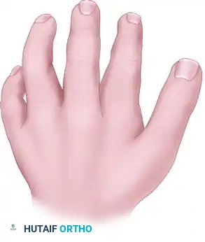
Syndactyly, colloquially referred to as "webbed fingers," represents a failure of the digits to separate during embryological development. To understand syndactyly, one must understand the precise timeline of human limb embryogenesis.
The upper limb bud begins to form at approximately 26 days of gestation. By the 6th week, the hand plate forms, and the digital rays become visible. The separation of these rays into individual fingers is driven by a highly regulated process of programmed cell death (apoptosis) in the interdigital necrotic zones. This process is orchestrated by signaling molecules, primarily from the Apical Ectodermal Ridge (AER) and the Zone of Polarizing Activity (ZPA).
Syndactyly results from an abnormal slowing of growth and a failure of this interdigital apoptosis during weeks 7 and 8 of gestation. Consequently, the mesenchymal tissue between the digits remains intact, leading to webbing.
Epidemiology and Genetic Factors
Syndactyly is the most common congenital anomaly of the hand, presenting with an incidence of approximately 1 in 2,000 live births. It occurs more frequently in males and is bilateral in about 50% of cases.
While the majority of syndactyly cases are sporadic, genetic factors play a significant role. Flatt's seminal epidemiological studies revealed a family history of syndactyly in 40% of his patients, strongly suggesting heredity as a primary etiological factor.
Genetically, several pedigrees have demonstrated an autosomal dominant inheritance pattern, particularly for syndactyly involving the long and ring fingers (the most common site of involvement). However, this genetic trait exhibits incomplete penetrance and variable expressivity, meaning that individuals carrying the gene may present with varying degrees of webbing, or occasionally, no clinical webbing at all. Syndactyly is also a hallmark feature of several complex craniofacial and systemic syndromes, most notably Apert syndrome and Poland syndrome.
Classification of Syndactyly
Accurate classification is paramount for surgical planning and prognosticating outcomes. Syndactyly is classified based on the extent of the webbing and the complexity of the involved tissues.
By Extent of Webbing:
- Complete Syndactyly: The digits are joined from the base of the web space all the way to the fingertips, often involving a shared paronychial fold and nail plate.
- Incomplete Syndactyly: The digits are joined from the web space to a point proximal to the fingertips.
By Tissue Complexity:
- Simple Syndactyly: The connection between the digits consists entirely of soft tissue (skin, fascia, and occasionally shared intrinsic muscle bands). The phalangeal bones are separate and distinct.
- Complex Syndactyly: The digits are joined by bone or cartilaginous fusions. This most commonly occurs at the distal phalanges but can involve any segment of the digit.
- Complicated Syndactyly: The most severe form, characterized by interposed accessory bones, disorganized neurovascular bundles, and severe tendinous anomalies. This is frequently seen in syndromic presentations (e.g., Apert syndrome).
Surgical Principles of Syndactyly Release
The surgical correction of syndactyly is not merely a matter of "splitting" the fingers. The circumference of two separate cylinders (the individual fingers) is mathematically greater than the circumference of one large cylinder (the conjoined fingers). Therefore, there is always a absolute deficiency of skin once the digits are separated.
Surgical Pearl: The golden rule of syndactyly release is to never release both sides of a single digit simultaneously. Doing so risks catastrophic devascularization of the finger. If a patient has multiple adjacent syndactylies (e.g., index-long, long-ring, ring-small), the releases must be staged.
Timing of Surgery
- Border Digits (Thumb-Index and Ring-Small): Because digits grow at different rates, a tethered border digit will rapidly develop a severe flexion and deviation contracture. Therefore, syndactyly of border digits should be released early, typically around 6 months of age.
- Central Digits (Long-Ring): These digits grow at similar rates, so tethering is less likely to cause angular deformities. Release is typically delayed until 12 to 18 months of age, allowing the hand to grow larger, which makes the microsurgical dissection of the neurovascular bundles safer.
Core Surgical Techniques
- Web Space Reconstruction: The foundation of a successful release is the creation of a deep, functional, and U-shaped web space. This is universally achieved using a broad, dorsally based rectangular or hourglass-shaped flap. Volar triangular flaps are also utilized to interdigitate with the dorsal flap, preventing linear scar contracture.
- Zig-Zag Incisions: Straight longitudinal incisions on the volar or dorsal aspects of the digits are strictly contraindicated, as they will inevitably lead to severe flexion contractures as the child grows. Bruner-style zig-zag incisions are utilized to separate the digits, ensuring that scars cross the flexion creases at oblique angles.
- Neurovascular Dissection: Using loupe magnification, the common digital nerve and artery must be meticulously identified and separated. In cases where the bifurcation of the nerve is distal to the planned web space, the nerve can be carefully teased apart longitudinally (intraneural neurolysis) to deepen the web. If the arterial bifurcation is distal, one digital artery may need to be ligated to achieve adequate depth, provided the digit has robust flow from its other digital artery.
- Skin Grafting: Because of the mathematical skin deficiency, the resulting bare areas on the lateral aspects of the newly separated digits must be covered with full-thickness skin grafts (FTSG). Split-thickness grafts are avoided due to their high propensity for secondary contracture. Donor sites for FTSG typically include the groin crease or the lower abdomen, which provide ample, pliable skin with minimal donor site morbidity.
Conclusion
The management of congenital hand anomalies requires a synthesis of biomechanical insight, meticulous preoperative planning, and precise microsurgical execution. The Broudy and Smith rotational osteotomy remains a powerful tool for restoring thumb opposition, relying on intelligent flap design to accommodate bony realignment. Similarly, the successful management of syndactyly demands a profound respect for the embryological origins of the deformity, utilizing strategic flap design and full-thickness skin grafting to restore independent digital function while preventing long-term contractures. Mastery of these techniques is essential for any reconstructive surgeon dedicated to optimizing the functional and aesthetic outcomes of the pediatric hand.
You Might Also Like