Dome Osteotomy and Excision of Vickers Ligament: Surgical Masterclass
Key Takeaway
The Carter and Ezaki technique for Madelung deformity combines a biplanar dome osteotomy with the excision of Vickers ligament. This procedure addresses the primary tethering force across the volar-ulnar distal radius while simultaneously correcting the multiplanar articular deformity. By performing a physiolysis and interposing a fat graft, surgeons can prevent premature physeal closure. This guide details the biomechanical rationale, step-by-step surgical approach, and postoperative rehabilitation protocols.
INTRODUCTION TO MADELUNG DEFORMITY AND THE CARTER-EZAKI PROCEDURE
Madelung deformity is a complex, multiplanar congenital or developmental anomaly of the wrist, characterized by a primary growth disturbance of the volar-ulnar aspect of the distal radial physis. This localized physeal arrest leads to a constellation of anatomic abnormalities: increased volar tilt and ulnar inclination of the distal radial articular surface, volar translation of the carpus, and relative dorsal subluxation of the distal ulna.
The Carter and Ezaki procedure—comprising the excision of Vickers ligament, physeal bar resection (physiolysis), and a biplanar dome osteotomy of the distal radius—remains the gold standard for symptomatic, skeletally immature patients presenting with this deformity. By addressing both the primary tethering pathology and the secondary osseous deformity, this technique halts disease progression, restores wrist biomechanics, and optimizes the remaining growth potential of the distal radius.
PATHOANATOMY AND BIOMECHANICS
To execute this procedure effectively, the operating surgeon must possess a profound understanding of the underlying pathoanatomy. Madelung deformity is frequently associated with Leri-Weill dyschondrosteosis, an autosomal dominant condition linked to mutations or deletions in the SHOX (Short Stature Homeobox) gene located on the pseudoautosomal region of the sex chromosomes.
The Role of Vickers Ligament
The hallmark of Madelung deformity is the presence of Vickers ligament, an anomalous, hypertrophic radiolunate ligament.
* Origin: Arises from the volar metaphysis of the distal radius.
* Course: Crosses the volar-ulnar physis and epiphysis.
* Insertion: Inserts onto the lunate.
This ligament acts as a rigid, unyielding tether. As the dorsal and radial aspects of the distal radial physis continue to grow normally, the tethered volar-ulnar corner is held back. This asymmetric growth creates a severe multiplanar deformity. The carpus follows the articular surface, sliding volarly and ulnarly, which wedges the lunate between the deformed radius and the prominent distal ulna.
Biomechanics of the Dome Osteotomy
Traditional closing-wedge or opening-wedge osteotomies possess inherent biomechanical flaws in the treatment of Madelung deformity. A closing-wedge osteotomy further shortens an already shortened radius, exacerbating ulnocarpal impaction. An opening-wedge osteotomy requires structural bone grafting and can create excessive tension on the volar soft tissues.
The biplanar dome osteotomy is biomechanically superior because it:
1. Preserves Radial Length: The center of rotation of angulation (CORA) is maintained within the osteotomy site, preventing further shortening.
2. Maximizes Bone Contact: The congruent spherical surfaces provide immense intrinsic stability and a massive surface area for rapid osteogenesis.
3. Allows Multiplanar Correction: The distal fragment can be rotated, extended, and supinated/pronated simultaneously along the dome's arc to correct the complex 3D deformity.
💡 Clinical Pearl
The true power of the dome osteotomy lies in its versatility. By utilizing curved osteotomes, the surgeon creates a "ball-and-socket" joint at the metaphyseal level, allowing infinite degrees of freedom to dial in the exact coronal and sagittal correction before definitive fixation.
INDICATIONS AND PREOPERATIVE PLANNING
Indications
- Skeletally Immature Patients: Typically girls aged 8 to 14 years with open physes and documented progression of the deformity.
- Symptomatic Presentation: Pain at the distal radioulnar joint (DRUJ), decreased grip strength, and restricted wrist range of motion (specifically supination and extension).
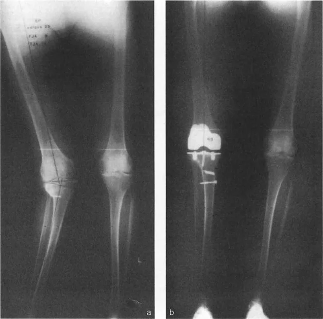
- Radiographic Evidence: Clear presence of a volar-ulnar physeal tether, increased volar tilt (>15 degrees), and increased ulnar inclination (>25 degrees).
Preoperative Imaging
- Standard Radiographs: PA, lateral, and oblique views of both wrists to quantify the deformity and assess skeletal maturity.
- Magnetic Resonance Imaging (MRI): Highly recommended to definitively identify the anomalous Vickers ligament, assess the extent of the physeal bar, and evaluate the integrity of the triangular fibrocartilage complex (TFCC).
- Computed Tomography (CT): 3D reconstructions are invaluable for preoperative templating, allowing the surgeon to calculate the exact radius of curvature required for the dome osteotomy.
SURGICAL ANATOMY AND EXPOSURE
The procedure utilizes the classic volar Henry approach to the distal radius, exploiting the internervous and intermuscular planes to safely access the volar metaphysis and physis.
- Superficial Interval: Between the flexor carpi radialis (FCR, innervated by the median nerve) and the radial artery.
- Deep Dissection: Involves the mobilization of the flexor pollicis longus (FPL) and the reflection of the pronator quadratus (PQ).
⚠️ Surgical Warning
The radial artery is highly vulnerable during the proximal extension of the Henry approach. It must be meticulously mobilized and retracted radially. Similarly, the palmar cutaneous branch of the median nerve, which lies ulnar to the FCR tendon, must be protected during the superficial incision.
STEP-BY-STEP SURGICAL TECHNIQUE (CARTER AND EZAKI)
1. Positioning and Setup
The patient is placed supine with the affected arm extended on a radiolucent hand table. A well-padded pneumatic tourniquet is applied to the proximal arm. Prophylactic intravenous antibiotics are administered. The arm is exsanguinated, and the tourniquet is inflated to the appropriate pediatric or adult pressure. Fluoroscopy (C-arm) must be positioned to allow seamless AP and lateral imaging throughout the case.
2. The Volar Approach
- Make a longitudinal incision over the distal volar forearm, following the course of the FCR tendon, extending approximately 6 to 8 cm proximally from the wrist crease.
- Incise the superficial fascia and open the FCR tendon sheath. Retract the FCR tendon ulnarward to protect the median nerve.
- Identify the radial artery and its venae comitantes. Carefully mobilize the neurovascular bundle and retract it radially.
- Develop the deep plane to expose the pronator quadratus muscle.
3. Elevation of the Pronator Quadratus
- Incise the pronator quadratus along its extreme radial border, leaving a small 1-2 mm cuff of tissue on the radius for later repair.
- Elevate the muscle subperiosteally from radial to ulnar. Retract the muscle belly ulnarward using a right-angle retractor. This exposes the distal radial metaphysis, the volar physis, and the epiphysis.
4. Identification and Excision of Vickers Ligament
- Carefully inspect the volar-ulnar aspect of the distal radius. Vickers ligament will appear as a distinct, thick, fibrous band originating from the metaphysis, crossing the physis, and diving distally toward the lunate.
- Using a #15 blade and fine tenotomy scissors, sharply reflect the ligament off the distal radius.
- Begin the release proximal to the physis on the metaphysis and continue the dissection distally.
- Completely release the ligament off the physis and the epiphysis. Ensure that the tether is entirely excised to prevent recurrence.
5. Physiolysis and Fat Interposition
- Once Vickers ligament is excised, inspect the underlying physis. In Madelung deformity, a localized bony or fibrous bar is typically present at the volar-ulnar corner.
- Using small curettes and a high-speed burr, meticulously remove any fibrous tissue or bone bridging the physis. The resection must continue until healthy, pearly-white physeal cartilage is visualized on all margins of the defect.
- Fat Interposition (Langenskiöld Principle): To prevent the reformation of the physeal bar, harvest a small piece of subcutaneous fat from the proximal margin of the incision. Pack this fat graft tightly into the physeal defect.
💡 Clinical Pearl
The fat graft acts as a biologic spacer. It is avascular and does not ossify, thereby preventing the osteogenic cells of the metaphysis and epiphysis from bridging the gap. This allows the remaining healthy physis to resume longitudinal growth.
6. The Biplanar Dome Osteotomy
- Identify the planned osteotomy site in the distal radial metaphysis, typically 1.5 to 2 cm proximal to the physis.
- Using a series of curved osteotomes (male and female), create a biplanar dome osteotomy. The convexity of the dome should ideally point proximally (though some surgeons prefer distal convexity depending on the specific deformity apex).
- The osteotomy must be completed circumferentially. Use a combination of curved osteotomes and a small oscillating saw if necessary, ensuring the dorsal periosteum is carefully managed to prevent displacement.
7. Deformity Correction and Fixation
- Once the osteotomy is mobile, insert a periosteal elevator or a specialized reduction tool into the osteotomy site.
- Correction Maneuver: Rotate the distal radial fragment by pronating it, extending it (dorsal tilt), and translating it radially. This complex maneuver corrects the volar tilt, ulnar inclination, and volar translation simultaneously.
- Assess the correction clinically and fluoroscopically. The articular surface should be restored to a neutral or slightly volar tilt (11 degrees) and normal ulnar inclination (22 degrees).
- Secure the osteotomy with one or two stout, smooth Steinmann pins (or heavy Kirschner wires) driven percutaneously from the radial styloid, across the osteotomy, and into the proximal ulnar cortex of the radial shaft.
- Note: Smooth pins are preferred over rigid plate fixation in skeletally immature patients to avoid tethering the remaining open physis and to allow for easy removal.
8. Contouring and Closure
- Following the rotation of the distal fragment, a prominent palmar step-off of the proximal metaphyseal fragment will often be present.
- Use a rongeur or a high-speed burr to smoothly resect this palmar step-off. This prevents postoperative flexor tendon irritation or rupture.
- Thoroughly irrigate the wound to remove all bone debris.
- Allow the tourniquet to deflate and achieve meticulous hemostasis.
- Repair the pronator quadratus over the osteotomy site using absorbable sutures. This provides a vital vascularized bed for bone healing and acts as a dynamic barrier between the osteotomy and the flexor tendons.
- Perform a routine layered closure of the subcutaneous tissue and skin.
POSTOPERATIVE CARE AND REHABILITATION
The postoperative protocol is designed to protect the osteotomy while it heals, given that smooth pin fixation provides less absolute stability than rigid plating.
Phase 1: Immediate Postoperative (Weeks 0-2)
- Immobilization: A bulky, well-padded long-arm splint is applied in the operating room. The forearm is positioned in neutral rotation or slight supination to maintain the correction.
- Edema Control: Strict elevation of the limb above the level of the heart is mandatory for the first 48-72 hours. Active range of motion of the fingers and thumb is encouraged immediately to prevent tendon adhesions and reduce swelling.
- Wound Check: At 2 weeks, the splint is removed, the wound is inspected, and sutures are removed.
Phase 2: Intermediate Healing (Weeks 2-6)
- Casting: The patient is transitioned to a long-arm cast or a rigid long-arm thermoplastic splint.
- Radiographic Monitoring: AP and lateral radiographs are obtained at 2 and 6 weeks to ensure maintenance of alignment and to assess callus formation at the dome osteotomy site.
Phase 3: Pin Removal and Mobilization (Weeks 6-8)
- Pin Removal: At 6 weeks, clinical and radiographic union is typically sufficient to allow for the removal of the Steinmann pins in the clinic.
- Transition: The patient is placed in a short-arm cast or a removable short-arm splint for an additional 2 to 4 weeks, depending on the robustness of the radiographic healing.
- Therapy Initiation: Gentle, active range of motion exercises for the wrist and forearm (pronation/supination) are initiated under the guidance of a certified hand therapist.
Phase 4: Strengthening and Return to Activity (Weeks 8-12+)
- Once the osteotomy is fully consolidated, progressive strengthening is introduced.
- Return to high-impact activities or contact sports is generally restricted until 4 to 6 months postoperatively, contingent upon complete radiographic remodeling and symmetric grip strength.
COMPLICATIONS AND PITFALLS
While highly effective, the Carter and Ezaki procedure carries specific risks that the orthopedic surgeon must anticipate and mitigate.
- Incomplete Excision of Vickers Ligament: Failure to completely resect the ligament and the associated physeal bar will inevitably lead to recurrence of the deformity as the patient grows. Meticulous visualization and the use of loupe magnification are critical.
- Loss of Reduction: Because the dome osteotomy is stabilized with smooth pins rather than rigid plates, loss of reduction can occur if the pins are too small, poorly placed, or if the patient is non-compliant with immobilization. Ensure pins engage the opposite thick cortical bone of the proximal radius.
- Flexor Tendon Rupture: If the palmar step-off of the proximal fragment is not adequately rongeured, or if the pronator quadratus is not securely repaired, the FPL or FDP tendons can fray against the raw bone edge, leading to delayed rupture.
- Pin Tract Infection: Superficial pin tract infections are common with percutaneous wires. They are typically managed successfully with oral antibiotics and local pin care. Deep infections requiring premature pin removal are rare but can compromise the osteotomy.
- Neuromuscular Injury: The palmar cutaneous branch of the median nerve and the radial artery are at risk during the approach. Retraction must be gentle, and the internervous planes must be strictly respected.
CONCLUSION
The dome osteotomy combined with the excision of Vickers ligament represents a sophisticated, biomechanically sound approach to the management of Madelung deformity. By directly addressing the pathoanatomic tether and utilizing a bone-preserving, multiplanar osteotomy, orthopedic surgeons can reliably restore wrist anatomy, alleviate pain, and optimize the functional trajectory of the pediatric patient's upper extremity. Mastery of the volar exposure, precise physeal management, and accurate 3D spatial correction are the cornerstones of success in this demanding but highly rewarding procedure.
You Might Also Like