Anteroposterior Compression Type II (APC II) Pelvic Ring Injuries: A Comprehensive Review of Epidemiology, Surgical Anatomy, and Biomechanics
Key Takeaway
An Anteroposterior Compression Type II (APC II) pelvic ring injury involves pubic symphysis diastasis (>2.5 cm) and anterior sacroiliac ligament disruption. It is rotationally unstable but vertically stable due to intact posterior sacroiliac ligaments. These injuries result from external rotation forces, often requiring prompt recognition and management to prevent long-term complications.
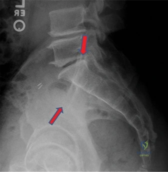
You are the registrar on-call. A 24-year-old male is brought into the trauma bay following a high-speed motorcycle collision. He is hemodynamically stable after initial resuscitation. The following radiograph is obtained.
Describe the radiographic findings and classify the injury according to the Young-Burgess system. What are the biomechanical implications of this specific pattern regarding pelvic stability?
Candidate: The radiograph shows a widened pubic symphysis, measuring approximately 4cm. This is an Anteroposterior Compression (APC) type II injury. It is rotationally unstable because the anterior sacroiliac ligaments are torn, but vertically stable because the posterior sacroiliac ligament complex remains intact, acting as a hinge.
Failing to mention the integrity of the posterior ligamentous complex. A candidate who simply says "it's an open-book fracture" without defining the stability or the Young-Burgess grade demonstrates a lack of understanding of the surgical implications for posterior fixation.
The candidate identifies the injury as an APC II based on the symphyseal diastasis (>2.5cm) and the "open-book" appearance. They explicitly state: "This is a rotationally unstable but vertically stable injury." Crucially, they identify that the posterior SI ligaments (the strongest ligamentous complex in the pelvis) are intact, differentiating it from an APC III, which would involve complete disruption of the SI joint and vertical instability. They mention that management focuses on restoring anterior rotational stability via symphyseal plating.
You have decided to proceed with open reduction and internal fixation (ORIF) of the pubic symphysis. During your pre-operative planning, what critical neurovascular structures are at risk, and how do you protect them during the approach?
Candidate: The key structures at risk are the bladder, the urethra, the spermatic cords (in males) or round ligaments (in females), and the obturator neurovascular bundle. I would ensure a Foley catheter is placed to decompress the bladder, dissect in the preperitoneal space, and retract the bladder superiorly. I would identify and protect the cords/round ligaments laterally.
Neglecting the role of the Foley catheter as a protective guide for the bladder, or failing to acknowledge the lateral location of the spermatic cords/round ligaments, which are easily injured if the surgeon dissects too far laterally during the Pfannenstiel approach.
Systematically categorize structures: 1) Bladder/Urethra: Decompress with Foley to reduce volume and define location; 2) Spermatic Cords/Round Ligaments: Protect by identifying them exiting the inguinal canal laterally; 3) Obturator Bundle: Risk during deep posterior retraction; 4) Vessels: The inferior epigastric vessels are at risk if the midline exposure is too broad. The candidate explains that dissection must remain in the preperitoneal plane, keeping the bladder protected behind the symphysis.
Post-operatively, the patient complains of persistent pain over the symphysis and difficulty with gait at 6 months. What are the potential causes, and how would you investigate this?
Candidate: Causes include hardware prominence, nonunion or malunion, or persistent occult sacroiliac joint instability. I would perform a clinical exam to localize pain, check for hardware tenderness, and obtain serial radiographs or a CT scan to check for hardware failure or nonunion.
Focusing only on the hardware (e.g., "the plate is too prominent"). A senior candidate must account for the fact that even with a perfect reduction, 20-40% of these patients have chronic pain due to the initial ligamentous insult to the posterior pelvic ring.
A structured, high-level approach: 1) Hardware: Is it prominent or failing? 2) Biological: Is there a nonunion or symptomatic heterotopic ossification? 3) Ligamentous: Is there residual occult posterior instability that was misdiagnosed as pure APC II? 4) Soft Tissue: Bursitis over the plate. The candidate suggests imaging (CT to assess union) and potentially a diagnostic block of the SI joint to rule out posterior pathology if the anterior site is consolidated.
