Pediatric Distal Radius Salter-Harris Type II Fracture: Diagnosis & Management Case Study
Key Takeaway
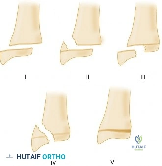
A pediatric Salter-Harris Type II distal radius fracture involves the distal radius metaphysis extending through the physis, often from a FOOSH injury. Diagnosis includes clinical examination for "dinner fork" deformity and localized tenderness, confirmed by PA, lateral, and oblique X-rays revealing the metaphyseal and physeal components, often with a "Thurston Holland fragment."
You are presented with this 10-year-old male who has sustained an injury following a fall onto an outstretched hand. Please describe the injury seen in these initial radiographs and detail your classification system.
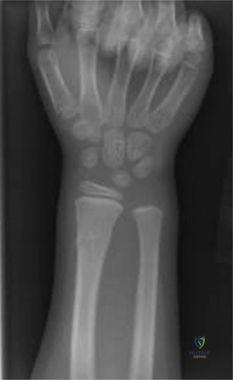
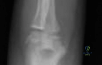
Candidate: The radiographs show a displaced fracture of the distal radius. There is a fracture line through the physis that extends into the metaphysis with a triangular fragment, which is a Salter-Harris Type II fracture. The displacement is dorsal with approximately 35 degrees of apex volar angulation.
Candidates often jump straight to "Salter-Harris II" without systematically describing the displacement or the presence of the Thurston Holland fragment. Failing to mention the maturity status or the risk of displacement in a 10-year-old shows a lack of clinical integration.
The candidate should state: "This is a displaced distal radial physeal fracture in a 10-year-old. Radiographically, it is a Salter-Harris Type II injury, evidenced by the fracture line traversing the physis and exiting through the metaphysis, creating a pathognomonic Thurston Holland fragment. There is significant sagittal deformity with 35 degrees of apex volar angulation and 75% dorsal translation. Given the patient’s age, this is a high-risk injury for secondary displacement, necessitating careful consideration for reduction and potential stabilization."
Given the radiographic appearance, you decide to perform a closed reduction. What are the biomechanical principles guiding your reduction maneuver, and how do you determine if the reduction is stable?
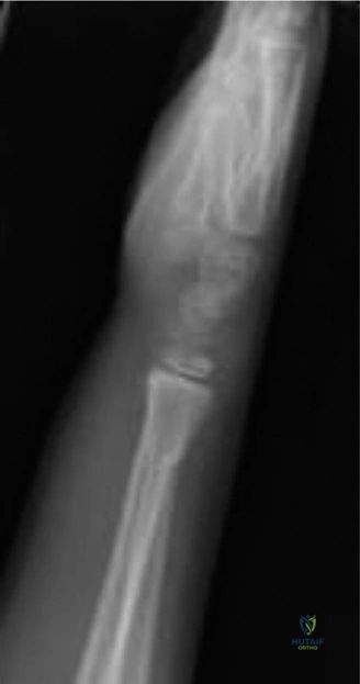
Candidate: I would perform Charnley’s maneuver: exaggerate the deformity to unlock the fragments, apply longitudinal traction to overcome muscle forces, then reduce the distal fragment volarly. Stability is determined by the cortical contact and the periosteal hinge integrity.
Failing to mention the role of the periosteum as a hinge or ignoring the "one and done" principle. Candidates often forget to discuss the neurovascular status post-reduction or fail to define what constitutes an "unstable" pattern (e.g., >50% translation).
Start with the biomechanics: 1) Exaggerate the deformity to disengage the metaphyseal spike from the periosteal hinge. 2) Apply longitudinal traction to fatigue the muscles. 3) Apply pressure to correct translation and angulation, then flex the wrist to engage the intact dorsal periosteum as a tension band. State that stability is assessed by the degree of initial displacement (>50% translation is inherently unstable), the presence of metaphyseal comminution, and the quality of the periosteal hinge. If stable after reduction, you should still consider K-wire fixation given the 75% translation risk of secondary shift.
The reduction is achieved and pinned. The patient is now 3 months post-op. What are the long-term concerns for a 10-year-old with a Salter-Harris II injury, and how would you monitor them?
Candidate: The main concern is premature physeal closure or growth arrest. I would monitor with serial radiographs to check for limb length discrepancy or angular deformity.
Being vague about the follow-up interval. Candidates often fail to mention the specific clinical signs of growth arrest (e.g., "Madelung-like" deformity) or forget that SH-II fractures have an excellent prognosis for growth because the germinal cells remain with the epiphysis.
Acknowledge that while Salter-Harris II fractures generally have a good prognosis because the germinal layer stays with the epiphysis, complications can arise from repeated reduction attempts or severe initial trauma. Clinical follow-up should involve monitoring for wrist pain, stiffness, or progressive deformity (e.g., volar tilt or radioulnar discrepancy). Radiographically, I would look for Harris growth arrest lines, physeal narrowing, or the development of a physeal bar. If a growth arrest is suspected, MRI is the gold standard for mapping the bar. Late intervention for physeal arrest includes bar resection or epiphysiodesis depending on remaining growth.
