Top Board-Certified Orthopedic Surgeons: Dallas & San Antonio
Key Takeaway
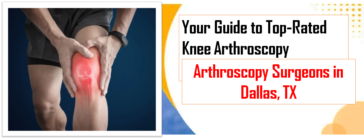
Your ultimate guide to Top Board-Certified Orthopedic Surgeons: Dallas & San Antonio starts here. To choose a knee arthroscopy surgeon, prioritize proven experience, excellent outcomes, and clear communication. Select a **boardcertified orthopedic surgeon** with specialized training in knee surgery and arthroscopy, who offers personalized treatment plans. Also, consider their bedside manner, cost, and insurance coverage to make an informed decision for your healthcare needs.
Introduction & Epidemiology
Knee arthroscopy, a minimally invasive surgical procedure, represents a cornerstone in the diagnosis and treatment of intra-articular knee pathologies. Originating in the early 20th century with Takagi and Watanabe in Japan, its widespread adoption in Western medicine occurred in the latter half of the century, revolutionized by advancements in optics, instrumentation, and video technology. This evolution has transformed orthopedics by enabling precise diagnosis and treatment of complex knee disorders with reduced morbidity, accelerated recovery, and superior cosmetic outcomes compared to open arthrotomy.
Epidemiologically, knee pain is a pervasive musculoskeletal complaint, and intra-articular derangements are a significant contributor. Meniscal tears and anterior cruciate ligament (ACL) injuries are among the most frequently diagnosed conditions necessitating arthroscopic intervention. The incidence of meniscal tears is estimated to be approximately 60-70 per 100,000 population per year, with a higher prevalence in active individuals and an increasing incidence in older adults due to degenerative changes. ACL ruptures occur at a rate of 30-78 per 100,000 athletes annually, predominantly in young, active populations involved in pivoting sports. Other common indications include chondral defects, loose bodies, synovial proliferation, and patellofemoral instability. The demographic profile of patients undergoing knee arthroscopy spans a broad range, from adolescent athletes to geriatric individuals, reflecting the diverse etiologies of knee pathology from acute trauma to chronic degenerative processes. The procedure significantly impacts patient quality of life, functional capacity, and return to activity.
Surgical Anatomy & Biomechanics
A thorough understanding of the complex anatomy and biomechanics of the knee joint is paramount for successful arthroscopic intervention and avoidance of iatrogenic injury.
Articular Anatomy
- Femoral Condyles: Distal femur presents medial and lateral condyles, articulating with the tibial plateau. The medial condyle is larger and more spherical, while the lateral condyle is flatter. The intercondylar notch houses the cruciate ligaments.
- Tibial Plateau: Proximal tibia features medial and lateral articular facets, separated by the intercondylar eminence (tibial spines). The medial plateau is concave, and the lateral is convex.
- Patella: Largest sesamoid bone, articulating with the trochlear groove of the femur. Critical for quadriceps lever arm efficiency.
-
Menisci:
C-shaped (medial) and O-shaped (lateral) fibrocartilaginous structures.
- Medial Meniscus: Broader posteriorly, firmly attached to the tibia via coronary ligaments and to the medial collateral ligament (MCL). Less mobile, making it more susceptible to tears.
- Lateral Meniscus: More mobile due to looser peripheral attachments and the hiatus for the popliteus tendon. Less frequently injured.
- Vascularity: Peripherally vascularized (red-red zone), becoming avascular centrally (white-white zone). This dictates healing potential after repair.
- Function: Load transmission (distribute contact stresses), shock absorption, joint stability, lubrication, proprioception.
Ligamentous Anatomy
-
Cruciate Ligaments:
- Anterior Cruciate Ligament (ACL): Originates from the posteromedial aspect of the lateral femoral condyle, inserts into the anteromedial intercondylar area of the tibia. Primary restraint to anterior tibial translation and secondary restraint to internal rotation. Consists of anteromedial (AM) and posterolateral (PL) bundles.
- Posterior Cruciate Ligament (PCL): Originates from the anterolateral aspect of the medial femoral condyle, inserts into the posterior intercondylar area of the tibia. Primary restraint to posterior tibial translation. Consists of anterolateral (AL) and posteromedial (PM) bundles.
-
Collateral Ligaments:
- Medial Collateral Ligament (MCL): Superficial and deep components. Originates from the medial femoral epicondyle, inserts into the medial tibia. Primary restraint to valgus stress.
- Lateral Collateral Ligament (LCL): Cord-like structure, originates from the lateral femoral epicondyle, inserts into the fibular head. Primary restraint to varus stress.
- Posterolateral Corner (PLC): Complex array of structures including the popliteofibular ligament, arcuate ligament complex, posterolateral capsule, and LCL. Critical for resisting varus and external rotation forces.
- Patellofemoral Ligaments: Medial patellofemoral ligament (MPFL) is a crucial stabilizer against lateral patellar translation.
Musculature & Tendons
- Quadriceps Tendon: Inserts into the patella, forming the extensor mechanism.
- Patellar Tendon: Extends from the patella to the tibial tuberosity.
- Hamstrings: Semimembranosus, semitendinosus, biceps femoris. Provide dynamic stability and contribute to knee flexion. Semitendinosus and gracilis are common autograft sources for ACL reconstruction.
- Popliteus Tendon: Originates from the lateral femoral epicondyle, inserts onto the posterior aspect of the tibia. Contributes to posterolateral stability and internal rotation of the tibia.
Neurovascular Anatomy
- Popliteal Artery and Vein: Located in the popliteal fossa, immediately posterior to the knee joint capsule. Vulnerable during posterior portal placement and procedures involving the posterior compartments.
- Common Peroneal Nerve: Courses laterally around the fibular neck. Susceptible to injury during lateral dissection, especially for LCL repair/reconstruction or procedures near the fibular head.
- Saphenous Nerve: Sensory nerve that courses medially. Injury can result in anesthesia or dysesthesia along the medial aspect of the leg.
- Infrapatellar Branch of the Saphenous Nerve (IPBSN): Frequently injured during anteromedial portal placement, leading to localized numbness.
Biomechanics
- Tibiofemoral Joint: A modified hinge joint, allowing flexion, extension, and limited rotation. "Screw-home mechanism" during terminal extension (external rotation of tibia).
- Patellofemoral Joint: Gliding articulation. Patellar tracking is influenced by quadriceps alignment, trochlear groove morphology, and ligamentous restraints (MPFL).
- Meniscal Biomechanics: Act as secondary stabilizers, especially after ligamentous injury. Distribute axial loads, increasing contact area and reducing peak stresses on articular cartilage.
- Ligamentous Biomechanics: Each ligament functions optimally at specific ranges of motion, providing primary and secondary restraints to prevent excessive translation and rotation.
Indications & Contraindications
Patient selection for knee arthroscopy requires careful consideration of the specific pathology, patient demographics, activity level, and comorbidities.
Indications for Knee Arthroscopy
-
Meniscal Pathology:
- Tears: Symptomatic tears causing mechanical symptoms (locking, catching, pain). Repairable tears (red-red, red-white zones, longitudinal, bucket-handle) in younger, active patients. Resection for unstable, irreparable tears or degenerative tears causing significant symptoms despite non-operative management.
- Cysts: Excision or decompression in conjunction with meniscal repair/resection.
-
Ligamentous Instability:
- ACL Rupture: Symptomatic instability in active individuals, often with associated meniscal or chondral injuries. Primary reconstruction.
- PCL Tears: Symptomatic instability, particularly in combined ligament injuries.
- Multi-ligamentous Knee Injuries: Arthroscopy for diagnostic purposes, concurrent meniscal/chondral work, and assistance with ligament reconstruction.
-
Chondral and Osteochondral Lesions:
- Debridement/Chondroplasty: For symptomatic lesions, particularly frayed or unstable cartilage.
- Microfracture: Full-thickness lesions < 2-4 cm² in younger patients.
- Osteochondral Autograft/Allograft Transfer (OATS/OCA): Larger, contained full-thickness defects.
- Autologous Chondrocyte Implantation (ACI) / Matrix-induced Autologous Chondrocyte Implantation (MACI): Bi-staged or single-stage procedures for large, symptomatic full-thickness defects.
- Loose Bodies: Symptomatic loose bodies causing pain, locking, or functional impairment.
-
Synovial Pathology:
- Synovitis: Diagnostic biopsy, synovectomy for chronic inflammatory conditions (e.g., rheumatoid arthritis, pigmented villonodular synovitis - PVNS).
- Plica Syndrome: Symptomatic plica causing impingement.
-
Patellofemoral Instability:
- Lateral Release: For lateral patellar hyperpression with lateral tilt (less common as primary procedure).
- Medial Patellofemoral Ligament (MPFL) Reconstruction: For recurrent patellar dislocation with MPFL insufficiency.
-
Infection:
- Septic Arthritis: Arthroscopic lavage and debridement is the preferred method for source control and debridement.
- Diagnostic Arthroscopy: When non-invasive imaging (MRI) is inconclusive for mechanical symptoms or suspected intra-articular pathology.
- Early Osteoarthritis (controversial): Debridement and lavage for mechanical symptoms, though efficacy for pain relief in mild-moderate OA is debated.
Contraindications for Knee Arthroscopy
-
Absolute Contraindications:
- Active infection (unless arthroscopy is for treating the infection, e.g., septic arthritis).
- Severe medical comorbidities precluding safe anesthesia and surgery (e.g., uncontrolled cardiac disease, severe pulmonary compromise).
- Ankylosed knee (technical difficulty in gaining access and performing maneuvers).
- Uncorrected coagulopathy.
-
Relative Contraindications:
- Severe osteoarthritis (often better managed with unicompartmental or total knee arthroplasty).
- Extensive soft tissue compromise or skin lesions around the knee joint.
- Lack of patient compliance for postoperative rehabilitation (critical for optimal outcomes, particularly in ligament reconstruction or meniscal repair).
- Extreme obesity (can make portal placement and visualization challenging).
Summary of Operative vs. Non-Operative Indications
| Pathology | Operative Indications (Arthroscopy) | Non-Operative Indications |
|---|---|---|
| Meniscal Tears | - Mechanical symptoms (locking, catching, giving way) | - Asymptomatic tears |
| - Repairable tears (vascularized zone, appropriate morphology) | - Degenerative tears (initial management with PT, injections) | |
| - Unstable tears unresponsive to non-op treatment | - Stable, small tears without mechanical symptoms | |
| ACL Rupture | - Symptomatic instability in active individuals | - Low demand individuals without instability |
| - Concomitant meniscal/chondral injuries | - Older, less active patients (though may still be indicated if symptomatic) | |
| PCL Tears | - Symptomatic instability (especially combined injuries) | - Isolated Grade I/II tears with stable gait |
| Chondral Defects | - Symptomatic full-thickness lesions (OATS, ACI/MACI) | - Asymptomatic lesions |
| - Unstable partial-thickness lesions (debridement, microfracture) | - Mild symptomatic lesions (PT, bracing, injections) | |
| Loose Bodies | - Symptomatic (locking, pain, effusions) | - Asymptomatic |
| Synovial Pathology | - Chronic synovitis unresponsive to medical therapy (PVNS, RA) | - Acute synovitis (rest, ice, NSAIDs, brief steroid course) |
| - Symptomatic plica syndrome | ||
| Patellofemoral Instability | - Recurrent patellar dislocation/subluxation (e.g., MPFL reconstruction) | - First-time dislocation (if no significant osteochondral injury and stable after reduction) |
| Septic Arthritis | - Diagnostic and therapeutic lavage/debridement | - Not applicable (surgical emergency) |
| Degenerative OA (controversial) | - Mechanical symptoms (locking, catching) in early OA (limited evidence for long-term benefit for pain or function) | - Primary management: PT, weight loss, NSAIDs, activity modification, intra-articular injections (steroids, hyaluronic acid) |
Pre-Operative Planning & Patient Positioning
Meticulous pre-operative planning and appropriate patient positioning are critical for optimizing surgical efficiency, minimizing complications, and achieving favorable outcomes.
Patient Evaluation
- History: Detailed account of injury mechanism, symptoms (pain, swelling, locking, giving way, instability), duration, prior treatments, and functional limitations. Medical comorbidities, allergies, and medication use (especially anticoagulants).
- Physical Examination: Comprehensive assessment including gait analysis, inspection for effusions/deformities, palpation for tenderness, range of motion (active and passive), ligamentous stability testing (Lachman, pivot shift, posterior drawer, varus/valgus stress), meniscal tests (McMurray, Apley), and patellofemoral examination. Neurovascular status.
-
Imaging:
- Radiographs: Weight-bearing anteroposterior, lateral, skyline (patellofemoral), and posteroanterior (Rosenberg view) views. Assess for degenerative changes, osteochondral defects, loose bodies, and alignment.
- Magnetic Resonance Imaging (MRI): Gold standard for soft tissue pathology (meniscus, ligaments, cartilage, synovium, bone marrow edema). Essential for pre-operative confirmation and identifying concomitant injuries.
- Computed Tomography (CT): Useful for detailed bone morphology, complex fractures, or planning osteotomy in malalignment cases. CT arthrogram for subtle chondral defects.
Anesthesia and Tourniquet
- Anesthesia: Can be general, spinal, or epidural. Often supplemented with peripheral nerve blocks (femoral nerve, adductor canal block, sciatic nerve block) for postoperative pain management.
- Tourniquet: Placed proximally on the thigh. Inflation typically to 250-300 mmHg or 100 mmHg above systolic blood pressure. Maximize inflation time is generally 2 hours, with reperfusion breaks as needed. Reduces blood loss, improves visualization.
Equipment Setup
- Arthroscopy Tower: Light source, camera control unit (CCU), video monitor (high-definition recommended), image capture system.
- Fluid Management System: Gravity-fed or automated pump for joint distension and irrigation. Saline (0.9% NaCl) is the standard irrigation fluid. Pump pressure typically set at 50-80 mmHg.
- Surgical Instruments: Arthroscopes (30° and 70° often used), probes, basket forceps, punches, shavers, graspers, scissors, cautery, meniscal repair devices (suture passers, all-inside devices, anchors), ACL reconstruction instrumentation (drills, guides, graft preparation station).
Patient Positioning
- Supine Position: Standard position for knee arthroscopy.
-
Leg Holder:
Essential for joint distraction and controlled manipulation.
- Standard Leg Holder: Non-sterile, positioned on the operating table, allowing the leg to hang freely or be supported.
- Pneumatic or Traction Leg Holder: Sterile, applied to the thigh, allows valgus/varus stress and flexion/extension. Commonly used for ACL reconstruction or complex procedures.
- Foot Control: Sterile arthroscopic boot or assistant providing manual control of the foot/ankle to apply varus/valgus stress, rotation, or flexion/extension. Crucial for opening joint compartments and visualizing specific structures.
- Contralateral Leg: Often placed in a well-padded stirrup or abducted on a leg support to prevent interference with instrument manipulation.
- Preparation and Draping: Thorough surgical prep from mid-thigh to mid-calf, ensuring adequate sterile field for potential extensions to open procedures or graft harvest.
Detailed Surgical Approach / Technique
A systematic approach to knee arthroscopy, from portal placement to specific operative techniques, is fundamental for comprehensive evaluation and effective treatment.
Portal Placement
Accurate and safe portal placement is paramount. Standard portals are established first, with accessory portals added as needed.
*
Anterolateral (AL) Portal:
Primary viewing portal. Typically located at the level of the joint line, lateral to the patellar tendon, immediately adjacent to the lateral border of the patella. Avoids IPBSN.
*
Anteromedial (AM) Portal:
Primary working portal. Located at the joint line, medial to the patellar tendon, adjacent to the medial border of the patella. Approximately 1 cm medial and 1 cm proximal to the joint line to avoid the IPBSN and saphenous nerve.
*
Superolateral/Superomedial Portals:
Used for visualization of the suprapatellar pouch, plica, or as outflow portals.
*
Posteromedial (PM) Portal:
Used for visualization and instrumentation of the posterior medial compartment (e.g., posterior horn medial meniscus tears, loose bodies, PCL tibial footprint). Located approximately 1 cm superior to the joint line and 1 cm posterior to the medial collateral ligament. Requires care to avoid saphenous neurovascular structures.
*
Posterolateral (PL) Portal:
Less frequently used, primarily for posterior lateral compartment pathology. Requires extreme caution due to proximity of common peroneal nerve and popliteal artery/vein. Typically placed under direct visualization through the intercondylar notch.
Diagnostic Arthroscopy (Systematic Examination)
After initial distension with saline and establishment of the primary portals, a systematic diagnostic arthroscopy is performed:
1.
Suprapatellar Pouch:
Evaluate synovium, plica, undersurface of quadriceps tendon, and patellofemoral articulation.
2.
Patellofemoral Joint:
Assess patellar tracking, trochlear groove, and articular cartilage of patella and femoral trochlea.
3.
Medial Gutter:
Examine for loose bodies, synovial pathology.
4.
Medial Compartment:
*
Medial Meniscus:
Evaluate anterior horn, body, and posterior horn using a probe. Assess stability, tear pattern, and vascularity.
*
Medial Femoral Condyle & Tibial Plateau:
Inspect articular cartilage.
*
MCL:
Evaluate integrity (indirectly).
5.
Intercondylar Notch:
*
ACL:
Assess integrity, fiber tension, tears. Probing is essential.
*
PCL:
Visualize and probe.
*
Intercondylar Notch Osteophytes:
Note any impingement.
6.
Lateral Gutter:
Examine for loose bodies, synovial pathology.
7.
Lateral Compartment:
*
Lateral Meniscus:
Evaluate anterior horn, body, and posterior horn, including popliteal hiatus. Assess tear pattern, stability.
*
Lateral Femoral Condyle & Tibial Plateau:
Inspect articular cartilage.
*
LCL:
Evaluate integrity (indirectly).
Meniscal Surgery
-
Partial Meniscectomy:
- Indicated for unstable, irreparable tears causing mechanical symptoms.
- Technique involves excising only the unstable, symptomatic portion of the meniscus while preserving as much healthy meniscal tissue as possible. Use basket forceps and arthroscopic shaver. The goal is to create a stable meniscal rim.
-
Meniscal Repair:
- Indicated for acute, longitudinal tears in the vascularized (red-red or red-white) zone, or unstable radial tears.
-
Techniques:
- All-Inside Repair: Uses preloaded devices that deploy anchors and sutures from within the joint capsule. Fast, avoids extra-articular incisions, but often more expensive and less robust than inside-out.
- Inside-Out Repair: Requires small posteromedial or posterolateral accessory incisions for suture retrieval. Gold standard for many tear types, robust fixation.
- Outside-In Repair: Used for tears in the anterior horn or body, involves passing sutures from outside the joint inward.
- Factors influencing repair success: Tear location, tear length, patient age, associated ACL injury (improves healing).
Anterior Cruciate Ligament (ACL) Reconstruction
-
Graft Harvest:
-
Autografts:
- Bone-Patellar Tendon-Bone (BTB): Strong, rigid fixation, bone-to-bone healing. Potential for anterior knee pain, patellar fracture.
- Hamstring Tendons (Semitendinosus and Gracilis - ST/G): Less donor site morbidity, good strength. Potential for hamstring weakness, less rigid fixation initially.
- Quadriceps Tendon: Growing popularity, robust, less anterior knee pain than BTB.
- Allografts: Avoids donor site morbidity, useful in revision or multi-ligament injuries. Concerns include slower incorporation, disease transmission risk (though rare), and cost.
-
Autografts:
-
Tunnel Placement:
Critical for isometric graft placement and avoiding impingement.
-
Femoral Tunnel:
Anatomic placement at the footprint of the native ACL on the lateral femoral condyle. Approaches include:
- Anteromedial (AM) Portal Technique: Allows accurate anatomic placement.
- Transtibial Technique: More technically straightforward but can lead to a non-anatomic (vertical) femoral tunnel, resulting in impingement and rotational instability.
- Outside-in Technique: Used less frequently, requires a lateral incision.
- Tibial Tunnel: Placed at the native ACL footprint on the tibial plateau, avoiding posterior translation of the graft and impingement in extension.
-
Femoral Tunnel:
Anatomic placement at the footprint of the native ACL on the lateral femoral condyle. Approaches include:
-
Graft Passage and Fixation:
Graft is passed through the tibial and femoral tunnels.
- Femoral Fixation: Suspensory devices (e.g., Endobutton), interference screws (bioabsorbable, titanium), cross-pins.
- Tibial Fixation: Interference screws, staples, posts, washers.
Chondral Lesion Management
- Debridement/Chondroplasty: Shaver and chondroplasty burr for unstable articular cartilage flaps.
- Microfracture: Induces bone marrow stimulation. Full-thickness chondral defects to subchondral bone, creating small perforations to allow mesenchymal stem cells to form fibrocartilage repair tissue. Indicated for defects < 2-4 cm².
- Osteochondral Autograft Transfer System (OATS): "Mosaicplasty." Transfer of osteochondral cylinders from a less weight-bearing area to the defect. Limited by donor site morbidity and size of defect.
- Osteochondral Allograft (OCA): For larger defects or revision cases. Requires precise sizing and matching.
- Autologous Chondrocyte Implantation (ACI) / Matrix-induced ACI (MACI): Bi-staged procedure. Chondrocytes are harvested, cultured, and then implanted into the defect under a periosteal flap (ACI) or on a bioabsorbable membrane (MACI). For larger defects, often > 4 cm².
Loose Body Removal
Graspers, probes, and shavers are used to locate and extract loose osteochondral fragments or synovial tissue.
Synovectomy
For chronic synovitis (e.g., PVNS, rheumatoid arthritis, synovial chondromatosis), extensive removal of inflamed or hypertrophied synovial tissue using a shaver.
Septic Arthritis Lavage & Debridement
Multiple portals are often used to ensure thorough irrigation and debridement of purulent material and fibrin debris. Biopsy of synovium for culture and sensitivity.
Complications & Management
Despite its minimally invasive nature, knee arthroscopy is not without potential complications. Proactive recognition and appropriate management are essential.
Intraoperative Complications
- Iatrogenic Chondral Damage: From instruments, camera, or excessive probing. Minimize contact and use blunt trocars. Salvage: Microfracture for small, focal defects.
-
Neurovascular Injury:
- Saphenous Nerve/IPBSN: Most common, typically sensory. Prevention: Precise portal placement. Management: Reassurance, rarely requiring neurolysis.
- Common Peroneal Nerve: At fibular head, especially with lateral compartment work or traction. Prevention: Careful dissection. Management: Observation, nerve grafting for severe transections.
- Popliteal Artery/Vein: Rare but devastating. Prevention: Careful posterior portal placement, avoid deep penetration. Management: Immediate open repair.
- Tourniquet Complications: Nerve palsies, compartment syndrome. Prevention: Minimize inflation time, appropriate pressure. Management: Release tourniquet, manage nerve palsy symptomatically, fasciotomy for compartment syndrome.
- Fluid Extravasation: Can lead to compartment syndrome of the thigh/calf, or abdominal distension. Prevention: Control pump pressure, use outflow portals, avoid excessive surgery time. Management: Cessation of irrigation, observation, fasciotomy if compartment syndrome develops.
- Instrument Breakage: Prevention: Careful handling, inspect instruments. Management: Arthroscopic retrieval or open arthrotomy if necessary.
Early Postoperative Complications
- Infection (Septic Arthritis): Incidence 0.1-0.5%. Presents with pain, swelling, erythema, fever, elevated inflammatory markers. Prevention: Strict sterile technique, prophylactic antibiotics. Management: Emergent arthroscopic lavage and debridement, intravenous antibiotics.
- Deep Vein Thrombosis (DVT) / Pulmonary Embolism (PE): Incidence low but potentially fatal. Prevention: Early mobilization, chemical prophylaxis (low molecular weight heparin) for high-risk patients or prolonged immobilization. Management: Anticoagulation.
- Hemoarthrosis: Excessive bleeding into the joint. Presents with painful swelling, limited ROM. Prevention: Adequate hemostasis, tourniquet release before closure. Management: Aspiration, compression, rest.
- Arthrofibrosis/Stiffness: Restricted range of motion. Prevention: Early, controlled rehabilitation. Management: Aggressive physical therapy, manipulation under anesthesia (MUA), arthroscopic lysis of adhesions (LOA).
- Reflex Sympathetic Dystrophy (RSD) / Complex Regional Pain Syndrome (CRPS): Rare, chronic pain syndrome. Prevention: Minimize surgical trauma, adequate pain control. Management: Multidisciplinary approach (PT, nerve blocks, medications).
- Numbness/Paresthesias: From nerve injury or traction. Management: Observation, gabapentin/pregabalin for neuropathic pain.
Late Postoperative Complications
- Persistent Pain: Multifactorial (incomplete treatment, progression of OA, complex regional pain syndrome). Management: Re-evaluation, imaging, PT, injections.
- Re-tear/Graft Rupture: Depends on initial pathology and activity level. Prevention: Adherence to rehabilitation, activity modification. Management: Revision surgery.
- Residual Instability: After ligament reconstruction, due to technical errors, graft failure, or inadequate rehabilitation. Management: Revision surgery.
- Progressive Osteoarthritis: Especially after meniscectomy or in knees with pre-existing chondral damage. Management: Conservative, lifestyle modification, eventually arthroplasty.
- Hardware Complications: Screw prominence, breakage, migration. Management: Hardware removal, revision.
Common Complications, Incidence, and Salvage Strategies
| Complication | Incidence (%) | Salvage Strategies |
|---|---|---|
| Iatrogenic Chondral Injury | 5-10 | Careful instrument handling; microfracture for small, full-thickness defects. |
| Nerve Injury (Sensory) | 1-3 (IPBSN) | Reassurance, observation; symptomatic treatment for neuropathic pain (gabapentin). |
| Nerve Injury (Motor) | <0.1 (Peroneal) | Observation, neurophysiology; nerve grafting if transection; foot drop orthosis. |
| Vascular Injury | <0.01 (Popliteal) | Immediate open surgical repair by vascular surgeon. |
| Septic Arthritis | 0.1-0.5 | Emergent arthroscopic lavage/debridement, IV antibiotics per culture. |
| DVT/PE | 0.1-1 (clinical DVT) | Anticoagulation. Prophylaxis for high-risk patients. |
| Hemoarthrosis | 1-3 | Aspiration, compression, rest. |
| Arthrofibrosis/Stiffness | 3-10 | Intensive PT, manipulation under anesthesia, arthroscopic lysis of adhesions. |
| Fluid Extravasation (Severe) | <0.1 | Immediate cessation of irrigation, observation; fasciotomy if compartment syndrome develops. |
| Graft Rupture (ACL) | 5-10 (re-tear) | Revision ACL reconstruction (often with different graft choice and tunnel placement). |
| Meniscal Re-tear | 5-20 (post-repair) | Revision meniscal repair or partial meniscectomy. |
| Persistent Pain | 5-15 | Thorough re-evaluation (imaging, exam), targeted PT, injections; consideration of further surgical intervention. |
Post-Operative Rehabilitation Protocols
Post-operative rehabilitation is as critical as the surgical procedure itself in determining long-term outcomes. Protocols vary significantly based on the specific pathology treated, the extent of the repair, and patient-specific factors. General principles include pain and swelling control, restoration of range of motion (ROM), progressive strengthening, proprioception training, and gradual return to activity.
General Principles
- RICE (Rest, Ice, Compression, Elevation): Immediately post-operatively to manage pain and swelling.
- Pain Management: Multimodal approach including NSAIDs, acetaminophen, opioids (short-term), nerve blocks.
- Early ROM: Crucial to prevent arthrofibrosis, but guided by surgical procedure.
- Progressive Weight-Bearing: Dictated by the stability of the repair and nature of the procedure.
- Strengthening: Gradual progression from isometric to isotonic/isokinetic exercises.
- Proprioception and Balance Training: Essential for regaining neuromuscular control and preventing re-injury.
- Functional Training: Sport-specific drills for athletes.
- Milestones-Based Progression: Rather than purely time-based, progression criteria should be met before advancing to the next phase.
Specific Protocols
1. Post-Meniscectomy
- Weight-Bearing: Immediate full weight-bearing as tolerated.
- ROM: Immediate full ROM as tolerated.
- Rehab Focus: Pain and swelling control, early quadriceps and hamstring strengthening, normalization of gait.
- Return to Activity: Usually 2-4 weeks for light activities, 4-6 weeks for full sport participation, depending on physical demands.
2. Post-Meniscal Repair
- Weight-Bearing: Highly variable based on tear type, location, and surgeon preference. Often protected weight-bearing (non-weight-bearing or touch-down weight-bearing) for 4-6 weeks.
- ROM: Restricted flexion initially (e.g., limit to 90° for 4-6 weeks) to protect the repair from shear forces. Gradual progression.
- Bracing: Often a hinged knee brace locked in extension for ambulation for 4-6 weeks.
- Rehab Focus: Protection of repair, pain/swelling control, gentle ROM, isometric strengthening. Progressive resistance training and functional activities after initial healing phase.
- Return to Activity: Gradual, often 4-6 months, with full contact sports at 6 months or later, provided specific criteria (strength, stability, ROM) are met.
3. Post-ACL Reconstruction
Typically a phased approach over 6-12 months.
*
Phase I (0-6 weeks): Protection and Early Motion
*
Goals:
Control pain and swelling, achieve full extension, protect graft.
*
Weight-Bearing:
Typically weight-bearing as tolerated with crutches, brace locked in extension for ambulation. Some surgeons use restricted weight-bearing protocols.
*
ROM:
Focus on regaining full extension immediately. Flexion progressed gradually (e.g., 0-90° by 2 weeks, 0-120° by 4-6 weeks).
*
Exercises:
Quadriceps sets, straight leg raises, hamstring curls (gentle for ST/G grafts), calf raises, neuromuscular electrical stimulation (NMES) for quadriceps.
*
Phase II (6-12 weeks): Controlled Strengthening
*
Goals:
Normalize gait, improve quadriceps and hamstring strength, restore full ROM.
*
Weight-Bearing:
Discontinue crutches and often brace (depending on surgeon).
*
Exercises:
Closed-chain exercises (squats, lunges, leg press), stationary bike, elliptical. Progress open-chain exercises cautiously (e.g., hamstring curls, leg extensions from 90-45 degrees to avoid anterior tibial shear force, especially for BTB grafts). Proprioceptive drills (balance board).
*
Phase III (3-6 months): Advanced Strengthening & Agility
*
Goals:
Return to sport-specific activities, enhance strength, power, agility, and endurance.
*
Exercises:
Higher-level closed-chain exercises, plyometrics (jumping, hopping), agility drills (cutting, weaving), light jogging progression.
*
Criteria for Progression:
Minimal pain/swelling, full ROM, no effusion, >80% quadriceps and hamstring strength compared to contralateral limb.
*
Phase IV (6-12 months): Return to Sport
*
Goals:
Safe return to sport, prevention of re-injury.
*
Exercises:
Sport-specific training, high-intensity plyometrics, full agility work.
*
Criteria for Return to Sport:
* Minimum 90% quadriceps and hamstring strength symmetry.
* No pain or swelling.
* Negative ligamentous stability tests.
* Successful completion of functional hop tests (single-leg hop, triple hop, crossover hop) with limb symmetry index >90%.
* Psychological readiness.
* Return to pivoting/contact sports is often delayed to 9-12 months to allow for graft maturation.
4. Post-Chondral Procedures (Microfracture, OATS, ACI/MACI)
- Weight-Bearing: Often protected for extended periods. Microfracture typically 6-8 weeks non-weight-bearing (NWB) or touch-down weight-bearing (TDWB). OATS/ACI/MACI can be NWB for 8-12 weeks or more.
- ROM: Controlled, often with continuous passive motion (CPM) machine post-operatively. Restrictions on deep flexion for femoral condyle lesions.
- Bracing: Hinged knee brace to protect.
- Rehab Focus: Graft protection, controlled ROM, gentle isometric exercises. Progress to light closed-chain strengthening after initial protective phase. Avoid impact loading for 6-12 months.
- Return to Activity: Very gradual, often 9-12 months for light activity, and impact sports may be contraindicated or highly restricted.
Summary of Key Literature / Guidelines
The field of knee arthroscopy is continuously evolving, supported by a robust body of literature and clinical practice guidelines that inform evidence-based practice.
Key Studies & Meta-Analyses
- Arthroscopy for Degenerative Meniscal Tears: Seminal studies like the Finnish Degenerative Meniscus Study (FIDELITY trial) (Sihvonen et al., 2013, NEJM) and numerous meta-analyses have challenged the routine use of arthroscopic partial meniscectomy (APM) for degenerative meniscal tears in patients without mechanical symptoms, demonstrating non-inferiority of exercise therapy compared to surgery. This has led to a paradigm shift towards non-operative management as a first-line treatment for this patient population.
- ACL Reconstruction Outcomes: Extensive literature supports ACL reconstruction for symptomatic instability in active individuals. Long-term studies indicate improved functional outcomes and return to sport with reconstruction, though a significant proportion may still develop early osteoarthritis. Graft choice (autograft vs. allograft, BTB vs. hamstring vs. quadriceps) remains an area of ongoing research, with recent meta-analyses showing comparable clinical outcomes but differences in donor site morbidity and re-rupture rates. Younger patients and those returning to high-demand sports often have lower re-rupture rates with BTB or quadriceps autografts.
- Meniscal Repair Techniques: Studies have demonstrated higher success rates for meniscal repair, particularly in the presence of a concomitant ACL reconstruction and for tears in the vascularized periphery. All-inside devices have shown comparable outcomes to inside-out repairs, with lower associated morbidity.
- Chondral Repair: Literature on microfracture, OATS, and ACI/MACI highlights the importance of patient selection, lesion size, and appropriate rehabilitation protocols. While these techniques can provide symptomatic relief, the quality of repair tissue often remains fibrocartilaginous, and long-term durability can vary.
Clinical Practice Guidelines & Consensus Statements
- American Academy of Orthopaedic Surgeons (AAOS): Publishes clinical practice guidelines on various knee conditions, including the management of meniscal tears, ACL injuries, and osteoarthritis. These guidelines provide evidence-based recommendations on diagnostic and treatment strategies.
- International Society of Arthroscopy, Knee Surgery and Orthopaedic Sports Medicine (ISAKOS): Offers consensus statements and recommendations on advanced arthroscopic techniques, rehabilitation, and outcomes reporting.
- Multicenter Arthroscopy of the Knee Study (MAKS) Group: Focuses on standardizing outcomes and research for various arthroscopic procedures.
Current Controversies & Future Directions
- Role of Arthroscopy in Degenerative OA: While historically common, the efficacy of arthroscopic debridement and lavage for symptomatic osteoarthritis without mechanical symptoms is widely debated, with strong evidence suggesting limited benefit over conservative management.
- Biologics in Cartilage Repair and Ligament Healing: The use of platelet-rich plasma (PRP), bone marrow aspirate concentrate (BMAC), and stem cells is an active area of research, with promising but often inconclusive results regarding their ability to enhance cartilage regeneration or accelerate ligament healing.
- Individualized ACL Reconstruction: Growing emphasis on patient-specific factors, including anatomy, activity level, and concomitant injuries, to tailor graft choice, tunnel placement, and rehabilitation protocols. Anatomic single-bundle reconstruction remains the standard, while the role of double-bundle reconstruction is still debated.
- Enhanced Imaging and Navigation: Advanced MRI techniques for cartilage assessment, and the potential integration of augmented reality and robotic assistance in arthroscopy, aim to improve precision and outcomes.
- Early Intervention for Patellofemoral Instability: Increasing evidence supports early MPFL reconstruction for recurrent patellar dislocations to prevent further cartilage damage and improve long-term stability.
In conclusion, knee arthroscopy remains a vital and evolving component of orthopedic surgery. Mastery requires a deep understanding of surgical anatomy, meticulous technique, nuanced patient selection, and rigorous adherence to evidence-based rehabilitation protocols. Continuous engagement with the scientific literature and clinical guidelines is imperative for all orthopedic surgeons dedicated to optimizing patient outcomes.
You Might Also Like
