Management of Delayed Union in Orthopaedic Trauma: Principles and Techniques
Key Takeaway
Delayed union represents a critical juncture in fracture management where healing has not progressed at the anticipated rate, typically between three to six months post-injury. This comprehensive guide details the pathophysiology, clinical evaluation, and evidence-based treatment algorithms for delayed union. From functional bracing and biophysical stimulation to advanced operative interventions like exchange nailing and compression plating with bone grafting, mastering these principles is essential for optimizing patient outcomes and preventing definitive nonunion.
INTRODUCTION TO DELAYED UNION
In the continuum of fracture healing, the distinction between a delayed union and a true nonunion is primarily a matter of degree and temporal progression. While the United States Food and Drug Administration (FDA) strictly defines a nonunion as a fracture that is a minimum of nine months post-injury with no radiographic signs of healing for three consecutive months, the timeline for a delayed union cannot be so arbitrarily set.
A delayed union is clinically defined as a state in which fracture healing has not advanced at the average, anticipated rate for the specific anatomical location, fracture morphology, and patient demographic. Typically, this manifests between three to six months post-injury. It represents a biological "stutter" in the osteogenic cascade—a critical window where the fracture retains the biological potential to heal, provided that the mechanical environment is optimized and biological impediments are addressed.
The management of delayed union requires a nuanced understanding of bone biology, biomechanics, and patient-specific socioeconomic factors. The orthopaedic surgeon must decide whether to persist with conservative modalities, employing functional bracing and biophysical adjuncts, or to intervene surgically to prevent the inevitable progression to a recalcitrant nonunion.
PATHOPHYSIOLOGY AND BIOMECHANICS
Fracture healing is a complex, highly orchestrated sequence of overlapping phases: inflammatory, soft callus formation, hard callus formation, and remodeling. Delayed union typically represents an arrest or severe deceleration during the transition from soft cartilaginous callus to hard woven bone.
Perren’s Strain Theory
Understanding delayed union requires a firm grasp of Perren’s Strain Theory. Strain is defined as the change in gap length divided by the original gap length ($\Delta L / L$).
* Primary Bone Healing: Requires absolute stability (strain < 2%).
* Secondary Bone Healing: Requires relative stability (strain between 2% and 10%), promoting callus formation.
* Delayed Union/Nonunion: Occurs when strain persistently exceeds 10%, leading to the destruction of the delicate microvascular network and preventing the differentiation of mesenchymal stem cells into osteoblasts. Instead, the gap fills with fibrous tissue or fibrocartilage.
Clinical Pearl: A delayed union is often the result of a "race" between the mechanical failure of the implant and the biological healing of the bone. If the biology is slow (delayed union), the implant will eventually experience fatigue failure unless intervention alters the mechanical environment.
Etiological Factors
The etiology of delayed union is multifactorial, generally categorized into host, injury, and iatrogenic factors:
* Host Factors: Nicotine use (causes profound peripheral vasoconstriction and inhibits osteogenesis), diabetes mellitus, malnutrition (low serum albumin/transferrin), osteoporosis, and chronic NSAID use (inhibits prostaglandin-mediated inflammatory phase).
* Injury Factors: High-energy trauma, severe soft tissue compromise (Gustilo-Anderson Type II and III open fractures), extensive periosteal stripping, and compromised vascular supply (e.g., scaphoid waist, talar neck, distal third tibial diaphysis).
* Iatrogenic Factors: Inadequate reduction leaving a gap > 2mm, excessive soft tissue stripping during open reduction, and inappropriate selection of fixation constructs leading to either excessive rigidity (preventing secondary healing) or excessive instability.
CLINICAL AND RADIOGRAPHIC EVALUATION
Clinical Assessment
Patients with a delayed union typically present with persistent pain at the fracture site, particularly upon axial loading or weight-bearing. Unlike a mature pseudoarthrosis, which may be painless due to the formation of a false joint capsule, a delayed union is almost universally symptomatic. Physical examination may reveal localized tenderness, swelling, and palpable motion or "give" at the fracture site.
Radiographic Evaluation
Serial orthogonal radiographs are the cornerstone of evaluation. Findings indicative of delayed union include:
* Persistent, visible fracture line beyond the expected healing timeframe.
* Lack of bridging trabeculae across the cortices.
* Inadequate or asymmetric callus formation.
* Early signs of hardware loosening (e.g., radiolucent halos around screws), indicating excessive micromotion.
Surgical Warning: Plain radiographs often underestimate the degree of non-bridging. If clinical suspicion remains high despite ambiguous radiographs, a fine-cut Computed Tomography (CT) scan with multiplanar reconstructions is the gold standard to definitively assess the percentage of cortical bridging.
CONSERVATIVE MANAGEMENT STRATEGIES
Delayed union can often be treated successfully without surgical intervention, provided the fracture alignment is acceptable and there is no interposed soft tissue. The goal of conservative management is to allow as much function as possible while optimizing the mechanical environment to stimulate osteogenesis.
Functional Bracing and Weight-Bearing
In the lower extremity, the application of Wolff’s Law is paramount. Controlled axial loading stimulates the piezoelectric effect within bone, promoting osteoblastic activity.
* Lower Extremity: Weight-bearing in a well-conforming walking cast or a patellar tendon-bearing (PTB) brace often hastens union in tibial diaphyseal fractures. The hydrostatic pressure generated by the surrounding musculature within a rigid cylinder stabilizes the fracture while allowing axial micromotion.
* Upper Extremity: Functional bracing (e.g., Sarmiento bracing for humeral shaft fractures) allows for early exercises of the adjacent joints (fingers, wrist, elbow, and shoulder). This prevents joint contractures and improves regional blood flow.
This conservative treatment can be continued for an additional 4 to 12 weeks. However, careful serial monitoring is required.
Biophysical Stimulation
If it appears that union will be delayed, nonoperative adjuncts may be considered:
* Low-Intensity Pulsed Ultrasound (LIPUS): Believed to stimulate mechanoreceptors on the cell membrane (integrins), upregulating the expression of COX-2 and promoting endochondral ossification.
* Pulsed Electromagnetic Fields (PEMF): Induces weak electrical currents within the bone, mimicking the natural piezoelectric effect and stimulating osteogenesis.
SURGICAL DECISION MAKING
If the fracture remains ununited after an extended trial of conservative management, a critical decision must be made: should conservative treatment be continued, or should the fracture be treated operatively?
Socioeconomic Considerations
When treating fractures of the upper extremity, the social and economic status of the patient must be heavily weighed. Prolonged immobilization (often 6 to 8 months after injury) may eventually result in union without surgery. However, the cost of prolonged convalescence, loss of income, and psychological toll on the patient may be unacceptable. In such cases, an operation is often justified in preference to risking prolonged disability, even if union might theoretically be achieved conservatively.
Indications for Operative Intervention
Surgical intervention for delayed union is strictly indicated under the following conditions:
1. Interposed Tissue: Soft tissue (muscle, fascia) trapped within the fracture gap preventing bony apposition.
2. Poor Reduction: Widely separated fragments or unacceptable angular/rotational malalignment.
3. Hardware Failure: Broken plates, bent nails, or backed-out screws indicating mechanical failure prior to biological union.
4. Atrophic Appearance: Radiographic evidence of avascular fracture ends with no biological attempt at callus formation.
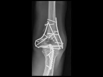
OPERATIVE PRINCIPLES AND TECHNIQUES
When the fracture has occurred in an area of bone where union is usually obtained easily (e.g., metaphyseal regions with rich cancellous bone), the techniques described for fresh fractures can often be utilized. However, diaphyseal delayed unions require specific strategies to alter both the mechanical and biological environment.
Preoperative Planning
Meticulous preoperative planning is mandatory. The surgeon must evaluate the current mechanical construct (is it too stiff or too flexible?) and the biological capacity of the host. Templating should include plans for hardware removal, the new fixation construct, and the source of bone graft (autograft vs. allograft/orthobiologics).
Plate Osteosynthesis and Bone Grafting
Open reduction is necessary to remove interposed fibrous tissue and to appose widely separated fragments.
Surgical Steps for Plating a Delayed Union:
1. Approach: Utilize standard extensile approaches. Expect altered anatomy due to scar tissue. Meticulous hemostasis and protection of neurovascular structures are critical.
2. Debridement: The fracture site must be opened and all interposed fibrous tissue excised. The sclerotic bone ends must be decorticated (shingled) using a sharp osteotome until punctate bleeding is observed (the "paprika sign"). This confirms viable, vascularized bone.
3. Reduction and Compression: The fragments must be anatomically reduced. For diaphyseal delayed unions, absolute stability is required. This is achieved using a dynamic compression plate (DCP) or locking compression plate (LCP) utilized in a compression mode.
4. Bone Grafting:
* Crucial Rule: Usually, bone grafts should be placed around the fracture if plates and screws are used when treating a delayed union.
* Autologous Iliac Crest Bone Graft (ICBG) remains the gold standard, providing osteoconductive scaffolding, osteoinductive growth factors (BMPs), and osteogenic cells. The graft should be packed meticulously into any residual defects and layered over the decorticated cortices (avoiding placement over the plate itself to prevent hardware burial).
Pitfall: Failing to achieve adequate compression across the fracture site during plating will leave the construct reliant entirely on the hardware, leading to inevitable fatigue failure before the bone graft can incorporate.
Intramedullary Nailing (IMN)
Intramedullary nailing is a highly effective treatment for diaphyseal delayed unions of the femur and tibia. The technique chosen depends on the presence of existing hardware and the need for open reduction.
Exchange Nailing
For delayed unions previously treated with an IM nail, exchange nailing is the treatment of choice.
1. Hardware Removal: The existing locking screws and nail are removed.
2. Over-reaming: The medullary canal is sequentially over-reamed by 1 to 2 millimeters larger than the previous nail.
3. Biological Benefit: This over-reaming process serves a dual purpose: it mechanically debrides the fibrous tissue within the canal and generates a rich slurry of autologous bone graft (reamings) that is deposited directly at the fracture site.
4. Nail Insertion: A larger diameter nail is inserted and statically locked to provide enhanced rotational and axial stability.
Closed vs. Open Nailing and Grafting Rules
The requirement for supplemental bone grafting during IM nailing depends entirely on the surgical approach:
* Closed Nailing: Grafts usually are not needed if a delayed union is treated with closed intramedullary nailing. The biological stimulation of reaming, combined with the load-sharing biomechanics of the nail, is typically sufficient to induce union.
* Open Nailing: If closed reduction cannot be achieved, or if interposed tissue must be manually excised, an open approach is required. If open nailing is done to achieve reduction, a bone graft usually is added. Opening the fracture site dissipates the fracture hematoma and the reaming slurry; therefore, supplemental autograft (e.g., ICBG) must be packed around the fracture site prior to closure.
Dynamization
In specific cases where an IM nail is statically locked and the fracture shows delayed union with a visible gap but no rotational instability, dynamization may be considered. This involves removing the locking screws from the longer segment of the bone (usually the dynamic slots), allowing the muscle forces and weight-bearing to axially compress the fracture site.
Surgical Warning: Dynamization should only be performed if the fracture is axially stable (e.g., transverse or short oblique). Dynamizing a comminuted or long spiral fracture will lead to catastrophic shortening and malrotation.
POSTOPERATIVE PROTOCOLS AND REHABILITATION
The postoperative management of a surgically treated delayed union must be tailored to the stability of the fixation construct and the biological quality of the bone.
Phase 1: Immediate Postoperative (Weeks 0-2)
- Immobilization: Depending on the construct, a temporary splint may be used for soft tissue rest.
- Edema Control: Elevation and cryotherapy.
- Range of Motion: Early active and active-assisted range of motion of adjacent joints is initiated to prevent stiffness and promote venous return.
Phase 2: Progressive Loading (Weeks 2-8)
- Weight-Bearing:
- IM Nails: Generally allow for early, progressive weight-bearing as tolerated, as the load-sharing device stimulates callus formation.
- Plates/Screws: Weight-bearing is typically restricted (toe-touch or partial) until early radiographic signs of graft incorporation and bridging callus are observed, as plates are load-bearing devices susceptible to fatigue failure.
- Monitoring: Clinical examination for pain at the fracture site and serial radiographs at 4-week intervals to assess the progression of the bone graft and callus formation.
Phase 3: Maturation and Strengthening (Weeks 8+)
- Once radiographic bridging is confirmed in at least three out of four cortices, full weight-bearing is permitted.
- Aggressive physical therapy focuses on restoring full muscular strength, proprioception, and functional biomechanics.
- Return to heavy labor or high-impact sports is restricted until complete radiographic remodeling is evident, which may take up to 12 to 18 months post-intervention.
CONCLUSION
The management of delayed union requires a high degree of clinical acumen, balancing the patience required for conservative, biologically driven modalities with the decisive action needed for surgical intervention. By adhering to the principles of mechanical stability, biological optimization, and meticulous surgical technique—particularly regarding the judicious use of bone grafting in open procedures—the orthopaedic surgeon can successfully navigate this complex complication, restoring function and preventing the morbidity of a definitive nonunion.
📚 Medical References
You Might Also Like