Advanced Diagnosis of SLAP Tears: Clinical Presentation, Examination & Imaging in an Athlete
Key Takeaway
Diagnosing athletic SLAP tears requires a detailed history of overhead shoulder pain. Clinical exam focuses on superior glenoid tenderness and positive tests (O'Brien's, Speed's, Kim's). While X-rays exclude bony issues, MRI or MR arthrography confirms Type II/IV labral tears with biceps anchor involvement, crucial for precise diagnosis.
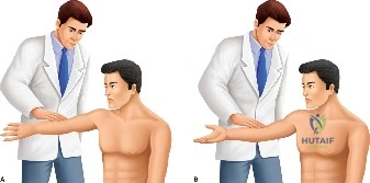
You are presented with a 32-year-old professional tennis player reporting a 9-month history of deep, aching shoulder pain and mechanical "clicking" with overhead serving. He has failed a structured, 3-month physiotherapy program. What is your differential diagnosis, and which specific clinical examinations would you perform to narrow this down?
Candidate: I would consider a SLAP lesion, rotator cuff tendinopathy, or biceps tendonitis. I would perform the O'Brien's test, Speed's test, and Yergason's test to check for labral or biceps pathology. I would also assess for impingement signs and test rotator cuff strength to rule out underlying cuff pathology.
Candidates often list tests without explaining their clinical utility or failing to address the "mechanical symptoms" aspect. A poor candidate forgets to perform a systematic screen for secondary instability or scapular dyskinesia, which is critical in an overhead athlete.
Structure the answer: 1. Differential: SLAP lesion (most likely), Biceps tendinopathy, GIRD (Glenohumeral Internal Rotation Deficit), or early cuff pathology. 2. Physical Exam: Perform a cluster of tests. Specifically, I would use the O'Brien’s active compression test (noting the relief with supination), Biceps Load II test, Anterior Slide test, and Compression-Rotation test. I would emphasize assessing for scapular dyskinesia and performing a GIRD measurement, as these are often the primary drivers in overhead athletes.
Your initial MRI was non-diagnostic. You ordered an MR arthrogram. Based on this image, identify the pathology and explain the significance of the findings in the context of the Snyder classification.
Candidate: The image shows contrast extravasation into the superior labrum. This is a SLAP lesion. Under the Snyder classification, this would be a Type II tear because the biceps anchor is involved.
Simply stating "it's a Type II" is insufficient. A weak candidate fails to explain *why* the MR arthrogram is necessary (contrast distension) and fails to sub-classify (Type IIA/B/C) which is vital for surgical planning.
The MR arthrogram demonstrates contrast extending between the superior labrum and the glenoid, confirming a detachment of the biceps anchor. This is a Snyder Type II SLAP lesion. Given the extension described, I would define it specifically (e.g., Type IIC for anterior and posterior involvement). This requires surgical intervention because the biceps anchor is unstable, which disrupts glenohumeral kinematics in an overhead athlete. I would also note the "peel-back" mechanism risk during surgery.
You have decided to proceed to arthroscopic repair. Describe your setup for the procedure and the critical intraoperative steps to ensure a successful outcome.
Candidate: I would place the patient in the lateral decubitus position. I would use a posterior viewing portal and an anterior-superior working portal. I'd debride the torn labrum, prepare the bone, and use two suture anchors to reattach the labrum to the glenoid.
Forgetting to mention the importance of decortication (bleeding bone bed) or the risk of anchor placement too medial on the glenoid neck. Also, failing to mention a diagnostic arthroscopy to rule out other pathology (e.g., hidden HAGL or cuff tears) is a common failure point.
I perform this in the lateral decubitus position. Key steps include: 1. Diagnostic arthroscopy to rule out concomitant pathology. 2. Labral mobilization with a blunt probe. 3. Decortication of the superior glenoid rim to a "bleeding bed" to enhance biological healing. 4. Anchor placement: Precise placement at the 11 o'clock and 1 o'clock positions (for a right shoulder), ensuring the anchor is at the articular margin (not too medial, to avoid subchondral bone loss). 5. Suture management: Ensuring the biceps anchor is properly tensioned without over-tightening, which can lead to postoperative stiffness.
