Embryology of the Upper Extremity and the Management of Congenital Transverse Deficiencies
Key Takeaway
Congenital transverse deficiencies of the upper extremity arise from disruptions in normal embryological development, specifically involving the apical ectodermal ridge or vascular insults during gestation. Understanding the intricate signaling centers—including the zone of polarizing activity and WNT pathways—is essential for orthopedic surgeons. This guide details the embryogenesis of the upper limb, the clinical presentation of transverse deficiencies, and evidence-based protocols for early prosthetic management and surgical intervention.
NORMAL EMBRYOLOGY OF THE UPPER EXTREMITY
The development of the upper extremity is a highly orchestrated, complex sequence of events that relies on precise spatial and temporal molecular signaling. For the orthopedic surgeon, a profound understanding of these embryological processes is not merely academic; it is the foundational basis for diagnosing, classifying, and treating congenital limb anomalies.
Chronological Development
The upper limb arises as a small, paddle-like bud of mesenchymal tissue on the lateral body wall, beginning precisely on day 26 of gestation. The upper extremity bud precedes the development of the lower extremity (leg bud) by approximately 24 to 48 hours.
Growth and development proceed in a strict proximal-to-distal fashion. By day 31 of gestation, the hand paddle is clearly present. Following the formation of the hand plate, a critical process of programmed cellular death (apoptosis) occurs within the interdigital necrotic zones. This fissuring of the hand paddle is completed by day 36. The central rays form first, rapidly followed by the preaxial (radial) and postaxial (ulnar) digits.
Following the establishment of the basic limb architecture, the formation of chondral elements and subsequent endochondral ossification begins. Joint cavitation, muscle migration, and vascular network development follow in rapid succession. The entire embryological process of upper limb formation is remarkably completed by the 8th week of gestation.
Molecular Signaling Centers
Limb development is governed by three primary signaling centers that dictate the three-dimensional growth of the extremity: the proximodistal, anteroposterior (radioulnar), and dorsoventral axes.
1. The Apical Ectodermal Ridge (AER) – Proximodistal Axis
The AER is a thickened layer of ectoderm located at the distal tip of the developing limb bud. It is the primary pacemaker for proximodistal growth.
* Mechanism: The AER secretes Fibroblast Growth Factors (primarily FGF-4 and FGF-8), which maintain the underlying mesenchyme in a highly proliferative, undifferentiated state known as the "progress zone."
* Pathology: Premature failure or disruption of the AER results in transverse deficiencies (congenital amputations).
2. The Zone of Polarizing Activity (ZPA) – Anteroposterior (Radioulnar) Axis
Radioulnar differentiation is controlled by a specialized group of mesodermal cells located in the posterior (postaxial/ulnar) margin of the limb bud, known as the ZPA.
* Mechanism: The ZPA produces a morphogen protein playfully named "Sonic Hedgehog" (SHH). The concentration gradient of SHH dictates radioulnar identity; high concentrations promote ulnar (postaxial) development, while low concentrations allow for radial (preaxial) development.
* Pathology: Alterations in SHH expression can lead to mirror-hand deformities (ulnar dimelia) or radial/ulnar clubhands. This shared signaling pathway explains why ulnar-deficient limbs frequently present with associated preaxial (radial) deficient hands.
3. The Wingless-Type (WNT) Signaling Center – Dorsoventral Axis
The dorsal ectoderm controls the dorsal differentiation of the underlying mesoderm through the WNT signaling pathway.
* Mechanism: Wnt-7a is expressed in the dorsal ectoderm and induces the expression of the transcription factor Lmx1b in the underlying dorsal mesenchyme, which specifies dorsal limb characteristics (e.g., nails, extensor tendons).
* Pathology: Disruption of Wnt-7a or Lmx1b results in ventralization of the dorsal limb (e.g., nail-patella syndrome).
Clinical Pearl: The signaling centers do not operate in isolation. A critical positive feedback loop exists between the AER (FGF) and the ZPA (SHH), mediated by the protein Gremlin. Disruption of this loop at any point can lead to catastrophic failure of limb formation, manifesting as severe transverse or longitudinal deficiencies.
PATHOGENESIS OF TRANSVERSE DEFICIENCIES
Transverse deficiencies, historically referred to as congenital amputations, are characterized by normal proximal development with a complete absence of skeletal elements distal to a specific transverse plane.
Etiology and Vascular Disruption
The primary embryological failure in a transverse deficiency is the premature cessation of the Apical Ectodermal Ridge (AER). While the exact trigger for AER failure is often idiopathic, it is widely believed to be secondary to a localized vascular infarct or hypoperfusion event during the critical window of limb development (weeks 4 to 8).
- Teratogenic Factors: The use of misoprostol (a prostaglandin E1 analogue) to induce abortion has been definitively shown to cause vascular disruption in utero. Infants surviving misoprostol exposure frequently present with transverse limb deficiencies and Moebius syndrome (cranial nerve VI and VII palsies).
- Chorionic Villus Sampling (CVS): Early CVS (performed before 10 weeks gestation) has been associated with an increased risk of transverse limb deficiencies, further supporting the vascular disruption hypothesis.
Genetic and Syndromic Associations
In the vast majority of cases, the usual unilateral transverse deficiency is a sporadic event with no underlying genetic basis. Parents can be reassured that the recurrence risk for future pregnancies is negligible.
However, rare bilateral or multiple transverse deficiencies may be inherited as an autosomal recessive trait (e.g., Adams-Oliver syndrome, though this typically presents with terminal transverse defects and aplasia cutis congenita).
Surgical Warning: While transverse deficiencies usually do not occur in association with major malformation syndromes, a thorough systemic evaluation is mandatory. Anomalies reported to occur in association with transverse deficiencies include hydrocephalus, spina bifida, myelomeningocele, clubfoot, radial head dislocation, and radioulnar synostosis.
CLINICAL EVALUATION AND PHENOTYPIC PRESENTATION
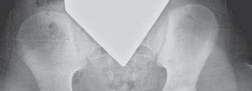
The clinical presentation of a transverse deficiency is dictated by the anatomical level of the defect. The most common level of deficiency is the proximal third of the forearm, followed by midcarpal amputations.
Anatomical Characteristics
A newborn presenting with a transverse deficiency typically exhibits a slightly bulbous, well-padded stump.
* Digital Nubbins: In more distal deficiencies (such as midcarpal or transmetacarpal levels), rudimentary, vestigial digital "nubbins" are common. These nubbins represent aborted attempts at digit formation following the vascular insult. They may contain tiny cartilaginous remnants or rudimentary nails but are universally nonfunctional.
* Muscle Hypoplasia: A critical diagnostic feature is the hypoplasia of the more proximal musculature. The muscles originating proximal to the amputation site but intended to insert distally will be atrophic or absent.
Differential Diagnosis: Amniotic Band Sequence
It is imperative to differentiate a true embryological transverse deficiency from an amputation caused by Congenital Constriction Band Syndrome (Amniotic Band Sequence).
* Transverse Deficiency: Characterized by proximal muscle hypoplasia, a smooth bulbous stump, and a predictable pattern of missing distal elements.
* Amniotic Band Sequence: Characterized by normal proximal muscle bulk, the presence of deep constriction rings, acrosyndactyly, and often an irregular, asymmetrical amputation stump.
Growth Prognosis and Biomechanics
Understanding the growth potential of the affected limb is crucial for long-term prosthetic planning.
* Upper Forearm Deficiency: In the most common upper forearm amputation, the forearm usually measures no more than 7 cm long at birth. Due to the absence of the distal radial and ulnar physes (which contribute 75% and 80% of forearm longitudinal growth, respectively), the stump can be expected to measure no more than 10 cm by skeletal maturity.
* Midcarpal Deficiency: In midcarpal amputations, the affected forearm will be relatively shorter than the contralateral normal side. However, because the proximal radioulnar joint and the interosseous membrane are largely intact, active pronation and supination are usually preserved. Preserving this rotational arc is a primary goal of management.
Clinical Pearl: Children with isolated transverse deficiencies generally possess normal intelligence and achieve standard developmental milestones. Psychological support should focus on empowering the child and mitigating parental guilt, emphasizing the sporadic nature of the anomaly.
PRINCIPLES OF PROSTHETIC MANAGEMENT
For the vast majority of patients with transverse deficiencies, extensive surgical reconstruction is neither required nor indicated. The cornerstone of treatment is early, structured prosthetic management.
Timing of Prosthetic Fitting
The integration of a prosthesis into the child's body schema is highly dependent on the timing of the initial fitting.
* Initial Passive Fitting: Treatment should consist of early prosthetic fitting of the deficient limb, preferably by the time the child is crawling (typically 6 to 9 months of age). At this stage, a passive prosthesis acts as a "prop" to assist with crawling, balance, and bimanual spatial awareness.
* Active Terminal Devices: By the time of independent ambulation and the development of cognitive cause-and-effect understanding (usually 15 to 18 months), the child can be transitioned to a body-powered prosthesis with an active terminal device (e.g., a voluntary-opening hook).
Developmental Milestones in Prehension
The child’s development of manual and bimanual skills progresses in an orderly and predictable pattern, which the prosthetist and occupational therapist must respect:
1. Birth to 9 Months: Prehension is achieved primarily by bilateral palmar grasp. The infant uses both upper limbs symmetrically to trap objects against the body. A passive prosthesis facilitates this gross bimanual trapping.
2. 9 to 18 Months: The child begins to develop a dominant hand and a non-dominant "assisting" hand. The normal limb assumes the dominant role for fine motor tasks (pincer grasp), while the prosthetic limb serves as the stabilizer.
3. 18+ Months: Refinement of bimanual coordination. The introduction of a cable-driven, body-powered prosthesis allows the child to actively grasp and release objects with the terminal device, utilizing biscapular abduction or glenohumeral flexion to operate the cable.
Advanced Prosthetic Options
As the child reaches school age and adolescence, myoelectric prostheses may be considered. These devices utilize surface electromyography (EMG) electrodes placed over the residual proximal musculature (e.g., biceps and triceps) to control motorized hands or hooks. While cosmetically superior and requiring less harnessing, myoelectric devices are heavier, more fragile, and lack the proprioceptive feedback provided by the tension in a body-powered cable system.
SURGICAL INTERVENTIONS AND STUMP OPTIMIZATION
While prosthetic management is the primary treatment modality, surgical intervention is occasionally required to optimize the residual limb for prosthetic wear or to maximize independent function.
Indications for Surgery
- Excision of Unstable Nubbins: Vestigial digital nubbins that are hypermobile, easily traumatized, or interfere with the intimate fit of a prosthetic socket should be excised. Care must be taken to ligate any rudimentary neurovascular bundles to prevent painful neuroma formation.
- Deep Clefts and Skin Invaginations: Deep skin clefts at the end of the stump can harbor moisture, leading to maceration and recurrent fungal or bacterial infections inside the prosthetic socket. Surgical revision to create a smooth, convex stump is indicated.
- Bony Overgrowth (Terminal Overgrowth): This is the most common and troublesome complication in pediatric amputations.
Management of Bony Overgrowth
Unlike adult amputations, where the bone is static, pediatric amputated bones continue to grow. The appositional growth at the distal end of the transected bone (most commonly the humerus, fibula, tibia, and radius) can outpace the growth of the overlying soft tissues. This results in a spike of bone piercing the skin, causing severe pain, bursa formation, and inability to wear a prosthesis.
- Pathophysiology: Bony overgrowth is not caused by the proximal physis pushing the bone through the skin; rather, it is a phenomenon of aggressive periosteal new bone formation at the transected distal end.
- Surgical Techniques:
- Stump Revision: Simple resection of the bony spike provides only temporary relief, with recurrence rates approaching 100% in young children.
- Autologous Cartilage Capping (Marquis Procedure): The most effective biological solution involves capping the transected bone end with an autologous osteochondral graft (often harvested from the iliac crest or a discarded fibula). The cartilage cap halts the aggressive periosteal bone formation.
- Synthetic Capping: The use of synthetic caps (e.g., silicone or high-density polyethylene) has largely been abandoned due to high rates of foreign body reaction, migration, and skin breakdown.
Pitfall: Never perform an epiphysiodesis of the proximal physis to treat distal bony overgrowth. This will permanently stunt the longitudinal growth of the already shortened residual limb, severely compromising the lever arm required for prosthetic control.
Advanced Reconstructive Options
In highly selected cases, particularly in bilateral transverse deficiencies, advanced reconstructive procedures may be considered to provide tactile, sensate prehension without the need for prostheses.
- The Krukenberg Procedure: Indicated for bilateral below-elbow deficiencies. This procedure separates the radius and ulna, converting the forearm into a sensate, pincer-like mechanism. The pronator teres is utilized to provide active pinch between the two bones. While cosmetically challenging for some cultures, the functional independence and preservation of tactile sensation it provides to a blind or bilaterally affected patient are unparalleled.
- Targeted Muscle Reinnervation (TMR): In older adolescents with proximal amputations, TMR can be utilized. Residual nerves (e.g., median, ulnar, radial) are surgically transferred to innervate distinct segments of the remaining proximal muscles. This creates new, amplified EMG signals that can intuitively control advanced multi-articulating myoelectric prostheses.
POSTOPERATIVE PROTOCOLS AND LONG-TERM REHABILITATION
The management of congenital transverse deficiencies requires a lifelong, multidisciplinary approach. The surgical and prosthetic interventions are only as successful as the rehabilitation program that accompanies them.
Multidisciplinary Care Team
- Orthopedic Surgeon: Monitors skeletal growth, manages bony overgrowth, and performs stump revisions as necessary.
- Prosthetist: Adjusts the socket fit to accommodate rapid pediatric growth spurts. Sockets typically need replacement every 12 to 18 months in growing children.
- Occupational Therapist (OT): Crucial for training the child in Activities of Daily Living (ADLs), prosthetic control, and bimanual integration. The OT focuses on maximizing the function of the residual limb, whether bare or prosthetically equipped.
- Psychologist/Social Worker: Provides vital support to the family, addressing parental guilt, peer integration, and the psychosocial challenges of living with a limb difference.
Rehabilitation Milestones
Postoperative rehabilitation following stump revision or nubbin excision focuses on rapid wound healing and immediate return to prosthetic wear. Prolonged periods without the prosthesis can lead to a loss of the child's established body schema and subsequent prosthetic rejection.
Desensitization techniques (massage, tapping, varied textures) should be initiated as soon as the surgical incisions are healed to prepare the stump for the shear forces of the prosthetic socket.
In conclusion, the successful management of congenital transverse deficiencies relies on a profound understanding of upper limb embryology, precise clinical evaluation, and a commitment to early, consistent prosthetic integration. Surgical intervention, while secondary, plays a critical role in optimizing the residual limb, ensuring that these children achieve their maximum functional potential and lead fully independent lives.
You Might Also Like