Multiple Hereditary Exostoses (MHE): Pathophysiology, Clinical Features, & Orthopedic Management
Key Takeaway
Multiple Hereditary Exostoses (MHE) is a genetic disorder forming osteochondromas. Key complications are skeletal deformities (e.g., genu valgum), pain, and neurovascular compression. The most serious risk is malignant transformation into a secondary peripheral chondrosarcoma. Lifelong monitoring and targeted surgical intervention are crucial for managing MHE patients effectively.
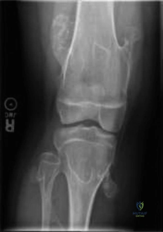
A 14-year-old boy with a known diagnosis of Multiple Hereditary Exostoses (MHE) presents to your clinic. His parents are concerned about a painless, firm lump on his distal femur that has been present for years but they feel it has become more prominent during his recent growth spurt. He has no neurovascular symptoms. How do you approach the initial assessment of this lesion?
Candidate: I would take a thorough history focusing on pain and interval growth. On examination, I would palpate the lesion for size, consistency, and mobility. I would then order orthogonal radiographs of the femur to evaluate the stalk, base morphology, and proximity to the physis. I would explain to the parents that most exostoses are benign, but we need to monitor for mechanical irritation or signs of malignant transformation.
Candidates often jump straight to "I'll excise it" or fail to mention the importance of physical examination findings regarding the cartilage cap (which might be palpable) and the patient's skeletal maturity status (since growth should cease at physeal closure).
The candidate must systematically frame the assessment: 1) Clinical history (specifically asking about new pain, which is the most reliable symptom of malignant transformation). 2) Physical exam (mapping the lesion and checking for tethering or neurovascular deficits). 3) Imaging (Orthogonal X-rays to classify the lesion—sessile vs. pedunculated). 4) Contextualizing the growth: "Given he is 14, he is likely near skeletal maturity; any growth at this stage must be closely scrutinized for malignant degeneration, although it is rare in adolescents."
Look at this radiograph. What are the key biomechanical considerations for the forearm in this patient, and how does the Masada classification guide your surgical decision-making?
Candidate: The forearm is at risk because the ulna is often tethered, leading to shortening, positive ulnar variance, and subsequent radial head subluxation or dislocation. The Masada classification categorizes the severity—Type I involves distal ulnar exostosis, Type II includes radial head dislocation. Treatment ranges from simple exostosis excision to ulnar lengthening and radial osteotomies to restore forearm mechanics.
Failing to mention the "Madelung-like" deformity or forgetting the specific surgical goal: that the ulnar lengthening is the priority to "re-center" the radial head.
The perfect answer addresses the pathomechanics: "The distal ulna is a significant longitudinal growth driver. Tethering leads to ulnar shortening and relative radial overgrowth/bowing, forcing the radial head to dislocate." The candidate should emphasize that Masada I/II requires ulnar lengthening (often using distraction osteogenesis for >2cm) to allow the radial head to reduce. Proactive excision of the distal ulnar tether is key to preventing further progression.
A 25-year-old patient with MHE presents with a new onset of deep, aching pain around his proximal thigh and a palpable mass. The mass has not grown significantly, but the pain is constant. How do you investigate this, and what is your threshold for suspicion of malignancy?
Candidate: In a skeletally mature patient, new onset pain is a red flag. I would immediately order an MRI. I am looking for the thickness of the cartilage cap; if it exceeds 1.5 cm, it is highly suspicious for malignant transformation to secondary chondrosarcoma. If confirmed, I would proceed with a biopsy and wide en-bloc resection.
Ignoring the "1.5 cm" cartilage cap rule or suggesting a simple marginal excision as the first step for a suspicious lesion.
The candidate must demonstrate oncologic awareness: 1) Adult + New Pain = Mandatory MRI. 2) Key biomarker is a cartilage cap > 1.5 cm on T2-weighted sequences. 3) The paradigm shifts from "excision" to "wide en-bloc resection" to avoid local seeding, which would be a catastrophic error if malignancy is present.
