Orthopaedic Management of Paralytic Deformities: Poliomyelitis & Myelomeningocele
Key Takeaway
The orthopedic management of paralytic deformities requires a profound understanding of altered biomechanics, muscle balancing, and joint stabilization. This comprehensive guide details evidence-based surgical interventions for poliomyelitis and myelomeningocele, encompassing tendon transfers, osteotomies, and arthrodesis across the upper and lower extremities. Designed for orthopedic surgeons, it provides step-by-step approaches to restoring function, correcting contractures, and achieving long-term stability in the flail or unbalanced limb.
Introduction to Paralytic Deformities
The orthopedic management of paralytic deformities—historically dominated by the sequelae of poliomyelitis and currently highly relevant in the context of myelomeningocele (spina bifida)—represents one of the most complex challenges in reconstructive surgery. While poliomyelitis presents as a pure lower motor neuron (LMN) flaccid paralysis due to anterior horn cell destruction, myelomeningocele presents a mixed, often progressive neurological picture complicated by upper motor neuron signs, tethered cord syndrome, and insensate skin.
The foundational principles of paralytic reconstruction were established by pioneers such as Steindler, Barr, and Mayer. The primary goals of surgical intervention are to correct fixed deformities, restore dynamic muscle balance, eliminate the need for cumbersome orthoses (where possible), and maximize independent function.
Clinical Pearl: Never perform a tendon transfer in the presence of a fixed joint contracture. The joint must be rendered supple through serial casting, soft tissue release, or corrective osteotomy prior to, or concurrent with, the transfer.
Core Biomechanical Principles of Tendon Transfers
Successful tendon transfer in the paralytic limb relies on strict adherence to biomechanical and physiological prerequisites:
* Adequate Strength: The donor muscle must be at least Medical Research Council (MRC) Grade 4, as it will typically lose one full grade of strength post-transfer.
* Similar Excursion: The donor tendon must possess an amplitude of excursion comparable to the paralyzed muscle it replaces.
* Straight Line of Pull: The tendon should be routed as directly as possible to its new insertion, avoiding acute angles that increase friction and reduce mechanical advantage.
* Synergistic Phase: Transferring a muscle that fires in the same phase of the gait cycle (e.g., swing vs. stance) yields superior functional results, though phase conversion can occur with extensive postoperative motor re-education.
* Tissue Bed Integrity: The tendon must be routed through a healthy, well-vascularized, and unscarred subcutaneous or interosseous bed to prevent adhesions.
Lower Extremity Reconstruction
Foot and Ankle Deformities
The foot and ankle are highly susceptible to paralytic deformities due to the complex interplay of invertors, evertors, dorsiflexors, and plantarflexors.
Paralytic Equinovarus and the Tibialis Posterior Transfer
When the anterior compartment (tibialis anterior, toe extensors) and lateral compartment (peroneals) are paralyzed, the unopposed pull of the tibialis posterior and Achilles tendon results in a rigid equinovarus deformity. The Barr procedure (transfer of the tibialis posterior to the dorsum of the foot) is the gold standard for restoring active dorsiflexion.
Surgical Technique:
1. Incision 1 (Medial Foot): Make a longitudinal incision over the navicular tuberosity. Detach the tibialis posterior tendon, preserving maximum length.
2. Incision 2 (Medial Calf): Make a longitudinal incision at the musculotendinous junction of the tibialis posterior. Withdraw the tendon into the calf wound.
3. Incision 3 (Anterior Leg): Make an incision lateral to the tibial crest. Create a generous window in the interosseous membrane.
4. Routing: Pass the tendon through the interosseous window into the anterior compartment. Ensure the window is large enough to prevent tethering during muscle contraction.
5. Insertion: Route the tendon subcutaneously to the dorsum of the foot. Fix it into the lateral cuneiform or cuboid (depending on the degree of varus/valgus imbalance) using a bio-tenodesis screw or pull-out wire, with the foot held in maximum dorsiflexion.
Arthrodesis of the Foot and Ankle
In cases of a completely flail foot or severe, rigid deformity where tendon transfers are unviable, stabilization via arthrodesis is indicated.
* Triple Arthrodesis: Fuses the subtalar, talonavicular, and calcaneocuboid joints. It corrects hindfoot varus/valgus and midfoot deformities while preserving ankle motion.
* Pantalar Arthrodesis: Described by Steindler and Leibolt, this involves fusion of the tibiotalar (ankle) joint in addition to the triple arthrodesis joints. It is indicated for a completely flail ankle and foot, providing a stable, plantigrade pillar for weight-bearing.
Surgical Warning: In myelomeningocele patients, arthrodesis must be approached with extreme caution. Insensate skin and poor bone stock increase the risk of non-union, Charcot arthropathy, and catastrophic postoperative ulceration.
Knee Deformities
Knee Flexion Contractures
Flexion contractures commonly result from unopposed hamstring activity or prolonged wheelchair positioning. Mild contractures (<20 degrees) may respond to posterior capsular release and hamstring lengthening. Severe contractures require osseous correction.
Supracondylar Femoral Extension Osteotomy:
1. Approach: Lateral approach to the distal femur.
2. Osteotomy: An anteriorly based closing wedge osteotomy is performed just proximal to the trochlea.
3. Fixation: Rigid internal fixation using a blade plate or locking compression plate.
4. Consideration: In severe cases, concomitant shortening of the femur may be necessary to prevent traction injury to the sciatic nerve and popliteal vessels.
Genu Recurvatum
Paralytic genu recurvatum occurs due to quadriceps weakness combined with a fixed equinus deformity or hamstring weakness.
* Soft Tissue Procedures: Perry's triple tenodesis utilizes the hamstrings and iliotibial band to create a posterior checkrein.
* Osseous Procedures: A proximal tibial flexion osteotomy (opening wedge posteriorly or closing wedge anteriorly) alters the mechanical axis to prevent hyperextension during the stance phase.
Hip and Pelvis Reconstruction
Paralytic Hip Dislocation
Hip instability in poliomyelitis and myelomeningocele is driven by muscle imbalance—specifically, strong flexors and adductors overpowering weak extensors and abductors. This leads to progressive coxa valga, acetabular dysplasia, and eventual dislocation.
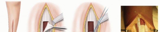
Mustard Iliopsoas Transfer:
Indicated for abductor weakness with a preserved gluteus maximus. The iliopsoas is detached from the lesser trochanter, routed through a window in the anterior ilium, and inserted into the greater trochanter to act as an abductor.
Abductor Paralysis and the External Oblique Transfer
A severe Trendelenburg gait (abductor lurch) consumes immense energy. The Thomas, Thompson, and Straub transfer utilizes the external abdominal oblique to restore pelvic stability.
Surgical Technique:
1. Harvest: An incision is made along the iliac crest. The aponeurosis of the external oblique is harvested, leaving the muscle belly intact.
2. Routing: The fascial strip is folded into a tube and routed distally over the iliac crest.
3. Insertion: The transfer is passed subcutaneously and fixed into the greater trochanter of the femur with the hip in maximum abduction.
4. Postoperative Protocol: The patient is immobilized in a 1.5 hip spica cast in wide abduction for 6 weeks to allow secure fascial healing.
Upper Extremity Reconstruction
While lower extremity surgery focuses on stability and weight-bearing, upper extremity reconstruction prioritizes spatial placement of the hand (shoulder/elbow) and prehension.
The Flail Shoulder
Paralysis of the deltoid and rotator cuff renders the arm functionally useless, even if hand function is preserved.
* Shoulder Arthrodesis: The procedure of choice for a flail shoulder with intact scapulothoracic musculature (trapezius, serratus anterior).
* Positioning (Rowe's Criteria): The glenohumeral joint is fused in 20-30 degrees of abduction, 20-30 degrees of flexion, and 20-30 degrees of internal rotation. This allows the hand to reach the mouth via scapulothoracic motion while permitting the arm to rest comfortably at the side.
Elbow Flexion Restoration
Restoration of active elbow flexion is critical for feeding and personal hygiene.
Steindler Flexorplasty
Indicated when the biceps and brachialis are paralyzed, but the wrist and finger flexors (supplied by the median and ulnar nerves) remain strong (MRC Grade 4+).
Surgical Technique:
1. Approach: A medial longitudinal incision over the elbow, identifying and protecting the ulnar nerve.
2. Osteotomy: The medial epicondyle is osteotomized, retaining the origin of the flexor-pronator mass.
3. Advancement: The epicondylar fragment is advanced proximally 5 to 7 cm along the anterior aspect of the humerus.
4. Fixation: The fragment is secured to the anterior humerus using a cortical screw or heavy non-absorbable sutures through drill holes.
5. Biomechanics: By moving the origin of the flexor-pronator mass proximally, its moment arm across the elbow joint is significantly increased, converting these muscles into primary elbow flexors.
Latissimus Dorsi Transfer
If the flexor-pronator mass is weak, a bipolar or unipolar latissimus dorsi transfer (Zancolli technique) can be utilized to restore either elbow flexion or extension (triceps paralysis), depending on the routing of the muscle pedicle.
Spinal Deformity in Myelomeningocele
Spinal deformities in myelomeningocele are nearly ubiquitous in high-level lesions (thoracic level). The deformities are characterized by rigid, progressive scoliosis, kyphosis, and severe pelvic obliquity, which compromise sitting balance and pulmonary function.
Surgical Management of Paralytic Scoliosis
- Preoperative Evaluation: MRI of the entire neuroaxis is mandatory to rule out a tethered cord, syringomyelia, or Arnold-Chiari malformation. Neurosurgical untethering must precede orthopedic correction if symptomatic.
- Surgical Approach: Due to absent posterior elements (spina bifida) and poor bone quality, isolated posterior fusions have an unacceptably high pseudarthrosis rate.
- Combined Anterior/Posterior Fusion: The gold standard involves an anterior release and interbody fusion, followed by a posterior instrumented fusion extending to the pelvis (Galveston technique or iliac screws) to correct pelvic obliquity and provide a stable sitting foundation.
Pitfall: Failure to extend the fusion to the pelvis in a non-ambulatory myelomeningocele patient with pelvic obliquity will inevitably lead to progressive seating imbalance, ischial pressure sores, and hardware failure.
Postoperative Protocols and Rehabilitation
The success of paralytic reconstructive surgery is heavily dependent on meticulous postoperative care.
1. Immobilization: Tendon transfers require rigid immobilization in a relaxed position for a minimum of 4 to 6 weeks to allow for biological healing at the tendon-bone interface.
2. Orthotic Management: Following cast removal, patients are transitioned to custom orthoses (e.g., AFO, KAFO) to protect the transfer during the initial rehabilitation phase.
3. Motor Re-education: Intensive physical therapy is required to train the brain to fire the transferred muscle in its new biomechanical role. Biofeedback and electromyography (EMG) can be highly effective adjuncts in phase conversion.
4. Long-Term Surveillance: Particularly in myelomeningocele, patients require lifelong follow-up to monitor for late complications such as Charcot arthropathy, hardware failure, and the recurrence of deformity due to neurological deterioration.
Conclusion
The operative management of poliomyelitis and myelomeningocele demands a master-level understanding of musculoskeletal biomechanics, neuroanatomy, and spatial deformity correction. Whether performing a complex tendon transfer to restore elbow flexion or a massive spinal fusion to achieve sitting balance, the orthopedic surgeon must meticulously plan each intervention. By adhering to the time-honored principles of joint suppleness, precise tendon routing, and rigid osseous stabilization, surgeons can profoundly alter the trajectory of a patient's life, restoring independence and dignity to the paralytic limb.
You Might Also Like