General Treatment Considerations in Neuromuscular Orthopaedics
Key Takeaway
The orthopaedic management of children with neuromuscular diseases requires a multidisciplinary approach focused on optimizing function, preventing deformity progression, and maximizing quality of life. Treatment encompasses the meticulous management of fragility fractures secondary to disuse osteopenia, the strategic application of spinal and lower extremity orthoses, and the implementation of customized seating systems to accommodate progressive postural collapse.
Introduction to Neuromuscular Orthopaedics
The orthopaedic management of patients with severe neuromuscular diseases represents one of the most complex and multidisciplinary challenges in modern surgical practice. Unlike idiopathic orthopaedic conditions, neuromuscular disorders are characterized by progressive muscle weakness, relentless joint contractures, spinal deformity, and profound disuse osteopenia. The overarching goal of orthopaedic intervention is rarely curative; rather, it is aimed at preventing the worsening of deformities, providing stability to the skeletal system, and fundamentally improving the quality of life for these children.
Although future advancements in gene therapy hold the promise of a definitive cure, meticulous orthopaedic treatment remains an absolute necessity to maximize function and comfort, regardless of the severity of the impairment. When establishing treatment algorithms, the surgeon must adhere to the hierarchy of patient priorities as famously delineated by Bleck. In order of paramount importance, these priorities are:
1. The ability to communicate with other people.
2. The ability to perform activities of daily living (ADLs).
3. Mobility (whether ambulatory or wheelchair-dependent).
Clinical Pearl: Surgical decision-making in neuromuscular orthopaedics must always be weighed against the anticipated functional gain. As demonstrated by Louis et al., who reported on 34 surgical procedures performed over a 12-year period in a select population with severe multiple impairments, interventions aimed at improving sitting posture, care, and comfort yield significant improvements in quality of life, with virtually no patients experiencing a worsening of their baseline functional status.
Pathophysiology and Classification of Muscular Dystrophies
A profound understanding of the genetic and biochemical basis of neuromuscular diseases is essential for the orthopaedic surgeon, as the natural history of the specific disease dictates the timing and nature of surgical intervention. The muscular dystrophies are a heterogeneous group of inherited disorders characterized by progressive muscle wasting and weakness.
The structural integrity of the muscle fiber is highly dependent on the dystrophin-glycoprotein complex, which bridges the inner cytoskeleton to the extracellular matrix. Mutations in the genes encoding these proteins lead to membrane instability, calcium influx, and subsequent myofiber necrosis.
Classification of Major Muscular Dystrophies
X-Linked Recessive Dystrophies
* Duchenne and Becker Muscular Dystrophy: Locus Xp21. These are caused by mutations in the gene encoding dystrophin. Duchenne muscular dystrophy (DMD) involves an absolute absence or severe truncation of dystrophin, leading to a rapid clinical decline. Becker muscular dystrophy (BMD) involves partially functional dystrophin, resulting in a milder phenotype.
* Emery-Dreifuss Dystrophy: Locus Xp28. Caused by mutations affecting emerin, a nuclear membrane protein. Characterized by early contractures (elbows, Achilles) and severe cardiac conduction defects.
Autosomal Dominant (AD) Dystrophies
* Myotonic Dystrophy: Locus 19q. Associated with the myotonin protein kinase.
* Facioscapulohumeral Dystrophy (FSHD): Locus 4q.
* Limb-Girdle Muscular Dystrophy (LGMD) Type 1: Includes LGMD-1A (5q) and LGMD-1B, characterized by proximal muscle weakness.
Autosomal Recessive (AR) Dystrophies
* Limb-Girdle Muscular Dystrophy (LGMD) Type 2: A complex group largely involving the sarcoglycan complex.
* LGMD-2A: 15q (Calpain)
* LGMD-2B: 2q
* LGMD-2C: 13q (γ-Sarcoglycan)
* LGMD-2D: 17q (α-Sarcoglycan)
* LGMD-2E: 4q (β-Sarcoglycan)
* LGMD-2F: 5q (δ-Sarcoglycan)
Congenital Dystrophies and Myopathies
* Congenital Muscular Dystrophy (AR): Locus 6q (Merosin).
* Fukuyama Disease (AR): Locus 9q13.
* Central Core Disease (AD): Locus 19q (Ryanodine receptor). Highly associated with malignant hyperthermia susceptibility.
* Nemaline Rod Disease (AD): Locus 1q22 (Tropomyosin).
Diagnostic Pitfall: Clinical phenotyping alone is insufficient for definitive diagnosis. Dystrophin testing via dystrophin immunoblotting from a muscle biopsy is a critical biochemical test. It is the gold standard for differentiating the severe Duchenne muscular dystrophy (absence of dystrophin) from the more indolent Becker muscular dystrophy (altered size/amount of dystrophin).
Fracture Management in Neuromuscular Disease
Fractures are exceedingly common in children with neuromuscular disease. The etiology is multifactorial, driven primarily by profound disuse osteoporosis, lack of mechanical loading on the skeleton, nutritional deficits, and frequent falls secondary to progressive lower extremity weakness and balance deficits.
Larson and Henderson demonstrated a significant decrease in bone mineral density (BMD) on dual-energy x-ray absorptiometry (DEXA) scans in boys with Duchenne muscular dystrophy, noting that a staggering 44% of these patients sustain fractures during their lifetime.
Principles of Fracture Care
The fundamental principle of fracture management in the neuromuscular patient is the rapid restoration of baseline mobility. Prolonged immobilization in this population is catastrophic; it leads to an irreversible loss of muscle strength, exacerbation of joint contractures, and a rapid acceleration of disuse osteopenia.
Metaphyseal Fractures
Most fractures in this population are nondisplaced or minimally displaced metaphyseal fractures. Due to the highly vascular nature of the metaphysis, these fractures heal rapidly.
* Treatment: Minimally displaced metaphyseal fractures of the lower limbs should be treated with functional splinting or lightweight fiberglass casting. The goal is to allow walking or weight-bearing transfers to be resumed as quickly as possible. If the patient already utilizes orthoses (e.g., Knee-Ankle-Foot Orthoses), these braces can often be modified or enlarged to accommodate the swollen, fractured limb, thereby acting as a custom splint that permits progressive weight-bearing.
Diaphyseal Fractures
Displaced diaphyseal fractures (particularly of the femur or tibia) present a more complex challenge.
* Conservative Management: Cast-braces may be utilized for minimally displaced fractures, provided they do not preclude the patient's ability to stand or transfer.
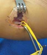
* Operative Management: Open reduction and internal fixation (ORIF) or minimally invasive submuscular plating is frequently indicated for displaced diaphyseal fractures. Surgical stabilization provides rigid fixation, eliminates the need for heavy, cumbersome spica casts, and allows for immediate mobilization and weight-bearing during the fracture healing phase.
Surgical Warning: When performing ORIF in neuromuscular patients, the surgeon must account for severe osteopenia. Standard cortical screws may suffer from poor purchase. The use of locking plate technology, longer plates to span the entire diaphysis (preventing peri-implant stress risers), and meticulous soft-tissue handling are mandatory.
Medical Management of Osteopenia
Orthopaedic surgeons must advocate for the medical optimization of bone health. The prophylactic and therapeutic medical treatment of disuse osteopenia is highly beneficial in decreasing the incidence of fragility fractures. This includes optimization of Vitamin D and calcium levels, and increasingly, the judicious use of intravenous bisphosphonates (e.g., pamidronate or zoledronic acid) to inhibit osteoclastic bone resorption and increase cortical thickness.
Principles of Orthotic Management
Orthoses play a critical role in the non-operative management of neuromuscular deformities. They are utilized to provide stability to weakened joints, prevent the rapid progression of contractures, and assist with positioning for both ambulation and seating.
Spinal Bracing
Progressive paralytic scoliosis is a hallmark of many neuromuscular diseases. As truncal weakness progresses, the spine collapses under the forces of gravity.
* Indications: Spinal bracing is occasionally used to assist with sitting balance and to free the upper extremities from having to support the trunk.
* Limitations: It is imperative to counsel parents that bracing may slow, but does not prevent, the ultimate progression of neuromuscular spinal deformity. It is a temporizing measure until the child reaches an appropriate age or size for spinal fusion.
* Design: Spinal bracing is typically accomplished using a polypropylene plastic shell lined with soft polyethylene foam to prevent pressure sores over osteopenic bony prominences. Common designs include:
* Bivalved Total-Contact Orthosis: An anterior and posterior shell design that allows for easy donning and doffing in a recumbent patient.
* Anterior-Opening Thoracolumbosacral Orthosis (TLSO): Custom-molded with precise lumbar lordotic contouring to maintain sagittal balance and optimize diaphragmatic excursion.
Lower Extremity Orthoses
- Knee-Ankle-Foot Orthoses (KAFOs): Indicated for patients with profound proximal muscle weakness (e.g., quadriceps weakness) who require mechanical locking of the knee to maintain an upright posture. If hip extensor or abductor weakness is severe, a pelvic band with hip and knee locks (HKAFO) can be added to provide multi-planar stability.
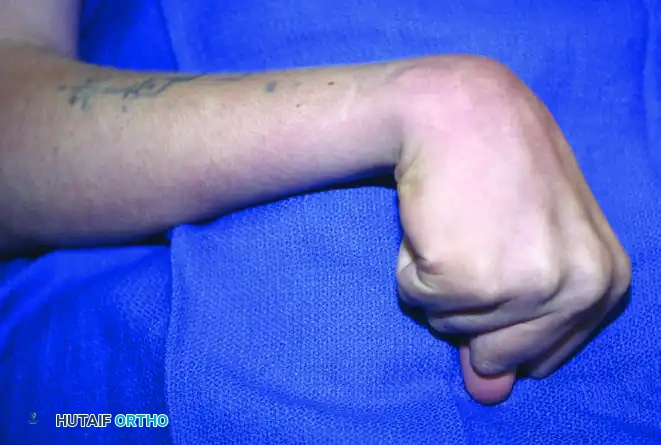
- Ankle-Foot Orthoses (AFOs): The most commonly prescribed orthosis in neuromuscular disease. AFOs are designed to position the ankle and foot in a plantigrade position. This is critical in an effort to prevent the progressive equinus and equinovarus deformities that result from the relentless contracture of the gastrocnemius-soleus complex and tibialis posterior muscles. Solid AFOs provide maximum control, while articulated AFOs may be used if sufficient eccentric calf control remains.
Advanced Seating Systems and Wheelchair Prescription
As neuromuscular disease progresses, walking becomes increasingly difficult, energy-consuming, and frustrating for the child. The transition to a wheelchair is an inevitable milestone in conditions like Duchenne muscular dystrophy and severe spinal muscular atrophy. The orthopaedic surgeon must view the wheelchair not merely as a mode of transport, but as a dynamic, custom orthosis for the entire body.
Ergonomics of the Wheelchair Receptacle
Whether the chair is manual or electric, it must be meticulously contoured to the patient's specific musculoskeletal anatomy. Poor seating leads to rapid progression of pelvic obliquity, scoliosis, and the development of decubitus ulcers.
- Pelvic Support: The foundation of seating is the pelvis. A narrow chair with a firm seat is mandatory. A "sling" seat must be strictly avoided, as it promotes internal rotation of the hips, pelvic obliquity, and a collapsing C-shaped scoliosis.
- Spinal Support: A firm backrest positioned in slight extension helps to support the spine, open the anterior chest wall, and maximize pulmonary vital capacity.
- Lateral Supports: Lateral thoracic spine supports built into the chair can significantly assist with sitting balance, allowing the child to use their hands for ADLs rather than for truncal support. However, similar to spinal bracing, these supports usually do not alter the natural history or progression of the underlying scoliosis.
Specialized Seating Clinics
The prescription of a neuromuscular wheelchair requires a multidisciplinary seating clinic involving the orthopaedic surgeon, physical therapist, and specialized orthotist/seating engineer. These clinics provide custom-fitted chairs with numerous modular options for daily use. Advanced custom-molded seating matrices can accommodate severe, rigid spinal deformities and fixed pelvic obliquities, ensuring that the pressure is distributed evenly across the buttocks and posterior thighs, thereby safeguarding the patient's skin integrity and maximizing their daily comfort.
Perioperative and Surgical Optimization
When conservative measures, orthoses, and seating systems can no longer manage the deformity or when fractures necessitate intervention, surgery becomes indicated.
Preoperative Considerations
Patients with neuromuscular disease are at high risk for perioperative complications.
* Cardiopulmonary Evaluation: Preoperative echocardiography and electrocardiography are mandatory, particularly in DMD and Emery-Dreifuss dystrophy, due to the high incidence of cardiomyopathy and conduction blocks. Pulmonary function tests (PFTs) dictate the need for postoperative ventilatory support.
* Anesthesia Risks: Patients with certain myopathies (e.g., Central Core Disease) are highly susceptible to Malignant Hyperthermia. Furthermore, the use of depolarizing muscle relaxants (succinylcholine) is absolutely contraindicated in muscular dystrophies due to the risk of massive potassium release, rhabdomyolysis, and fatal cardiac arrest.
Intraoperative Positioning and Execution
- Positioning: Extreme care must be taken during intraoperative positioning. The severe contractures and profound osteopenia make these patients highly susceptible to iatrogenic fractures and nerve palsies during positioning on the operating table. All bony prominences must be heavily padded.
- Surgical Efficiency: Minimizing blood loss and operative time is critical. In spinal deformity surgery, the use of pedicle screw constructs with pelvic fixation (e.g., Galveston technique or iliac screws) is standard to correct pelvic obliquity and provide rigid fixation that eliminates the need for postoperative bracing.
Postoperative Protocols
The postoperative protocol must emphasize rapid mobilization. Prolonged bed rest is detrimental. Following lower extremity surgery (e.g., contracture releases or fracture fixation), patients should be mobilized to their custom seating systems or initiated on weight-bearing protocols within 24 to 48 hours. Aggressive chest physiotherapy and incentive spirometry are vital to prevent atelectasis and pneumonia, which remain the leading causes of postoperative morbidity in the neuromuscular population.
You Might Also Like