Orthopedic Osteoid Osteoma: Epidemiology, Spinal Anatomy & Biomechanics Review
Key Takeaway
Osteoid osteoma is a benign bone tumor often causing severe nocturnal pain, dramatically relieved by NSAIDs. It's common in young adults, frequently affecting long bones and the spine. Spinal lesions, often in posterior elements, can cause painful scoliosis or mimic disc pain. Understanding its epidemiology, precise surgical anatomy, and biomechanics is crucial for effective management and preventing instability.
Introduction & Epidemiology
Osteoid osteoma (OO) is a benign, osteoblastic tumor characterized by a small, radiolucent nidus (typically less than 1.5-2 cm in diameter) surrounded by a zone of reactive sclerotic bone. This lesion constitutes approximately 10-12% of all benign bone tumors and 2-3% of all primary bone tumors. It is most commonly diagnosed in children and young adults, with a peak incidence in the second and third decades of life (ages 10-25 years). There is a notable male predilection, with a male-to-female ratio of approximately 2:1.
The most characteristic clinical presentation is insidious onset localized pain, which is typically worse at night and dramatically relieved by non-steroidal anti-inflammatory drugs (NSAIDs) such as aspirin. This classic symptomology is attributed to the high concentration of prostaglandin E2 (PGE2) and prostacyclin within the nidus, produced by proliferating osteoblasts and associated inflammatory cells. These prostaglandins sensitize nociceptors and lead to increased vascularity, contributing to the intense nocturnal pain.
While osteoid osteomas can occur in any bone, they are most frequently found in the long bones, particularly the diaphysis and metaphysis of the femur and tibia. Spinal involvement accounts for 10-20% of all osteoid osteomas, often presenting with axial back pain. Within the spine, the posterior elements (pedicles, laminae, articular facets, and spinous processes) are affected in 75-80% of cases, with the lumbar spine being the most common segment, followed by the cervical and thoracic regions. Vertebral body involvement is less frequent. Spinal osteoid osteomas can be diagnostically challenging, often mimicking mechanical back pain, discogenic pain, facet arthropathy, or even more aggressive neoplastic processes. In children, they may present as painful scoliosis, with the curve concave towards the side of the lesion, believed to be a compensatory mechanism due to muscle spasm. Neurological symptoms such as radiculopathy or myelopathy are rare but can occur if the nidus is strategically located near neural structures or causes significant reactive bone formation impinging on the spinal canal or neural foramen.
Surgical Anatomy & Biomechanics
Understanding the intricate surgical anatomy of the spine is paramount for the safe and effective management of spinal osteoid osteomas. The posterior elements, where most spinal osteoid osteomas reside, are composed of the pedicles, laminae, transverse processes, spinous processes, and the superior and inferior articular processes forming the facet joints.
- Pedicles: These short, stout cylindrical processes project posteriorly from the vertebral body, connecting it to the posterior elements. They define the medial and lateral boundaries of the spinal canal and superior and inferior boundaries of the neural foramen. Lesions within the pedicle can directly impinge upon traversing nerve roots or the spinal cord itself, necessitating precise localization and careful resection.
- Laminae: These flat plates extend posteriorly and medially from the pedicles, meeting in the midline to form the spinous process. A nidus here may cause localized pain or, rarely, encroachment on the spinal canal.
- Articular Facets (Zygapophyseal Joints): These synovial joints are formed by the superior and inferior articular processes. Lesions within or adjacent to the facet joint can cause localized joint pain and potentially lead to early degenerative changes if not managed effectively. Extensive resection of a facet joint, particularly bilaterally, can lead to iatrogenic spinal instability.
- Transverse Processes: These lateral projections serve as attachment points for muscles and ligaments. Lesions here are generally less problematic from a neural or stability perspective unless large or associated with significant reactive changes.
- Spinous Process: The most posterior projection, also serving for muscle attachment. Lesions here are often more amenable to straightforward resection.
Biomechanics:
The spine's stability relies on the integrity of its three-column structure (anterior, middle, posterior). Resection of a spinal osteoid osteoma, especially if involving significant portions of the pedicle, lamina, or facet joint, can compromise spinal stability. The specific biomechanical implications depend on the size and location of the nidus, and the extent of bone resection required for complete excision.
*
Scoliosis:
As mentioned, pediatric spinal osteoid osteomas can induce a painful scoliotic deformity. The mechanism is believed to be persistent muscle spasm on the side of the lesion, which over time can lead to structural changes. Excision of the nidus typically resolves the pain and allows for spontaneous correction of the scoliosis in many cases, especially if performed before significant structural changes occur.
*
Neural Compression:
Even small nidi, if strategically located, can cause radicular symptoms due to nerve root compression within the neural foramen (e.g., pedicle or lateral vertebral body lesions) or myelopathy (e.g., intraspinal lesions).
*
Iatrogenic Instability:
Excessive removal of the posterior elements, particularly bilateral facetectomies or extensive unilateral facetectomy with pedicle involvement, can compromise the tension band effect of the posterior ligamentous complex and lead to segmental instability. Pre-operative assessment of the anticipated resection volume and consideration for prophylactic instrumentation and fusion are crucial, especially in the lumbar spine.
Internervous Planes: For posterior approaches to the spine, the primary internervous plane is typically between the erector spinae group (innervated by posterior rami of spinal nerves) and deeper muscles like the multifidus (also innervated by posterior rami). Subperiosteal dissection directly off the spinous processes and laminae minimizes muscle damage and preserves vascularity.
Indications & Contraindications
The management of osteoid osteoma has evolved significantly with the advent of percutaneous techniques, primarily radiofrequency ablation (RFA) and cryoablation (CNA). Open surgical excision is now typically reserved for specific indications where percutaneous methods are less suitable or have failed.
Operative Indications
- Intractable Pain: Persistent, severe pain despite a trial of maximal non-operative management, including consistent NSAID use and activity modification. This is the most common indication for any intervention.
- Neurological Deficits: Presence of radiculopathy, myelopathy, or other neurological symptoms directly attributable to the lesion or its reactive sclerosis impinging on neural structures.
- Progressive Spinal Deformity: Development or progression of painful scoliosis or kyphosis secondary to the osteoid osteoma, particularly in skeletally immature patients.
- Atypical Location/Deep Lesions: Lesions that are technically challenging or unsafe for percutaneous ablation due to proximity to critical neurovascular structures (e.g., spinal cord, major nerve roots, major vessels) or complex anatomy.
- Large Lesions: While no strict size cutoff exists, very large nidi or extensive reactive sclerosis may be less effectively treated by percutaneous methods.
- Diagnostic Uncertainty: When imaging is inconclusive or atypical features raise suspicion for a more aggressive tumor, open biopsy and excision provide definitive histopathological diagnosis.
- Failure of Percutaneous Ablation: Recurrence of symptoms or persistent pain after one or more attempts at RFA or CNA.
- Lesions within or adjacent to Growth Plates: While RFA can be used carefully, open excision may allow for more precise removal with reduced risk of physeal damage.
- Pathological Fracture Risk: Rare, but if a lesion significantly compromises bone integrity, surgical stabilization may be considered along with excision.
Non-Operative Indications
- Mild, Intermittent Pain: Symptoms that are well-controlled with oral NSAIDs and do not significantly impair quality of life or function.
- Asymptomatic Lesions: Very rare, usually incidental findings on imaging for unrelated conditions. Observation is appropriate.
- Children Nearing Skeletal Maturity: Given the rare potential for spontaneous resolution, observation with NSAID management may be considered if symptoms are mild, though spontaneous resolution is less common in spinal lesions.
- High Surgical Risk: Patients with significant co-morbidities that make any invasive procedure high-risk, where the benefits of intervention do not clearly outweigh the risks, and symptoms are tolerable with conservative measures.
Contraindications
-
Absolute Contraindications:
- Active local or systemic infection.
- Uncorrected severe coagulopathy.
- Patient refusal.
-
Relative Contraindications:
- Uncontrolled severe systemic medical conditions (e.g., cardiac, pulmonary) that significantly increase surgical risk.
- Lack of clear radiographic evidence of an osteoid osteoma, necessitating further diagnostic workup.
- Patient preference for percutaneous ablation if technically feasible and safe.
Operative vs. Non-Operative Indications Summary
| Indication Type | Operative Management | Non-Operative Management |
|---|---|---|
| Pain | Intractable, NSAID-refractory pain | Mild, intermittent, NSAID-responsive pain |
| Neurological | Radiculopathy, myelopathy, or other focal deficit | Absence of neurological compromise |
| Spinal Deformity | Progressive painful scoliosis/kyphosis | Stable spine, no significant or progressive deformity |
| Lesion Location | Near critical neurovascular structures, high risk for RFA | Accessible to percutaneous ablation, low risk |
| Size/Type | Large lesions, atypical presentation, diagnostic uncertainty | Typical presentation, small size, clear diagnosis |
| Prior Treatment | Failure of percutaneous ablation | Initial management strategy for mild/controlled symptoms |
| Growth Plates | Lesions adjacent to or involving physes in select cases |
Pre-Operative Planning & Patient Positioning
Thorough pre-operative planning is critical to ensure precise localization of the nidus, minimize surgical morbidity, and anticipate potential complications, particularly in the complex anatomy of the spine.
Pre-Operative Planning
-
Diagnostic Imaging Review:
- Computed Tomography (CT) Scan: This is the gold standard for diagnosing and localizing osteoid osteomas. A high-resolution CT scan with thin axial and sagittal cuts is essential. It clearly delineates the central lucent nidus, the surrounding reactive sclerosis, and its precise relationship to cortical bone, medullary cavity, and vital adjacent structures (spinal canal, neural foramen, vessels). Reviewing these images in detail allows the surgeon to conceptualize the 3D anatomy and plan the safest and most direct surgical corridor.
- Magnetic Resonance Imaging (MRI): While less effective than CT for visualizing the nidus directly, MRI is valuable for assessing soft tissue inflammation, peri-nidal edema, neural impingement (e.g., nerve root compression, dural sac effacement), and for differentiating osteoid osteoma from other entities such as infection, stress fracture, or other tumors. T2-weighted sequences often show significant edema around the nidus.
- Plain Radiographs: Often normal or show subtle sclerosis. May show lucency or nidus in specific views. Limited utility for precise localization in the spine.
- Bone Scintigraphy (Technetium-99m): Highly sensitive for osteoid osteoma ("double-density sign" or "hot spot") but lacks specificity and anatomical detail. Useful for identifying the general area of involvement when clinical suspicion is high but initial radiographs are equivocal.
-
Nidus Localization Strategy:
- Intraoperative Fluoroscopy/C-arm: Essential for verifying the correct spinal level and guiding initial bone exposure.
- Intraoperative O-arm or Navigation System: Increasingly utilized for complex spinal cases. Fusion of pre-operative CT data with intraoperative imaging allows for real-time, highly accurate 3D guidance to the nidus, minimizing dissection and improving resection accuracy.
- Pre-operative CT-Guided Wire Localization: For very small or deeply situated nidi, especially in unusual locations, a radiologist can percutaneously place a localization wire or small metallic marker directly into the nidus under CT guidance prior to surgery. This serves as an intraoperative target.
- Gamma Probe: If bone scintigraphy showed intense uptake, a gamma probe can theoretically be used intraoperatively to detect higher radiation counts over the nidus, but this is less common than CT-guided techniques in spinal surgery.
- Anticipation of Instability: Based on the location and estimated extent of resection (e.g., involving a significant portion of the pedicle, lamina, or facet joint), the surgeon must pre-plan for potential iatrogenic instability and prepare for spinal instrumentation and fusion if deemed necessary. This involves having appropriate pedicle screws, rods, and bone graft material available.
- Informed Consent: Detailed discussion with the patient regarding the nature of the condition, proposed surgical procedure, potential benefits, risks (including neurological injury, infection, bleeding, incomplete resection/recurrence, spinal instability, persistent pain), and alternative treatment options.
- Antibiotic Prophylaxis: Standard broad-spectrum intravenous antibiotic (e.g., cefazolin) administered within 60 minutes prior to skin incision.
Patient Positioning
The patient is typically positioned prone on a specialized spinal surgical frame, such as a Jackson table, Wilson frame, or similarradiolucent frame. This position offers several advantages:
- Minimizes Abdominal Compression: Allows the abdomen to hang freely, reducing intra-abdominal pressure. This decreases epidural venous bleeding, which significantly improves visualization in the surgical field.
- Optimizes Spinal Alignment: Depending on the frame, the spine can be positioned to achieve optimal lordosis or a neutral alignment, which may facilitate access and aid in maintaining physiological spinal curves.
- Access for Imaging: A radiolucent table permits unimpeded use of intraoperative fluoroscopy or O-arm imaging.
- Prevents Pressure Injuries: Meticulous attention to padding pressure points (face, chest, iliac crests, knees, ankles, arms) is essential to prevent peripheral nerve palsies (e.g., ulnar, brachial plexus, peroneal nerves) and skin breakdown. The head is typically positioned in a foam donut, ensuring neutral cervical spine alignment and protecting the eyes.
- Securing the Patient: The patient must be securely strapped to the table to prevent movement during the procedure, especially during imaging or when instruments are applied.
Detailed Surgical Approach / Technique
The goal of open surgical excision of an osteoid osteoma is complete removal of the nidus. This provides definitive pain relief and histological confirmation. The specific approach depends heavily on the anatomical location of the nidus within the spine. Generally, a posterior midline approach is employed for lesions in the posterior elements.
1. Incision and Exposure
- Skin Incision: A posterior midline skin incision is made, centered over the vertebral level identified during pre-operative planning and verified intraoperatively with fluoroscopy. The length of the incision should be adequate for exposure but as minimal as possible.
- Subcutaneous Dissection: Dissection proceeds through the subcutaneous tissue to the lumbodorsal fascia.
- Fascial Incision: The lumbodorsal fascia is incised longitudinally, typically just lateral to the spinous processes to minimize muscle stripping.
- Subperiosteal Dissection: Using electrosurgery and periosteal elevators, the paraspinal muscles (erector spinae) are meticulously stripped subperiosteally off the spinous processes and laminae. This dissection proceeds laterally to expose the facet joints and transverse processes as needed for adequate visualization. Self-retaining retractors are carefully placed to maintain exposure without causing undue pressure on surrounding soft tissues.
- Internervous Planes: The subperiosteal dissection itself utilizes an internervous plane. The erector spinae muscles (multifidus, longissimus, iliocostalis) are innervated by the posterior rami of the spinal nerves. By staying subperiosteal, nerve and muscle damage are minimized.
2. Intraoperative Nidus Localization
This is arguably the most critical step to ensure complete resection and avoid excessive bone removal.
- Fluoroscopy/C-arm: Initial localization to confirm the correct vertebral level is performed with fluoroscopy after exposure of the bony elements. A metal marker placed on the spine can be used to compare with pre-operative imaging.
- Advanced Navigation (O-arm / Intraoperative CT): For precise 3D localization, an intraoperative O-arm scan or portable CT scanner can be performed. The images are fused with pre-operative planning data, allowing real-time, highly accurate guidance of surgical instruments (e.g., K-wires, drills, osteotomes) directly to the nidus. This minimizes the extent of bone resection and reduces the risk to neural structures.
- Gamma Probe (less common): If pre-operative bone scintigraphy shows intense uptake, a sterile gamma probe can be used intraoperatively to identify areas of increased metabolic activity corresponding to the nidus.
- Visual/Tactile Clues: Sometimes, the nidus and reactive sclerosis can be palpated as a firm, irregular area, or visually identified by a change in bone texture or color, but this is unreliable for definitive localization.
3. Nidus Resection
Once precisely localized, the nidus is carefully resected. The goal is complete en bloc removal of the nidus to minimize recurrence, while preserving as much normal bone and spinal stability as possible.
-
Resection Techniques:
- Osteotome and Mallet: For superficial lesions or those in non-critical areas, a small osteotome can be used to excise the nidus with a small margin of surrounding reactive bone.
- High-Speed Burr: A high-speed burr with continuous irrigation is excellent for precise and controlled bone removal, especially for lesions within the pedicle or close to neural elements. It allows for gradual shaving of bone until the nidus is identified and removed.
- Curettes: Small curettes can be used to scoop out the central nidus once the surrounding sclerotic bone has been breached.
- En Bloc Excision: For small, well-circumscribed lesions in less critical areas (e.g., spinous process, transverse process), a small block of bone containing the nidus can be excised. This often provides the best chance of complete removal.
-
Special Considerations for Spinal Locations:
- Pedicle Lesions: Require careful removal of the nidus from within the pedicle. A burr is often preferred. The medial pedicle cortex is often thinned, and neural structures lie immediately adjacent. Decompression of the nerve root may be necessary if impingement is present.
- Lamina/Facet Lesions: Resection should aim to preserve the structural integrity of the lamina and facet joints. If a significant portion of the facet joint needs to be removed (e.g., >50% of unilateral facetectomy), intraoperative assessment for instability and consideration for prophylactic instrumentation and fusion are crucial, especially in the lumbar spine.
- Intraspinal Lesions (rare): If the nidus has grown into the spinal canal or directly impinges on the dura, a laminectomy or hemilaminectomy may be required to expose and safely remove the lesion and decompress the neural structures.
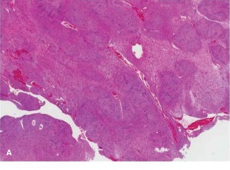
- Specimen Submission: The resected nidus and surrounding reactive bone must be sent for histopathological confirmation. The nidus typically appears as a reddish-brown, soft, granular tissue. Pathological analysis will reveal haphazardly arranged osteoid trabeculae and woven bone with prominent osteoblasts and a vascular fibrous stroma.
4. Spinal Stability Assessment and Reconstruction (if needed)
- After nidus resection, the surgeon must carefully assess the remaining bone structure for iatrogenic instability. If significant portions of the posterior elements (e.g., bilateral pars interarticularis, extensive facetectomy) have been removed, or if the lesion itself has weakened the vertebral segment, spinal instrumentation and fusion may be indicated.
- For example, if a significant pedicle lesion required substantial pedicle bone removal, or if an extensive laminectomy was necessary, pedicle screw fixation at the affected level and potentially adjacent levels, with rod placement and bone grafting, may be performed to restore stability and prevent progressive deformity. This is particularly important in younger patients or those with pre-existing spinal issues.
5. Hemostasis and Closure
- Meticulous hemostasis is achieved using bipolar cautery, bone wax, and hemostatic agents (e.g., Gelfoam, Surgicel). The epidural space must be carefully inspected for any ongoing bleeding, especially if neural decompression was performed.
- A suction drain may be placed, particularly if significant dead space exists or if extensive bleeding was encountered.
- The wound is closed in layers: deep fascia, subcutaneous tissue, and skin.
Complications & Management
While open surgical excision of osteoid osteoma is generally safe and highly effective, potential complications must be recognized and managed appropriately.
| Complication | Incidence | Salvage / Management Strategy |
|---|---|---|
| Incomplete Resection / Recurrence | 5-10% for open excision, higher for RFA if technical issues (up to 20%) | Persistent pain post-op. Re-imaging (CT scan) is crucial to confirm residual nidus. If confirmed, repeat excision or percutaneous ablation (RFA/CNA) may be necessary. Referral to a specialized orthopedic oncology center may be warranted for complex recurrences. |
| Neurological Injury | <1% (nerve root or spinal cord injury) | Paresis, paresthesia, sensory loss, or bladder/bowel dysfunction. Immediate post-operative imaging (CT/MRI) to rule out hematoma, retained foreign body, or hardware impingement (if instrumentation performed). Urgent neurosurgical consultation. High-dose methylprednisolone pulse may be considered. Surgical exploration and decompression are indicated if a compressive lesion is identified. |
| Postoperative Hematoma | 2-5% | Swelling, increased pain, signs of neurological compromise (rare, but emergent). Monitor vital signs and neurological status closely. For small, stable hematomas, observation and drain management suffice. For large, expanding, or symptomatic hematomas (especially with neural compression), urgent surgical evacuation, irrigation, and inspection for bleeding source. |
| Infection | <1-2% (superficial or deep) | Superficial: Wound erythema, drainage, pain. Managed with oral antibiotics and local wound care. Deep: Fever, severe pain, wound dehiscence, purulent discharge. Requires urgent surgical debridement, irrigation, IV antibiotics (culture-guided), and potential removal of spinal instrumentation if present and infected. |
| Spinal Instability | Varies significantly based on extent of resection and location | Progressive back pain, deformity, or neurological symptoms. Assessed pre-operatively. If significant posterior element removal (e.g., extensive facetectomy, pedicle resection) was unavoidable, prophylactic spinal fusion should be considered. If symptomatic instability develops post-op, surgical fusion (pedicle screws and rods) is typically required. |
| Persistent Pain (non-recurrence) | Varies; can be due to residual reactive sclerosis, post-operative scar tissue, or unrelated spinal issues | Pain persisting despite confirmed complete nidus resection. Conservative management with NSAIDs, physical therapy, and pain management strategies. Rule out other pain generators. Referral to a pain specialist may be beneficial. |
| Nonspecific Post-operative Pain | Common, typically resolves over weeks/months | Generalized back pain from surgical dissection. Managed with multimodal analgesia (NSAIDs, acetaminophen, short-term opioids), physical therapy, and activity modification. Educate patient on normal recovery trajectory. |
| Hardware-related Complications (if fusion) | Up to 15-20% for screw malposition; lower for symptomatic malposition | Pain, nerve irritation, or pseudarthrosis. Intraoperative O-arm/navigation minimizes malposition. If symptomatic and confirmed malposition, revision surgery for hardware repositioning or removal may be necessary. Pseudarthrosis may require revision fusion. |
| Dural Tear / CSF Leak | Rare, usually associated with laminectomy/decompression | Headache, clear wound drainage, meningismus. Intraoperative repair with sutures, patch graft (fascia, fat), and sealant. Post-op management includes strict bed rest, head elevation, and lumbar drain if necessary. If persistent, surgical re-exploration and repair. |
Post-Operative Rehabilitation Protocols
Post-operative rehabilitation following spinal osteoid osteoma excision aims to restore function, alleviate pain, prevent recurrence, and protect spinal stability. Protocols are individualized based on the extent of surgical resection, spinal stability, and whether fusion was performed.
General Principles
- Pain Management: Multimodal analgesia (NSAIDs, acetaminophen, neuropathic agents, judicious use of opioids) is crucial to facilitate early mobilization and participation in therapy.
- Wound Care: Meticulous wound care to prevent infection.
- Early Mobilization: Unless specific contraindications (e.g., significant instability, fusion requiring rigid bracing) exist, early mobilization is encouraged to prevent deconditioning, reduce the risk of thromboembolic events, and promote recovery.
Phase 1: Immediate Post-Operative (Day 0 - Week 2)
- Goals: Pain control, wound healing, prevention of complications, independent transfers and ambulation.
-
Activities:
- Day 0-1: Out of bed to chair, short walks with assistance. Emphasis on log-rolling technique for spinal precautions.
- Day 1-7: Progressive increase in ambulation distance and frequency. Avoid prolonged sitting, standing, or walking. Gentle range of motion for extremities.
- Spinal Precautions (for non-fusion cases with minimal instability risk): Avoid BLT (bending, lifting heavy objects, twisting) beyond normal activities of daily living.
- Physical Therapy: Initial assessment, patient education on posture, body mechanics, and activity modification.
Phase 2: Early Recovery (Weeks 2 - 6)
- Goals: Reduce pain, improve core stability, gentle strengthening, return to light daily activities.
-
Activities:
- Pain Management: Transition from opioids to non-opioid analgesics.
-
Physical Therapy:
- Core Stabilization: Gentle isometric contractions of transverse abdominis and multifidus muscles. Pelvic tilts.
- Postural Re-education: Emphasize neutral spine posture during sitting, standing, and walking.
- Walking Program: Gradually increase duration and intensity.
- Light Aerobics: Stationary bike or elliptical (low impact) can be introduced cautiously.
- Restrictions: Continue to avoid heavy lifting (>5-10 lbs), aggressive twisting, or high-impact activities. Avoid prolonged static postures.
Phase 3: Intermediate Strengthening (Weeks 6 - 12)
- Goals: Progressive strengthening, improved endurance, restoration of functional movement patterns.
-
Activities:
-
Physical Therapy:
- Progressive Core Strengthening: Planks, bird-dog exercises, gentle bridging, side planks.
- Spinal Mobility: Gentle, controlled lumbar and thoracic range of motion exercises (flexion, extension, rotation) if stability permits.
- Resistance Training: Introduction of light weights for upper and lower extremities, ensuring proper spinal mechanics.
- Balance and Proprioception Exercises.
- Gradual Return to Work: For sedentary jobs, return is often possible during this phase. Manual labor jobs will require longer.
-
Physical Therapy:
Phase 4: Advanced Rehabilitation & Return to Activity (Months 3+)
- Goals: Optimize strength, power, agility, and return to full work, recreational, and sporting activities.
-
Activities:
-
Physical Therapy:
- Sport-Specific Training: If applicable, gradual introduction of movements and drills relevant to desired sports or activities.
- Advanced Core and Total Body Strengthening: Incorporate functional movements.
- Impact Loading: Gradually introduce higher-impact activities as bone healing and muscle strength improve.
- Surgeon Clearance: Return to full unrestricted activities, especially contact sports or heavy labor, requires surgeon clearance based on clinical assessment and, if fusion was performed, radiographic evidence of fusion.
-
Physical Therapy:
Special Considerations
-
Spinal Fusion Cases:
- Bracing: A rigid thoracolumbosacral orthosis (TLSO) or lumbar orthosis (LSO) may be prescribed for 6-12 weeks to provide external support and promote fusion, depending on the extent of fusion and surgeon preference.
- Activity Restrictions: More stringent restrictions on bending, lifting, and twisting. Ambulation is still encouraged early.
- Longer Recovery: Overall recovery and return to full activity will be significantly longer, typically 6-12 months, pending solid fusion.
- Pediatric Patients: Monitor for resolution of scoliosis and potential for growth disturbances. Rehabilitation focuses on restoring normal play activities and participation in sports appropriate for age.
- Recurrence: Patient education on symptoms of recurrence (return of classic pain) and prompt follow-up if suspected.
Summary of Key Literature / Guidelines
The management of osteoid osteoma, particularly spinal osteoid osteoma, is guided by a robust body of literature and has seen significant evolution. Current consensus emphasizes a diagnostic approach rooted in high-resolution imaging and a treatment algorithm prioritizing minimally invasive options.
-
Diagnostic Imaging Gold Standard:
- CT scan remains the cornerstone for diagnosis and precise localization of osteoid osteoma. Its ability to clearly visualize the central lucent nidus and surrounding reactive sclerosis is unparalleled, making it indispensable for surgical planning or guiding percutaneous procedures.
- MRI plays a crucial adjunctive role, especially in spinal lesions, to assess soft tissue inflammation, potential neural compression, and to rule out other differential diagnoses (e.g., infection, stress fracture, other tumors). Bone scintigraphy, while highly sensitive, lacks specificity and anatomical detail, serving primarily as a screening tool.
-
Percutaneous Ablation as First-Line Treatment:
- Numerous studies, including large case series and systematic reviews, have established radiofrequency ablation (RFA) as the preferred first-line treatment for most accessible osteoid osteomas, including many in the spine. RFA offers high success rates (typically 80-95%), minimal invasiveness, lower morbidity, shorter recovery times, and reduced costs compared to open surgery. Cryoablation (CNA) is an effective alternative with similar success rates, particularly useful in locations where heat dissemination from RFA might be risky (e.g., near nerves).
- Success rates for RFA in spinal lesions are slightly lower than appendicular lesions, ranging from 70-90%, primarily due to anatomical complexity and proximity to neural structures.
-
Indications for Open Surgical Excision:
-
Current guidelines reserve open surgical excision for specific scenarios where percutaneous methods are contraindicated, unsafe, or have failed. Key indications include:
- Spinal lesions with actual or impending neurological compromise: When the nidus or reactive sclerosis is directly impinging on the spinal cord or nerve roots.
- Lesions in critical anatomical locations: Such as those very close to major nerves, vessels, or growth plates, where the thermal injury risk of RFA is unacceptable.
- Large or atypical lesions: Where complete ablation by percutaneous methods is uncertain, or when diagnostic uncertainty necessitates histological confirmation through open biopsy and excision.
- Progressive spinal deformity: Especially scoliosis in skeletally immature patients.
- Failure of percutaneous ablation: Persistent symptoms after one or more percutaneous attempts.
- Lesions requiring associated spinal stabilization: When the planned resection will significantly compromise spinal stability.
-
Current guidelines reserve open surgical excision for specific scenarios where percutaneous methods are contraindicated, unsafe, or have failed. Key indications include:
-
Outcomes of Open Excision:
- Open surgical excision, when complete, offers a very high success rate (>95%) for definitive cure and pain relief. Recurrence rates are low (<5%) following complete en bloc excision.
- The literature consistently demonstrates that the resolution of pain and, in pediatric cases, the correction of associated scoliosis, are excellent following complete removal of the nidus.
-
Key Literature Sources:
- Landmark studies by authors such as Assoun et al. (1997), Rosenthal et al. (1998), and subsequent reviews have detailed the efficacy and safety of RFA.
- Orthopedic oncology texts (e.g., Campbell's Operative Orthopaedics, Enneking's Musculoskeletal Tumor Surgery) provide comprehensive discussions on surgical approaches and pathological features.
- Specialized journals such as Journal of Bone and Joint Surgery (Am & Br) , Clinical Orthopaedics and Related Research , Spine , The Bone & Joint Journal , and Skeletal Radiology regularly publish research on osteoid osteoma.
- Professional societies like the American Academy of Orthopaedic Surgeons (AAOS) and the North American Spine Society (NASS) offer educational resources and guidelines for management.
In summary, while percutaneous ablation remains the primary modality for most osteoid osteomas due to its minimally invasive nature and high efficacy, open surgical excision retains a critical role for spinal lesions, those associated with neurological compromise, large or atypical presentations, or in cases where percutaneous methods have failed or are contraindicated. Precise pre-operative planning, accurate intraoperative localization, and meticulous surgical technique are paramount to achieving a successful outcome and minimizing complications.
You Might Also Like