Intramedullary Nails and External Fixators: Advanced Biomechanics, Design Principles, and Clinical Performance
Key Takeaway
Intramedullary nails (IMNs) and external fixators (ExFix) are fundamental orthopedic solutions. IMNs are load-sharing devices placed centrally, offering axial support and rotational stability via interlocking screws. ExFix provide external stabilization using pins and frames, allowing versatile reduction and fixation. Both leverage material science, geometry, and specific design principles to optimize bone healing and mechanical stability in trauma management.
Nails and External Fixators: Design, Load & Performance Explained
Introduction & Epidemiology
Intramedullary nails (IMNs) and external fixators (ExFix) represent fundamental biomechanical solutions in orthopedic traumatology and reconstructive surgery. Their evolution has been driven by a deeper understanding of fracture biology, biomechanics, and soft tissue considerations. IMNs, first extensively popularized by Küntscher for femoral shaft fractures, provide internal splinting and load-sharing capabilities, facilitating early mobilization. External fixators, with historical roots tracing back to Malgaigne, have undergone significant advancements from simple unilateral frames to complex circular and hybrid constructs, invaluable in damage control orthopedics (DCO), open fractures, and deformity correction.
Epidemiologically, long bone fractures, particularly those of the femur and tibia, represent a substantial burden, often managed definitively with IMNs due to their high union rates and functional outcomes. IMNs are the gold standard for most diaphyseal fractures of the femur, tibia, and humerus. ExFix, conversely, sees extensive application in high-energy trauma, such as Gustilo-Anderson Type II and III open fractures, pelvic ring disruptions, and periarticular injuries as either temporary or definitive stabilization. Their use in polytrauma patients for DCO has dramatically reduced morbidity and mortality associated with early total care in unstable patients. The choice between IMN and ExFix, or their sequential application, hinges on myriad factors including fracture pattern, soft tissue envelope integrity, patient physiology, and surgeon expertise, all underpinned by principles of mechanical load transmission and biological response.
Surgical Anatomy & Biomechanics
Understanding the design, load characteristics, and performance of IMNs and ExFix requires a thorough grasp of their underlying biomechanical principles and interaction with bone.
Intramedullary Nails (IMNs)
IMNs are load-sharing devices designed to bear a portion of the axial load while providing angular and rotational stability. Their efficacy stems from their central placement within the medullary canal, close to the neutral axis of the bone, minimizing bending moments.
Design Principles
- Material Science: IMNs are typically manufactured from stainless steel (e.g., 316L) or titanium alloys (e.g., Ti-6Al-4V). Titanium alloys offer superior biocompatibility, lower elastic modulus (closer to bone, reducing stress shielding), and improved fatigue resistance, though stainless steel remains a cost-effective and strong option.
-
Geometry:
- Solid vs. Cannulated: Cannulated nails allow for guidewire insertion, facilitating accurate placement and reaming. Solid nails are stronger for a given diameter but require precise reaming.
- Straight vs. Bowed: Nails for long bones (femur, tibia) are anatomically pre-bent to conform to the natural anterior femoral bow and anterior tibial bow, reducing cortical impingement and iatrogenic fracture risk.
- Diameter and Wall Thickness: A larger diameter nail generally provides greater stiffness. Wall thickness influences strength, particularly in cannulated designs.
- Interlocking Screw Holes: Strategically placed holes allow for the insertion of transverse screws to prevent shortening (static locking) and control rotation. Dynamization, allowing for controlled axial micromotion, can be achieved by removing one screw or utilizing slotted holes in certain designs, encouraging callus formation.
-
Locking Mechanisms:
- Proximal/Distal Locking: Essential for achieving rotational and axial stability, particularly in comminuted or segmental fractures. Locking screws are typically cortical screws.
- Static Locking: At least two screws per fragment, preventing axial collapse and rotation.
- Dynamic Locking: Allows for controlled axial micromotion, promoting secondary bone healing through callus formation. Often used in specific fracture patterns or delayed unions.
- Recon Nails: Specific designs for proximal femoral fractures, incorporating larger diameter lag screws or helical blades for head-neck segment fixation, providing enhanced resistance to cut-out.
Load Sharing and Performance
- Axial Load: The IMN's central location allows it to share axial compressive loads with the bone, in contrast to plates which bear load eccentrically. This promotes Wolff's Law, encouraging bone remodeling and reducing stress shielding effects often seen with plates.
- Rotational Stability: Primarily achieved by interlocking screws. The number and spacing of screws significantly influence rotational stability.
- Bending Stiffness: The nail's diameter and material properties determine its bending stiffness. A larger diameter and higher modulus material increase stiffness.
-
Mechanical Environment:
- Reaming vs. Unreaming: Reaming the medullary canal prepares a larger diameter canal, allowing for insertion of a larger, stiffer nail. This can enhance mechanical stability but may temporarily compromise endosteal blood supply. Unreamed nails preserve endosteal blood supply but provide less mechanical stability due to smaller diameter.
- Fit-and-Fill: Optimal fit-and-fill (nail diameter close to canal diameter) maximizes contact with the endosteum, enhancing stability and load transfer.
- Stress Shielding: While less pronounced than with plates, some degree of stress shielding can occur if the nail is excessively rigid, potentially delaying bone remodeling.
External Fixators (ExFix)
External fixators provide external stabilization through pins inserted into bone, connected by an external frame. They are versatile, allowing for reduction, compression, distraction, and dynamization.
Design Principles
-
Frame Configuration:
- Uniplanar: Pins in one plane, connected by one or two bars. Offers stability in one plane, limited rotational control.
- Biplanar/Multiplanar: Pins in multiple planes, connected by multiple bars, significantly increasing rigidity.
- Circular Fixators (Ilizarov, Taylor Spatial Frame): Rings connected by threaded rods, allowing for complex multiplanar deformity correction and limb lengthening. Offers superior stability and precise control.
- Hybrid Fixators: Combine pins (often into metaphysis/epiphysis) and wires (often into diaphysis) with rings and bars. Useful for complex periarticular fractures.
-
Pin Design:
- Schanz Pins: Threaded pins (full, half, or conical threads) providing purchase in cortical bone. Various diameters (e.g., 4mm, 5mm, 6mm) and tip designs (trocar, diamond). The thread design influences pull-out strength.
- Wires (Kirschner wires, Ilizarov wires): Smaller diameter, tensioned wires providing fixation, typically in circular frames.
- Hydroxylapatite (HA) Coated Pins: Designed to improve bone-pin interface strength and reduce pin loosening, though clinical efficacy is debated.
- Material: Connecting rods and clamps are commonly made of carbon fiber (radiotranslucent, lighter) or aluminum (strong, autoclavable).
- Clamps and Connecting Rods: Clamps secure pins to rods; rods connect clamps to form the frame. Their design allows for adjustability and rigidity.
Load Bearing and Performance
-
Stiffness and Rigidity:
The primary function of an ExFix is to provide a stable mechanical environment. Frame rigidity is influenced by several factors:
- Pin Diameter and Number: Larger diameter pins and more pins increase stiffness.
- Pin Spacing: Pins placed farther apart within the same bone segment increase stability.
- Pin Configuration: Placing pins in multiple planes (biplanar) provides significantly more rigidity than uniplanar.
- Distance from Bone: The closer the frame to the bone, the stiffer the construct.
- Frame Geometry: Triangle or quadrilateral frames are inherently stiffer than linear uniplanar frames.
- Connecting Rods: Larger diameter and shorter connecting rods increase stiffness. Carbon fiber rods are generally more flexible than stainless steel or aluminum for the same diameter.
-
Pin-Bone Interface:
The interface between the pin and bone is critical. Factors influencing it include:
- Thermal Necrosis: Excessive heat generation during pin insertion can cause osteonecrosis, leading to early pin loosening. Slow, controlled drilling with irrigation is essential.
- Stress Risers: Pin holes create stress risers in the bone, potentially leading to iatrogenic fracture upon removal, especially in osteopenic bone.
- Pin Tract Infection: The most common complication, affecting mechanical stability and potentially leading to osteomyelitis.
-
Mechanical Environment:
ExFix can provide a spectrum of mechanical environments:
- Static: Highly rigid frames, often for initial stabilization of unstable fractures.
- Dynamic: Controlled micromotion can be introduced by adjusting frame components (e.g., dynamizing specific rods in circular fixators, removing components in modular frames) to stimulate callus formation and accelerate healing.
- Load Bearing: ExFix bears the entire load externally, protecting the fracture site. This is crucial for initial stabilization of severely comminuted or open fractures where internal fixation is contraindicated or deferred. However, it can lead to significant stress shielding of the bone.
Indications & Contraindications
The selection of an IMN or an ExFix depends on a complex interplay of patient factors, fracture characteristics, soft tissue status, and surgeon preference.
Intramedullary Nails (IMNs)
Indications:
*
Diaphyseal Fractures:
*
Femur:
Most diaphyseal femoral fractures (Type A, B, C) in adults. Pathological fractures of the femur. Ipsilateral femoral neck and shaft fractures (often require recon nails).
*
Tibia:
Most diaphyseal tibial fractures (Type A, B, C), including segmental fractures.
*
Humerus:
Diaphyseal humeral fractures (often antegrade nailing, sometimes retrograde).
*
Metaphyseal Fractures:
* Certain subtrochanteric femoral fractures (stable patterns, often with recon nails).
* Select supracondylar femoral fractures (retrograde nailing in specific patterns).
* Select proximal tibial fractures (though often require plating due to wide metaphysis).
*
Impending Pathological Fractures:
Prophylactic stabilization in metastatic disease.
*
Malunion/Nonunion:
As a revision strategy, often with exchange nailing.
Contraindications:
*
Active Infection:
Absolute contraindication in the presence of active, uncontrolled infection at the fracture site or in the medullary canal. Relative contraindication if controlled infection (e.g., after debridement, antibiotics, and temporary ExFix).
*
Severely Comminuted Segmental Defects:
Where insufficient bone contact or large segmental loss prevents stable interlocking, or where canal cannot be adequately reamed.
*
Unacceptably Wide Medullary Canal:
May lead to inadequate fit-and-fill, requiring a larger nail potentially exceeding bone dimensions.
*
Very Proximal/Distal Fractures:
Where insufficient bone stock proximal or distal to the fracture does not allow for stable interlocking screw placement (e.g., short articular segments, very distal metaphyseal extension).
*
Skeletal Immaturity:
Risk of physeal injury, especially in distal femur or proximal tibia. Growth plate sparing techniques (e.g., elastic stable IMN or flexible nails) are used in pediatric patients.
*
Severe Open Fractures (Gustilo Type IIIC):
With significant vascular injury requiring repair and where IMN risks infection or vascular compromise. Initial ExFix is often preferred.
External Fixators (ExFix)
Indications:
*
Open Fractures:
Gustilo-Anderson Type II and III open fractures as primary definitive stabilization or as a temporizing measure for DCO.
*
Polytrauma/Damage Control Orthopedics (DCO):
For rapid, temporary stabilization of unstable long bone, pelvic, or periarticular fractures in hemodynamically unstable patients, allowing for resuscitation and definitive fixation later.
*
Pelvic Ring Injuries:
Unstable pelvic fractures (e.g., APC II/III, LC II/III, vertical shear) for rapid stabilization.
*
Periarticular Fractures:
As provisional fixation (e.g., high-energy pilon, tibial plateau, distal radius, elbow fractures) to allow soft tissue swelling to subside before definitive internal fixation. Sometimes definitive fixation for comminuted periarticular fractures in patients with poor soft tissues.
*
Severe Soft Tissue Compromise:
Fractures with extensive soft tissue damage, burns, or degloving injuries where internal fixation is deemed unsafe or premature.
*
Limb Lengthening and Deformity Correction:
Using circular or rail fixators.
*
Infected Nonunions/Osteomyelitis:
For debridement, stabilization, bone transport, and subsequent reconstruction.
*
Arthrodesis:
Joint fusion.
Contraindications:
*
Patient Non-compliance:
Relative contraindication, as pin care and frame management require active patient participation.
*
Severe Osteopenia:
Increased risk of pin loosening and pull-out.
*
Deep-seated Infection:
If pin placement cannot bypass the infected zone or would spread infection.
*
Poor Soft Tissue Coverage over Pin Sites:
Can increase pin tract infection risk.
Table: Operative vs. Non-Operative Indications for Fractures Amenable to Either
| Fracture Type | General Operative Indications (Often IMN or ExFix) | General Non-Operative Indications (Casting/Bracing) |
|---|---|---|
| Femoral Shaft | Unstable, displaced, comminuted, pathological. | Extremely rare in adults; select stable, non-displaced in children (flexible nails preferred over cast). |
| Tibial Shaft | Displaced (>50% cortical apposition loss), unstable, comminuted, open fractures, segmental. | Stable, non-displaced, closed fractures without significant soft tissue injury (e.g., boot, cast, brace). |
| Humeral Shaft | Displaced (>20° angulation, >3cm shortening, >30° rotation), segmental, pathological, open, nonunion. | Stable, non-displaced, minimally angulated closed fractures (e.g., coaptation splint, functional brace, hanging cast). |
| Pelvic Ring | Unstable (APC II/III, LC II/III, Vertical Shear), open, persistent bleeding. | Stable (APC I, LC I), minimally displaced. |
| Distal Radius | Displaced intra-articular (>2mm step-off, >2mm gap), unstable extra-articular (dorsal/volar angulation >20°, radial shortening >5mm). | Stable, minimally displaced extra-articular, elderly with low demand. |
| Pilon (Tibial Plafond) | Any displaced, intra-articular fracture, open fractures, significant soft tissue injury. | Very rare; extremely stable, non-displaced, non-articular (e.g., small avulsion). |
Pre-Operative Planning & Patient Positioning
Meticulous pre-operative planning and appropriate patient positioning are critical for successful outcomes with both IMNs and ExFix, minimizing complications and optimizing surgical efficiency.
Pre-Operative Planning
-
Patient Assessment:
- ATLS Review: For trauma patients, adherence to ATLS protocols is paramount. Assess hemodynamic stability, associated injuries, and comorbidities.
- Comorbidity Assessment: Evaluate medical comorbidities (diabetes, peripheral vascular disease, immunosuppression) that may affect bone healing or increase infection risk.
- Medication Review: Anticoagulants, anti-platelets, steroids, bisphosphonates.
-
Imaging:
- Standard Radiographs: AP and lateral views of the affected bone, including joints above and below. Traction views can aid in assessing fracture reducibility.
- Computed Tomography (CT) Scans: Essential for complex periarticular fractures (e.g., pilon, tibial plateau, femoral condyle, pelvic), providing detailed 3D anatomy, fracture mapping, and assessment of articular involvement.
- Magnetic Resonance Imaging (MRI): Seldom used for acute fracture planning unless soft tissue injury (ligamentous, meniscal) is suspected and impacts management.
-
Templating:
-
Intramedullary Nails:
- Length: Measure the contralateral uninjured bone on AP radiographs (true length may require magnification adjustment or use of ruler). Alternative: measure from greater trochanter to knee joint line.
- Diameter: Measure the medullary canal at the narrowest isthmus on AP and lateral views. Plan for appropriate reaming.
- Curvature: Select a nail with appropriate pre-bend to match anatomical curvature (e.g., femoral bow) to avoid cortical impingement.
- Locking Screws: Pre-plan entry and exit points for interlocking screws to ensure adequate bone purchase.
-
External Fixators:
- Pin Placement Zones: Identify safe corridors for pin insertion, avoiding neurovascular structures, joint capsules, and fracture lines. Utilize anatomical landmarks and fluoroscopy.
- Frame Configuration: Sketch the intended frame (e.g., uniplanar, biplanar, delta, circular), anticipating stability requirements based on fracture pattern and patient needs.
- Reduction Strategy: Plan how fracture reduction will be achieved (manual, joystick pins, ligamentotaxis, intra-operative ExFix adjustment).
-
Intramedullary Nails:
-
Implant Selection:
- Select the appropriate implant system (manufacturer, nail type, pin size) based on fracture characteristics, surgeon familiarity, and hospital inventory.
- Ensure all necessary instruments, jigs, and accessories are available.
Patient Positioning
Correct patient positioning is crucial for surgical access, fracture reduction, and fluoroscopic visualization.
-
General Considerations:
- Anesthesia: General anesthesia is typically required. Regional blocks can be adjuncts for post-operative pain control.
- Fluoroscopy: Position the C-arm for unobstructed AP and lateral views of the entire fracture and relevant joint segments. Ensure the operating table allows free movement of the C-arm.
- Patient Stability: Secure the patient to the table to prevent movement during manipulation or instrumentation.
-
Intramedullary Nailing:
-
Femoral Nailing:
- Supine on Traction Table: Standard for most femoral shaft fractures. Allows for closed reduction via continuous traction, controlled rotation, and contralateral limb access. Requires specific traction boots and perineal post.
- Supine on Radiolucent Table (Lateral Decubitus for Entry): Alternative for cases where a traction table is not suitable, or for more proximal fractures requiring greater trochanteric entry (often requires an assistant to hold reduction).
- Lateral Decubitus: Less common, but can provide optimal access for trochanteric entry portal.
- Tibial Nailing: Typically supine on a radiolucent table. Knee flexed (often to 90 degrees) with support under the thigh to allow access to the proximal tibia entry portal and for gravity-assisted reduction of apex anterior angulation.
-
Humeral Nailing:
- Supine: For antegrade nailing, often with beach chair position or modified supine with arm abducted.
- Lateral Decubitus: For retrograde nailing, or for antegrade approaches where lateral access is preferred.
-
Femoral Nailing:
-
External Fixation:
- Supine: Most common for long bone fractures (femur, tibia, humerus) and pelvis. Allows for bilateral limb assessment and easier C-arm access.
- Semi-Fowler/Beach Chair: For upper extremity frames.
- Lateral Decubitus: Less common, but may be used for specific pelvic pin placement.
- Traction: Often applied manually or via a traction table to aid in preliminary reduction before frame assembly.
Detailed Surgical Approach / Technique
The surgical technique for IMNs and ExFix involves distinct principles of access, reduction, and fixation, each with specific considerations for biomechanical stability and biological preservation. This section will generalize the principles applicable across common fracture sites.
Intramedullary Nailing - General Principles
IMN insertion generally follows a systematic sequence: patient positioning, entry portal creation, fracture reduction, reaming (if chosen), nail insertion, and proximal/distal locking.
-
Anatomical Entry Portal:
-
Femur:
- Piriformis Fossa: Traditional entry point, through gluteus medius, just medial to the greater trochanteric tip. Requires careful protection of the superior gluteal neurovascular bundle.
- Greater Trochanteric Tip: Increasingly popular, through the vastus lateralis and gluteus medius. Less risk to superior gluteal neurovascular bundle, potentially easier access. May be associated with higher rates of hip pain.
- Pear-shaped Reamer: Used to widen the entry portal to match the nail's curvature and prevent iatrogenic fracture.
-
Tibia:
- Patellar Tendon Splitting: Incision directly over the patellar tendon, splitting it longitudinally. Entry just medial to the mid-patellar tendon, at or slightly proximal to the tibial tubercle. Risk of anterior knee pain.
- Medial/Lateral Parapatellar: Incision adjacent to the patellar tendon, either medially or laterally, retracting the tendon.
- Suprapatellar/Transpatellar: Entry through the quadriceps tendon and patella, allowing for nail insertion with the knee in less flexion. Reduces risk of apex anterior malreduction in some cases but introduces patellar violation.
-
Humerus:
- Antegrade: Deltoid splitting approach. Entry through the greater tuberosity, just medial to the supraspinatus insertion.
- Retrograde: Via olecranon fossa (trans-olecranon or paratricipital) for more distal fractures.
- Internervous Planes: While specific entry portals traverse different planes, the principle is to minimize muscle stripping and neurological injury. For example, the piriformis entry involves splitting gluteus medius fibers, respecting its innervation.
-
Femur:
-
Fracture Reduction:
- Indirect Reduction: The cornerstone of IMN. Achieved via continuous traction, external manipulation (manual, external fixator as a reduction aid), joystick pins, or cerclage wires (sparingly, considering blood supply). Avoid opening the fracture site unless absolutely necessary (e.g., irreducible).
- Fluoroscopic Control: Constant monitoring in AP and lateral planes to confirm alignment, length, and rotation.
- Rotational Alignment: Clinical assessment of rotation (e.g., foot rotation relative to patella for tibia, arm rotation for humerus) is crucial and often more accurate than fluoroscopy for the femur.
-
Reaming vs. Unreaming:
- Reaming: Gradual increase in reamer size (0.5mm increments) to create a cylindrical canal slightly larger than the chosen nail diameter. Provides a larger, stiffer nail and better bone-nail contact, enhancing stability. Potential compromise of endosteal blood supply, but often transient.
- Unreaming: Preserves endosteal blood supply, potentially beneficial in open fractures or compromised soft tissues. However, limits nail diameter and stiffness.
-
Nail Insertion:
- Carefully insert the nail over the guidewire, avoiding excessive force to prevent iatrogenic fracture or malreduction.
- Monitor position with fluoroscopy. Ensure the nail passes the fracture site without difficulty.
-
Proximal and Distal Locking:
- Proximal Locking: Typically performed first. Utilizes a targeting jig or freehand technique with fluoroscopy. At least two screws per fragment are desired for rotational stability.
- Distal Locking: Often the most challenging due to free-hand technique without a jig. Requires accurate fluoroscopic targeting (perfect circles, "gun barrel" view). Two screws are preferred for complete rotational control.
- Static vs. Dynamic: Static locking prevents any shortening or rotation. Dynamic locking allows for controlled axial micromotion, which can stimulate healing, often achieved by removing one screw or using a slotted hole.
External Fixation - General Principles
ExFix application emphasizes rapid, reproducible stabilization with minimal soft tissue stripping, prioritizing pin placement in safe zones.
-
Pin Placement and Safe Zones:
- General Principle: Place pins as far as possible from the fracture site, perpendicular to the bone, through minimal soft tissue.
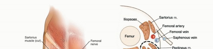
- Safe Zones: Strict adherence to known anatomical safe corridors is paramount to avoid neurovascular structures (e.g., superficial peroneal nerve in the distal tibia, radial nerve in the humerus, femoral artery/nerve in the proximal femur, sciatic nerve in the pelvis).
- Drilling Technique: Make small skin incisions, spread soft tissues to bone, pre-drill with a drill bit 0.5-1.0mm smaller than the pin diameter, use a drill guide, and cool with saline to prevent thermal osteonecrosis. Manual insertion of self-drilling/self-tapping pins may be preferred by some.
- Cortex Engagement: Ensure bicortical purchase for optimal pull-out strength, especially in the diaphysis.
-
Frame Assembly:
- Initial Pins: Insert the most critical pins first (e.g., supra-acetabular for pelvis, most stable fragment for long bones).
- Connectors/Clamps: Attach clamps to the pins, then connect with appropriate rods.
- Frame Construction: Build a frame configuration that provides adequate stability for the fracture type (e.g., uniplanar for provisional, biplanar/delta for definitive).
-
Rigidity:
Optimize frame rigidity by:
- Increasing pin diameter and number.
- Increasing pin spread within a bone segment.
- Placing pins in multiple planes.
- Reducing frame-to-bone distance.
- Using larger diameter, shorter connecting rods.
-
Fracture Reduction:
- Manual Reduction: Often achieved manually by applying traction and aligning segments.
- Joystick Pins: Temporary pins inserted into fragments can be used to manipulate and align them.
- Articulation: Many modern fixator clamps allow for fine-tuning of reduction in multiple planes after initial frame assembly.
- Fluoroscopic Control: Continuously monitor reduction in AP and lateral views.
-
Frame Adjustments and Dynamization:
- Dynamization: For specific fracture types (e.g., comminuted tibia), the frame can be dynamized by removing a connecting rod or loosening specific clamps, allowing controlled axial micromotion to stimulate healing. This is typically done after initial callus formation.
- Deformity Correction: Circular fixators allow for complex multiplanar deformity correction and limb lengthening through precise adjustments of threaded rods.
Complications & Management
Complications associated with IMNs and ExFix can be significant, necessitating a thorough understanding of their incidence, prevention, and salvage strategies.
Intramedullary Nailing Complications
| Complication | Incidence (Approx.) | Salvage Strategies |
|---|---|---|
| Nonunion / Delayed Union | 2-10% | Dynamization, exchange nailing (larger diameter nail), bone grafting, plate augmentation, revision to external fixation. |
| Malunion (Rotation/Angulation) | 5-15% (rotational) | Corrective osteotomy and refixation (often with plating or another nail). |
| Infection (Superficial/Deep) | 1-5% (open fractures higher) | Debridement, irrigation, antibiotics. Retain nail if stable and limited infection; exchange nailing with antibiotic-coated nail; explantation and staged reconstruction (ExFix, antibiotics, then re-nailing). |
| Hardware Failure (Nail/Screw Breakage) | <2% (often secondary to nonunion) | Address underlying nonunion, explantation, revision nailing/plating. |
| Iatrogenic Fracture | 1-3% | Extend fixation, specific plating, careful reaming and nail insertion. |
| Nerve/Vascular Injury | <1% | Exploration, repair, neurolysis. |
| Fat Embolism Syndrome | Rare but severe | Supportive care (ventilation), prevention (careful reaming). |
| Entry Portal Pain (Femur) | 10-25% (greater trochanteric) | Hardware removal (nail/screws), physical therapy, corticosteroid injection. |
| Distraction at Fracture Site | <5% | Dynamization, compression via nail (if possible), bone grafting. |
| Compartment Syndrome | <1% | Emergency fasciotomy. |
External Fixation Complications
| Complication | Incidence (Approx.) | Salvage Strategies |
|---|---|---|
| Pin Tract Infection | 5-50% (most common) | Meticulous pin care (daily cleaning). Oral antibiotics (superficial). Debridement, IV antibiotics, pin removal/replacement (deep/abscess). |
| Loose Pins / Pin Pull-out | 5-20% | Pin replacement in new bone, frame revision, consider definitive internal fixation if appropriate. |
| Neurovascular Injury | <1% | Careful pin placement in safe zones. Exploration, repair if identified. |
| Nonunion / Delayed Union | 5-15% | Frame dynamization, bone grafting, conversion to internal fixation. |
| Malunion | 5-10% | Frame adjustment (if possible), corrective osteotomy and refixation. |
| Compartment Syndrome | <1% | Emergency fasciotomy (monitor closely, ExFix may mask swelling). |
| Frame Impingement/Patient Discomfort | Common | Frame adjustment, padding. |
| Stress Risers / Fracture at Pin Site | <2% (upon removal) | Protection after frame removal (e.g., cast, brace), manage as new fracture. |
| Joint Stiffness | Variable | Early ROM, physical therapy, frame modification to allow joint motion. |
Post-Operative Rehabilitation Protocols
Post-operative rehabilitation is integral to achieving optimal functional outcomes following IMN or ExFix. Protocols are tailored to the specific implant, fracture stability, bone healing progression, and patient factors.
General Principles
- Pain Management: Multimodal analgesia to facilitate early mobilization and rehabilitation.
- Early Mobilization: As permitted by fracture stability and implant, to prevent complications such as stiffness, deep vein thrombosis (DVT), and muscle atrophy.
- Weight-Bearing Progression: Based on radiographic signs of healing, fracture pattern, and stability of the implant.
- Wound/Pin Site Care: Crucial for infection prevention.
Intramedullary Nailing
Due to their load-sharing nature and inherent stability, IMNs typically allow for earlier weight-bearing and functional rehabilitation.
-
Weight-Bearing:
- Femur: Full weight-bearing as tolerated is often initiated immediately post-op for stable diaphyseal fractures, especially with statically locked nails. For very comminuted or segmental fractures, partial weight-bearing progressing to full over 6-12 weeks based on callus formation.
- Tibia: Partial weight-bearing with crutches or walker is often initiated early (e.g., touch-down weight-bearing) and progressed to full weight-bearing by 6-12 weeks, depending on fracture comminution and signs of union. Statically locked nails generally allow earlier progression.
- Humerus: Typically non-weight-bearing (upper extremity), but early active and passive range of motion (ROM) is encouraged for the shoulder and elbow.
-
Range of Motion (ROM):
- Adjacent Joints: Early active and passive ROM exercises for joints proximal and distal to the fracture (e.g., hip/knee for femur, ankle/knee for tibia, shoulder/elbow for humerus) to prevent stiffness.
- Gradual Progression: Increase intensity and load as fracture healing progresses.
-
Strengthening:
- Isometrics can begin early. Progressive resistive exercises are introduced once sufficient bone healing is evident radiographically.
- Focus on core stability and strength of muscles surrounding the injured bone.
-
Fracture Healing Assessment:
- Regular radiographic follow-up (AP/Lateral) at 4-6 week intervals to assess callus formation, bridging, and cortical integrity. Clinical assessment of pain and stability.
- Nail dynamization may be considered for delayed unions if statically locked.
-
Hardware Removal:
- Generally not routine unless symptomatic (e.g., prominent screw heads, trochanteric pain, infection).
- Typically performed 1-2 years post-op, once bone is fully consolidated.
External Fixation
Rehabilitation with ExFix often involves a more cautious approach, especially regarding weight-bearing, due to the external nature of the frame and potential for pin-bone interface issues.
-
Weight-Bearing:
- Initial Phase: Often non-weight-bearing or touch-down weight-bearing for 4-8 weeks, particularly for long bone and periarticular fractures.
- Progression: Gradual progression to partial and then full weight-bearing based on radiographic evidence of callus formation, clinical stability, and the ability to dynamize the frame. This is crucial in deformity correction (e.g., Ilizarov) where controlled axial load stimulates osteogenesis.
-
Pin Site Care:
- Daily cleaning with sterile saline or antiseptic solution.
- Monitor for signs of infection (erythema, swelling, drainage, pain).
- Patient education on self-care is vital.
-
Range of Motion (ROM):
- Early active and passive ROM for unrestricted joints. The frame may limit motion of adjacent joints, requiring compensatory exercises.
- For circular frames, joint movement may be incorporated into the frame design (e.g., hinges).
-
Strengthening:
- Isometrics for muscles within the affected segment.
- Progressive strengthening exercises as healing progresses and frame stability allows.
-
Frame Adjustment/Dynamization:
- As healing progresses, the frame may be adjusted to introduce controlled axial micromotion (dynamization) to stimulate callus formation, particularly in limb lengthening and nonunion management.
- This requires careful instruction and patient compliance.
-
Frame Removal:
- Timing is critical, based on robust radiographic and clinical evidence of union. Premature removal can lead to re-fracture.
- Post-removal, the limb often requires protection (e.g., cast, brace) for several weeks due to stress risers at pin sites and initial weakness of the healed bone. Gradual return to full activity.
Summary of Key Literature / Guidelines
The landscape of IMN and ExFix is continually refined by robust clinical research and guidelines, shaping best practices in orthopedic trauma.
- Reamed vs. Unreamed Nailing: Meta-analyses and prospective studies (e.g., the Canadian Orthopaedic Trauma Society (COTS) studies) have generally shown no significant difference in rates of nonunion or infection for closed tibial shaft fractures, but reamed nails often allow for a larger, stiffer implant and may accelerate union. For open fractures, unreamed nails were historically preferred to preserve endosteal blood flow, though modern reaming techniques with low-pressure, high-volume irrigation have minimized this concern, making reamed nails acceptable in many Gustilo I/II open fractures.
- Timing of Definitive Fixation in Polytrauma: The concept of "Damage Control Orthopedics" (DCO) versus "Early Total Care" (ETC) is well-established. DCO, typically involving ExFix for initial stabilization, is indicated for hemodynamically unstable polytrauma patients (e.g., ISS > 40, base deficit > 6, lactate > 2.5, hypothermia, coagulopathy). ETC (often IMN) is preferred for stable patients, reducing the risks associated with prolonged ExFix.
- Open Fracture Management: The Gustilo-Anderson classification guides management. Type I/II open fractures are often amenable to early definitive internal fixation (IMN or plate) after debridement and antibiotics. Type III fractures, especially IIIB/C with significant soft tissue loss and vascular injury, usually require initial DCO with ExFix, serial debridement, and plastic surgery involvement, with delayed definitive fixation. Guidelines emphasize emergent debridement within 6 hours, broad-spectrum antibiotics, and tetanus prophylaxis.
- Femoral Shaft Fractures: IMN is the gold standard for nearly all adult diaphyseal femoral fractures, demonstrating superior union rates, faster rehabilitation, and fewer complications compared to plating or conservative management. Retrograde IMN is a viable option for distal femur fractures and polytrauma patients with associated ipsilateral lower extremity injuries.
- Tibia Shaft Fractures: IMN is the preferred definitive treatment for most unstable diaphyseal tibial fractures. Careful attention to rotation and alignment is paramount.
- Proximal Femoral Fractures (Subtrochanteric): Recon nails (cephalomedullary nails) have largely supplanted plates for many subtrochanteric fractures, offering biomechanical advantages by load-sharing and providing better fixation in osteopenic bone, reducing failure rates.
- Circular External Fixators: The Ilizarov method revolutionized the management of complex nonunions, osteomyelitis, and limb length discrepancies. The Taylor Spatial Frame (TSF) provides highly precise, computer-assisted deformity correction, expanding on Ilizarov's principles. These methods rely on controlled osteogenesis and specific biomechanical frame configurations.
- Evolution of Pin Design: Research continues into pin coatings (e.g., HA, antibiotic), material science, and insertion techniques to minimize pin tract infection and optimize bone-pin interface longevity, although definitive superiority of novel designs remains under active investigation.
These guidelines and evidence-based practices underpin the rational application and continuous advancement of intramedullary nails and external fixators in modern orthopedic surgery.
You Might Also Like