Comprehensive Orthopedic Review: Surgical Anatomy, Biomechanics, and Interventions for Knee Pain
Key Takeaway
Effective surgical management of knee pain requires a deep understanding of its epidemiology, intricate surgical anatomy (bony, ligamentous, meniscal, musculotendinous, neurovascular), and complex biomechanics. This knowledge is crucial for orthopedic surgeons, residents, and students to judiciously apply operative and non-operative strategies, ensuring successful outcomes and patient relief.
Effective Treatment of Knee Pain: Your Path to Lasting Relief
Introduction & Epidemiology
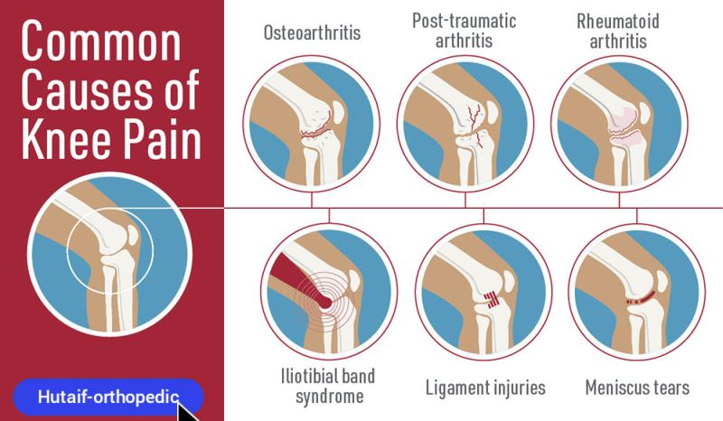
Knee pain represents a ubiquitous musculoskeletal complaint globally, profoundly impacting patient quality of life and healthcare systems. The American Academy of Orthopaedic Surgeons (AAOS) reports that approximately one-third of the American population experiences knee pain at some juncture in their lives. This prevalence spans all age demographics, from pediatric and adolescent populations affected by growth-related conditions or sports injuries to the elderly contending with degenerative processes. Etiologies are diverse, encompassing acute traumatic injuries, chronic degenerative conditions, inflammatory arthropathies, and overuse syndromes. A comprehensive understanding of the underlying pathology, coupled with judicious application of non-operative and operative strategies, is paramount for effective management. This academic review aims to provide a high-yield reference guide for orthopedic surgeons, residents, and medical students, elucidating the surgical anatomy, biomechanics, indications, techniques, complications, and rehabilitation protocols pertinent to common operative interventions for knee pain.
Surgical Anatomy & Biomechanics
A profound understanding of the knee's intricate anatomy and complex biomechanics is foundational to successful surgical intervention.
Bony Anatomy
The knee joint comprises the articulation of the distal femur, proximal tibia, and patella.
*
Distal Femur:
Features medial and lateral condyles, separated posteriorly by the intercondylar notch. The condyles articulate with the tibial plateau and, anteriorly, form the trochlear groove for patellar articulation.
*
Proximal Tibia:
Comprises the medial and lateral tibial plateaus, separated by the intercondylar eminence (tibial spines). The plateaus are typically slightly sloped posteriorly.
*
Patella:
A sesamoid bone embedded within the quadriceps tendon, articulating with the femoral trochlea. Its posterior surface is covered by thick articular cartilage.
Ligamentous Anatomy
The knee's stability is maintained by a complex network of intra-articular and extra-articular ligaments.
*
Cruciate Ligaments:
*
Anterior Cruciate Ligament (ACL):
Originates from the posterior aspect of the lateral femoral condyle and inserts into the anteromedial aspect of the tibial intercondylar eminence. Primary restraint to anterior tibial translation and secondary restraint to internal rotation.
*
Posterior Cruciate Ligament (PCL):
Originates from the anterior aspect of the medial femoral condyle and inserts into the posterior aspect of the tibial intercondylar eminence. Primary restraint to posterior tibial translation.
*
Collateral Ligaments:
*
Medial Collateral Ligament (MCL):
Superficial and deep layers. Originates from the medial femoral epicondyle and inserts onto the medial aspect of the tibia. Primary restraint to valgus stress.
*
Lateral Collateral Ligament (LCL):
Cord-like structure, originating from the lateral femoral epicondyle and inserting onto the fibular head. Primary restraint to varus stress.
*
Posterolateral Corner (PLC):
A complex of structures including the LCL, popliteofibular ligament, popliteus tendon, and arcuate complex. Critical for resisting varus, external rotation, and posterior translation.
*
Patellofemoral Ligaments:
Primarily the Medial Patellofemoral Ligament (MPFL), originating from the medial femoral epicondyle and inserting onto the superomedial patella. Crucial for patellar stability, resisting lateral dislocation.
Meniscal Anatomy
The medial and lateral menisci are C-shaped (medial) and O-shaped (lateral) fibrocartilaginous structures, acting as shock absorbers, load distributors, and secondary stabilizers.
*
Attachments:
Horns attach to the tibial plateau. Periphery attaches to the joint capsule. Medial meniscus is more rigidly attached (coronary ligaments) and intimately connected to the deep MCL. Lateral meniscus is more mobile, separated from the LCL by the popliteus tendon.
*
Vascularity:
The outer 10-30% (red-red zone) is vascularized by the genicular arteries, supporting repair. The inner 70-90% (white-white zone) is avascular, posing challenges for healing. The transition is the red-white zone.
Musculotendinous Anatomy
- Extensor Mechanism: Quadriceps femoris muscle, quadriceps tendon, patella, patellar tendon (ligament) inserting onto the tibial tuberosity.
- Flexors/Rotators: Hamstrings (semimembranosus, semitendinosus, biceps femoris), gastrocnemius, popliteus.
Neurovascular Anatomy
The popliteal fossa contains the vital neurovascular bundle.
*
Artery:
Popliteal artery (continuation of superficial femoral artery), branching into genicular arteries.
*
Vein:
Popliteal vein.
*
Nerves:
Tibial nerve (medial), common peroneal nerve (lateral), saphenous nerve (sensory, medial knee). Surgical approaches must meticulously protect these structures.
Biomechanics
- Kinematics: The knee exhibits a combination of rolling and gliding movements during flexion and extension. The "screw-home mechanism" involves terminal external rotation of the tibia on the femur during the last degrees of extension, locking the knee.
- Load Bearing: The menisci distribute axial loads across the tibial plateau, reducing contact stress. Meniscectomy significantly increases contact pressure and predisposes to osteoarthritis.
- Stability: Ligaments provide static stability, while muscles (quadriceps, hamstrings) provide dynamic stability. Proprioception, mediated by mechanoreceptors within ligaments and joint capsule, is critical for coordinated movement and injury prevention.
- Patellofemoral Tracking: Complex interplay of bony morphology (trochlear depth), ligamentous restraints (MPFL), and muscle forces (vastus medialis obliquus). Maltracking can lead to pain and instability.
Indications & Contraindications
The decision for operative intervention is predicated on a thorough clinical assessment, imaging correlation, and failure of appropriate non-operative management.
General Principles
Surgical indications typically include:
* Progressive, unremitting pain or functional limitation refractory to non-operative treatment (e.g., physical therapy, NSAIDs, activity modification, injections).
* Mechanical symptoms (locking, catching, giving-way) indicative of internal derangement or instability.
* Radiographic evidence of significant structural damage (e.g., end-stage arthritis, unstable fracture).
* Potential for further joint damage or accelerated degeneration if untreated.
General contraindications include active systemic or local infection, severe uncontrolled medical comorbidities precluding safe anesthesia or recovery, extreme obesity (relative), severe peripheral vascular disease, and non-compliance with post-operative protocols.
Specific Conditions & Procedures
| Indication Category | Operative Management | Non-Operative Management |
|---|---|---|
| Meniscal Pathology | Symptomatic meniscal tears causing mechanical symptoms (locking, catching, effusions); tears in vascularized zones (red-red, red-white) for repair; unstable tears; radial/root tears (repair preferred); displaced bucket-handle tears. | Asymptomatic tears; stable tears in avascular zones without mechanical symptoms; degenerative tears responsive to conservative care (RICE, NSAIDs, PT). |
| Ligamentous Instability (e.g., ACL) | Functional instability, recurrent giving-way, high-demand athletes requiring return to pivoting sports, combined ligamentous injuries, significant knee laxity on examination. | Sedentary patients, stable knee despite rupture, willingness to modify activity, successful functional rehabilitation, low-impact activity preference. |
| Osteoarthritis (OA) | Severe, end-stage pain and functional impairment unresponsive to comprehensive non-operative treatment (medications, PT, injections, bracing); significant radiographic changes (Kellgren-Lawrence Grade III-IV) with structural deformity. | Mild-moderate symptoms, early-stage arthritis, patients amenable to lifestyle modification, weight loss, NSAIDs, physical therapy, intra-articular injections (corticosteroid, hyaluronic acid, PRP). |
| Patellofemoral Instability | Recurrent patellar dislocations despite diligent conservative management; significant patellofemoral malalignment with contributing factors (e.g., trochlear dysplasia, tibial tubercle-trochlear groove distance >20mm); associated chondral injury. | First-time dislocation, stable patella after reduction, amenable to physical therapy focusing on quadriceps strengthening, VMO activation, gait retraining; patellar stabilizing brace. |
| Angular Deformity (e.g., Varus/Valgus) | Symptomatic unicompartmental arthritis in younger, active patients with correctable deformity (e.g., high tibial osteotomy for medial compartment OA with varus deformity; distal femoral osteotomy for lateral compartment OA with valgus deformity) to delay arthroplasty. | Asymptomatic deformity, mild pain managed with conservative measures, bicompartmental or tricompartmental OA (arthroplasty indicated). |
Pre-Operative Planning & Patient Positioning
Meticulous pre-operative planning and appropriate patient positioning are critical for minimizing complications and optimizing outcomes.
Pre-Operative Assessment
- History and Physical Examination: A comprehensive medical history, including comorbidities, medications, and previous surgeries. A detailed knee examination assesses gait, alignment, range of motion (ROM), effusion, tenderness, stability (Lachman, pivot shift, anterior/posterior drawer, varus/valgus stress tests), patellar tracking, and neurovascular status.
-
Imaging Studies:
- Radiographs: Standard series includes weight-bearing AP, lateral, and Merchant/skyline views of the patella. Long-leg alignment views are essential for assessing mechanical axis and planning osteotomies or arthroplasty.
- Magnetic Resonance Imaging (MRI): Gold standard for soft tissue evaluation (menisci, ligaments, cartilage, bone edema), crucial for internal derangement.
- Computed Tomography (CT): Useful for complex fractures, detailed bony morphology, trochlear dysplasia assessment, and pre-operative planning for revision arthroplasty or patient-specific instrumentation.
- Bone Scintigraphy: Rarely used but may identify occult infections or metabolic activity.
- Templating: For arthroplasty, pre-operative templating using radiographs or CT scans helps determine implant size, guide component positioning, and predict limb alignment.
- Medical Optimization: Address and optimize any underlying medical conditions (e.g., diabetes, cardiac disease) to reduce surgical risk. Pre-operative physiotherapy and education can enhance recovery.
Patient Positioning
-
Supine Position:
Most common for knee arthroscopy, total knee arthroplasty (TKA), and unicompartmental knee arthroplasty (UKA).
- Lower Extremity Preparation: The operative leg is prepared circumferentially from the hip to the foot. A tourniquet is typically applied to the proximal thigh to provide a bloodless field, inflated to supra-systolic pressures (e.g., 250-300 mmHg) or according to limb occlusion pressure.
- Leg Holder/Positioner: For arthroscopy, the knee can be placed in a leg holder (e.g., knee stabilizer with a well-padded boot) or suspended via a traction tower (e.g., for ligament reconstruction) to facilitate valgus/varus stress and flexion/extension. The contralateral leg is often placed in a well-padded stirrup or frog-leg position.
- Draping: Standard sterile knee draping, allowing for manipulation of the limb and access to graft harvest sites if needed.
- Anesthesia: General anesthesia, regional anesthesia (spinal/epidural), or a combination. Peripheral nerve blocks (femoral, adductor canal, sciatic, popliteal) are frequently employed for post-operative analgesia.
- Prophylaxis: Deep vein thrombosis (DVT) prophylaxis is crucial, including mechanical (intermittent pneumatic compression devices) and often chemical (anticoagulants) measures. Perioperative antibiotics are administered according to institutional protocols.
Detailed Surgical Approach / Technique
This section will detail commonly performed surgical interventions for knee pathology.
1. Arthroscopic Meniscectomy and Meniscal Repair
General Principles of Knee Arthroscopy
- Portals: The standard portals are anterolateral (lateral to patellar tendon, superior to lateral meniscus) and anteromedial (medial to patellar tendon, superior to medial meniscus). Accessory portals (e.g., superomedial, superolateral, posteromedial, posterolateral) may be used for specific pathologies or visualization.
- Diagnostic Arthroscopy: A systematic evaluation of the knee joint components is performed, including the patellofemoral joint, medial compartment, lateral compartment, and intercondylar notch. Chondral surfaces, menisci, and ligaments are inspected.
- Fluid Management: Continuous inflow via gravity or pump, maintaining joint distension and clear visualization.
Partial Meniscectomy
- Indications: Unstable meniscal tears in the avascular zone, degenerative tears causing mechanical symptoms, or tears deemed irreparable.
-
Technique:
- Visualization: Arthroscope in one portal, instrument (e.g., probe, basket forceps, shaver) in the other.
- Assessment: The tear is probed to assess its stability, location, and pattern.
- Resection: Unstable or irreparable fragments are meticulously resected using basket forceps and shavers. The goal is to remove only the damaged tissue while preserving as much functional meniscal tissue as possible to minimize increased contact pressures.
- Sculpting: The remaining meniscal rim is contoured smoothly, avoiding ragged edges that could cause persistent symptoms.
Meniscal Repair
- Indications: Longitudinal tears, radial tears, root avulsions, or bucket-handle tears located in the vascularized (red-red, red-white) zones in younger, active patients.
-
Technique (Example: All-Inside Repair):
- Preparation: The torn edges of the meniscus and the capsular rim are débrided and rasped to stimulate a healing response and create a fresh bleeding edge.
- Tear Reduction: The torn meniscal fragment is reduced into its anatomical position using a probe.
- Suture Device Deployment: An all-inside meniscal repair device (e.g., Fast-Fix, Meniscal Cinch) is deployed across the tear, placing sutures perpendicularly. Multiple sutures are typically placed 3-5 mm apart to achieve a stable repair.
- Knot Tying (if applicable): For some devices, the knots are tied intra-articularly or extra-articularly.
- Stability Check: The repair is gently probed and stressed to ensure stability.
-
Other Techniques:
- Inside-Out Repair: Requires a posterior incision and careful neurovascular protection to retrieve sutures passed from inside the joint to the capsule. Often used for posterior horn tears.
- Outside-In Repair: Sutures passed from outside the knee, through the meniscus, and retrieved inside the joint with a needle passer. Useful for anterior horn tears.
2. Arthroscopic Anterior Cruciate Ligament (ACL) Reconstruction
Graft Choices
-
Autograft (Preferred):
- Bone-Patellar Tendon-Bone (BPTB): Strong, rigid fixation, good osteointegration. Potential for patellofemoral pain, kneeling pain, patellar fracture.
- Hamstrings (Semitendinosus/Gracilis - ST/G): Less donor site morbidity, potentially faster rehabilitation. Risk of hamstring weakness, graft stretching.
- Quadriceps Tendon (QT): Increasingly popular, strong, less patellofemoral pain than BPTB.
- Allograft: Avoids donor site morbidity, readily available. Concerns include disease transmission (extremely rare), slower incorporation, and potentially higher failure rates in younger, active patients.
Surgical Steps (using ST/G autograft as an example)
-
Graft Harvest:
- A small incision is made over the pes anserinus insertion.
- The sartorius fascia is incised, and the semitendinosus and gracilis tendons are identified.
- A tendon stripper is used to harvest the tendons proximally.
- The harvested tendons are prepared on the back table, folded into a quadruple-strand graft, and whip-stitched at the ends. The length and diameter are measured.
- Portal Placement and Diagnostic Arthroscopy: Standard anterolateral and anteromedial portals.
- Notch Preparation: Any ACL stump remnants or hypertrophic synovium in the intercondylar notch are débrided to provide clear visualization and prevent impingement.
-
Femoral Tunnel Drilling:
- Anteromedial (AM) Portal Technique: The arthroscope is placed in the anterolateral portal. A guide pin is inserted through the AM portal, aiming for the anatomical footprint of the ACL on the lateral femoral condyle (typically at the 10:30 position in the right knee, 1:30 in the left, midway between anterior and posterior cartilage margins). An appropriate reamer drills the femoral tunnel.
- Transtibial Technique (less common for anatomical reconstruction): A guide pin is drilled from the tibia, through the ACL footprint, and then into the femur. Offers less independent control of femoral tunnel position.
-
Tibial Tunnel Drilling:
- A tibial guide is used to place a guide pin on the tibial plateau, aiming for the center of the ACL footprint (just anterior to the PCL, midway between the tibial spines). The pin should exit the tibia approximately 2-3 cm distal to the joint line, just medial to the tibial tuberosity.
- The tibial tunnel is reamed to the appropriate diameter.
- Graft Passage: A suture loop is passed through the tibial and femoral tunnels. The prepared graft is then pulled through the tunnels into the joint.
-
Graft Fixation:
- Femoral Fixation: Cortical button (e.g., EndoButton) is often used, flipped over the lateral femoral cortex. Interference screws (titanium or bioabsorbable) can also be used, particularly in the AM portal technique.
- Tibial Fixation: An interference screw is typically placed alongside the graft in the tibial tunnel. Further fixation may include a post screw or staple.
- Tensioning: The graft is tensioned with the knee in 20-30 degrees of flexion, and internal rotation, to restore stability without overtightening.
- Closure: Portals are closed with sterile strips or sutures.
3. Total Knee Arthroplasty (TKA)
Incision and Exposure
- Incision: A midline longitudinal skin incision, typically extending from 3-4 cm proximal to the patella to the tibial tuberosity.
- Arthrotomy: Most commonly, a medial parapatellar arthrotomy is performed. The patella is then everted laterally to expose the femoral condyles and tibial plateau.
- Capsular Incision: The medial capsule and synovium are incised, taking care to preserve the VMO attachment if possible.
Bone Resection
- Distal Femoral Resection: A distal femoral cutting guide is placed, typically referencing the epicondylar axis or posterior condylar axis, to resect bone perpendicular to the mechanical axis of the femur (commonly 5-7 degrees of valgus from the anatomical axis).
- Tibial Resection: A proximal tibial cutting guide is used to resect bone perpendicular to the mechanical axis of the tibia. A posterior slope of 3-7 degrees is often incorporated to improve flexion and kinematics.
- Femoral Condylar Resections: Anterior and posterior femoral cutting blocks are used to resect the anterior, posterior, and chamfer cuts, creating space for the femoral component.
- Patellar Resurfacing (Optional): The posterior surface of the patella is resected, and a patellar button component is cemented if resurfacing is performed.
Soft Tissue Balancing
- Gap Balancing: The goal is to create symmetric and rectangular flexion and extension gaps.
- Releases: For varus deformity, medial soft tissue releases (e.g., superficial MCL, posterior oblique ligament) are performed sequentially. For valgus deformity, lateral releases (e.g., LCL, popliteus, posterolateral capsule) are performed.
- Trial Components: Trial components are inserted to assess patellar tracking, knee stability, and range of motion. Adjustments to bone cuts or soft tissue releases may be required.
Final Component Implantation
- Bone Preparation: The bone surfaces are meticulously cleaned and dried.
- Cementation: Methyl methacrylate cement is applied to the bone surfaces and the back of the prosthetic components (femoral, tibial, patellar). The components are then impacted into place.
- Cement Curing and Excess Removal: Excess cement is removed after components are seated, and the cement is allowed to cure.
- Closure: The arthrotomy is closed in layers, typically with absorbable sutures. The patella is reduced, and the retinaculum, deep fascia, and skin are closed. A drain may be placed.
Complications & Management
Despite advancements, orthopedic knee surgeries are not without potential complications. Proactive recognition and timely management are crucial.
| Complication | Incidence (Approx.) | Salvage Strategies / Management |
|---|---|---|
| Infection | Arthroscopy: 0.1-0.5%; ACLR: 0.5-1%; TKA: 0.5-2% | Superficial: Oral/IV antibiotics. Deep (acute): Irrigation and debridement (I&D), hardware retention (if stable), IV antibiotics. Deep (chronic/TKA): Two-stage revision arthroplasty (excision arthroplasty, antibiotic spacer, delayed re-implantation); chronic suppressive antibiotics; arthrodesis; amputation (rare). |
| Neurovascular Injury | <1% (all knee surgeries, popliteal artery/tibial/peroneal nerves) | Immediate surgical exploration, vascular repair/grafting by vascular surgeon, nerve repair/neurolysis, fasciotomy for compartment syndrome. |
| Deep Vein Thrombosis (DVT) / Pulmonary Embolism (PE) | DVT: 5-20% (clinical), PE: <1% (symptomatic) without prophylaxis | DVT: Anticoagulation (LMWH, DOACs). PE: Anticoagulation; thrombolysis for massive PE; IVC filter for contraindications to anticoagulation. |
| Arthrofibrosis / Stiffness | ACLR: 5-10%; TKA: 2-10% | Aggressive physical therapy, manipulation under anesthesia (MUA) for TKA or arthroscopy-related stiffness, arthroscopic lysis of adhesions (LOA) for intra-articular causes, cyclops lesion excision for ACLR, revision arthroplasty for persistent severe stiffness. |
| ACL Graft Failure / Re-tear | 5-10% (primary ACLR) | Conservative: For low-demand patients. Revision ACL Reconstruction: Often a two-stage procedure if tunnel widening or malposition, consideration of additional knee pathology (e.g., meniscal repair, ALL reconstruction). |
| TKA Instability (Flexion/Extension Gap Mismatch) | 1-2% | Revision TKA with more constrained components (e.g., posterior-stabilized, varus-valgus constrained), careful soft tissue releases/rebalancing. |
| Periprosthetic Fracture (TKA) | 0.5-2% | Femoral: ORIF with plates/screws, revision TKA with long stems, total femoral replacement (severe cases). Tibial/Patellar: ORIF, partial/total patellectomy. |
| Patellofemoral Complications (TKA) | 5-10% (maltracking, fracture, pain, clunk syndrome) | Lateral retinacular release, patellar resurfacing revision, patellectomy (rare), component realignment. |
| Meniscal Re-tear (post-repair) | 15-25% | Conservative: Asymptomatic. Surgical: Repeat repair, partial meniscectomy, meniscal allograft transplantation (selected cases for symptomatic meniscal deficiency). |
| Hardware-related pain (Osteotomy/ACLR) | 5-10% | Hardware removal after bony union for osteotomies or after graft incorporation for ACLR (if symptomatic). |
| Nonunion / Malunion (Osteotomy) | 5-10% | Revision osteotomy with bone grafting, hardware revision; conversion to TKA if intractable. |
Post-Operative Rehabilitation Protocols
Structured and individualized post-operative rehabilitation is as critical as the surgical procedure itself for optimizing functional outcomes and facilitating a safe return to activity. Protocols vary based on the procedure, patient factors, and surgeon preference.
General Principles
- Early Mobilization: Unless specifically contraindicated (e.g., certain meniscal repairs), early controlled range of motion (ROM) is encouraged to prevent stiffness and promote cartilage health.
- Pain Management: Multimodal analgesia including regional blocks, NSAIDs, and opioid sparingly.
- Progressive Weight-Bearing: Gradual increase in weight-bearing status based on surgical stability and healing.
- Strengthening: Focus on quadriceps, hamstrings, gluteal, and calf musculature.
- Proprioception and Neuromuscular Control: Essential for restoring balance and dynamic joint stability.
- Patient Education: Crucial for adherence and understanding precautions.
1. ACL Reconstruction Rehabilitation Protocol
This is typically a phase-based protocol:
*
Phase 1 (Protective Phase: Weeks 0-6):
*
Goals:
Protect graft, control pain/swelling, achieve full knee extension, regain 0-90° flexion, activate quadriceps.
*
Interventions:
Knee brace (locked in extension for initial ambulation), crutches (PWB progressing to FWB), passive ROM (CPM possibly), quadriceps setting, straight leg raises (SLR), gentle hamstring stretches.
*
Phase 2 (Intermediate Phase: Weeks 6-12):
*
Goals:
Full ROM, normalize gait, restore basic strength and proprioception.
*
Interventions:
Discontinue brace/crutches (as tolerated), closed-chain strengthening (wall squats, leg press, mini-squats), single-leg balance, stationary cycling, elliptical.
*
Phase 3 (Advanced Strengthening & Return to Activity Phase: Months 3-6):
*
Goals:
Progress strength, power, agility; initiate sport-specific drills.
*
Interventions:
Open-chain strengthening (caution with isolated hamstrings or quadriceps at specific angles), plyometrics, agility drills (cutting, jumping), gradual return to jogging, functional testing.
*
Phase 4 (Return to Sport Phase: Months 6-12+):
*
Goals:
Achieve maximal strength and power, return to full unrestricted sport.
*
Interventions:
High-level sport-specific training, advanced plyometrics, dynamic stability exercises.
*
Criteria for Return to Sport:
Typically involves a minimum of 9 months post-op, resolution of pain/swelling, full ROM, no apprehension, symmetrical quadriceps and hamstring strength (>90% compared to contralateral limb), successful completion of functional hop tests, and psychological readiness.
2. Meniscal Repair Rehabilitation Protocol
More conservative than meniscectomy, prioritizing protection of the repair.
*
Initial Phase (Weeks 0-4):
*
Goals:
Protect repair, control pain/swelling, limited weight-bearing, restricted flexion.
*
Interventions:
Hinged knee brace (locked in extension for ambulation, restricted flexion 0-60° or 0-90°), crutches (NWB or PWB), gentle ROM within limits, quadriceps activation, ankle pumps.
*
Intermediate Phase (Weeks 4-12):
*
Goals:
Gradual increase in ROM and weight-bearing, progressive strengthening.
*
Interventions:
Wean from crutches/brace, progress to FWB, closed-chain strengthening (gentle squats), stationary cycling, core strengthening.
*
Advanced Phase (Months 3-6+):
*
Goals:
Restore full strength, agility, return to activity.
*
Interventions:
Full ROM, advanced strengthening, proprioceptive drills, sport-specific training. High-impact activities generally delayed until 4-6 months, contact sports 6-9 months.
3. Total Knee Arthroplasty (TKA) Rehabilitation Protocol
Focus on early ROM and weight-bearing.
*
Acute Post-operative Phase (Days 0-7):
*
Goals:
Control pain, minimize swelling, initiate early ambulation, achieve 0-90° ROM.
*
Interventions:
CPM (controversial, but used in some centers), early out-of-bed ambulation with assistive devices, quadriceps sets, ankle pumps, active-assistive ROM (flexion/extension), pain management. DVT prophylaxis.
*
Sub-Acute Phase (Weeks 1-6):
*
Goals:
Achieve independent ambulation, increase ROM to 0-110°+, improve strength and endurance.
*
Interventions:
Progression of weight-bearing and ambulation distance, stair climbing, light resistance exercises (seated knee extension, hamstring curls), stationary bike.
*
Chronic Phase (Weeks 6-12+):
*
Goals:
Maximize strength, functional independence, return to light leisure activities.
*
Interventions:
Advanced strengthening, balance training, endurance activities. Avoid high-impact activities.
Summary of Key Literature / Guidelines
Evidence-based guidelines and landmark studies continually shape contemporary orthopedic practice for knee pathology. Staying abreast of current literature is paramount.
-
ACL Reconstruction:
- Graft Choice: Extensive literature supports similar clinical outcomes for autologous BPTB and hamstring grafts, though each has distinct donor site morbidities. QT autograft is gaining traction with favorable early results. Allografts show higher failure rates in younger, active populations.
- Tunnel Placement: Anatomic single-bundle reconstruction, particularly using an anteromedial portal technique for femoral tunnel placement, is favored over transtibial techniques to more accurately reproduce the native ACL footprint and biomechanics. The role of double-bundle reconstruction remains debated, with no clear superior clinical outcome demonstrated over well-performed single-bundle anatomical reconstruction for most patients.
- Concomitant Injuries: Management of associated meniscal tears or anterolateral ligament (ALL) injuries influences outcomes and rehabilitation. Evidence suggests concomitant ALL reconstruction may improve rotational stability and reduce graft failure rates in high-risk patients (e.g., those with hyperlaxity, revision ACLR).
- Return to Sport: AAOS guidelines emphasize objective criteria, including strength symmetry, functional hop tests, and a minimum 9-month post-operative period, to minimize the risk of re-injury.
-
Meniscal Surgery:
- Repair vs. Meniscectomy: Strong evidence supports meniscal repair over meniscectomy when feasible, due to the critical role of the meniscus in knee joint health and the increased risk of osteoarthritis following meniscectomy. Tears in the red-red and red-white zones, larger tears, and tears in conjunction with ACL reconstruction have higher success rates for repair.
- Degenerative Meniscal Tears: For older patients with degenerative meniscal tears without mechanical locking, Level I evidence (e.g., from studies by Sihvonen et al.) indicates that arthroscopic partial meniscectomy is often no more effective than sham surgery or physical therapy. Non-operative management is the first-line treatment.
- Meniscal Root Tears: These are considered equivalent to total meniscectomy in terms of load transmission and are increasingly recognized as critical injuries requiring repair to preserve joint function.
-
Total Knee Arthroplasty (TKA):
- Long-Term Outcomes: TKA is a highly successful procedure for end-stage knee osteoarthritis, with implant survival rates exceeding 90% at 10-15 years.
- Alignment: While mechanical axis alignment has been the gold standard, emerging concepts like kinematic alignment and functional alignment are being investigated, aiming to restore individual patient knee kinematics and potentially improve patient satisfaction, though long-term implant survival implications are still being studied.
- Patellar Resurfacing: The decision to resurface the patella remains controversial, with varied practices among surgeons. Current literature suggests similar functional outcomes with or without resurfacing, but patellar complications may be slightly higher without resurfacing in some patient subsets.
- UKA: Unicompartmental knee arthroplasty offers faster recovery and potentially better kinematics for highly selected patients with isolated unicompartmental osteoarthritis and intact ligaments. Strict patient selection criteria are essential to achieve comparable long-term outcomes to TKA.
-
Osteotomies: High Tibial Osteotomy (HTO) and Distal Femoral Osteotomy (DFO) are established procedures for delaying TKA in younger, active patients with unicompartmental osteoarthritis and correctable varus or valgus malalignment, respectively. Outcomes are highly dependent on patient selection, surgical technique, and proper correction of alignment.
The landscape of knee surgery is continually evolving with ongoing research into biologics (e.g., PRP, stem cells for cartilage repair), robotic assistance, and advanced imaging for pre-operative planning. Adherence to established guidelines from organizations like AAOS, ESSKA, and ISAKOS, combined with a critical review of new evidence, ensures optimal patient care.
You Might Also Like