Find Relief After Years of Dealing: 15 Knee Replacement Stories
Key Takeaway
Your ultimate guide to Find Relief After Years of Dealing: 15 Knee Replacement Stories starts here. Knee replacement surgery significantly improves patients' quality of life, allowing them to overcome severe osteoarthritis and persistent knee pain. After **years of dealing** with limited mobility, many individuals successfully regain the ability to enjoy activities such as hiking, dancing, and daily tasks without discomfort. These inspiring stories emphasize that the benefits of surgery often outweigh the recovery challenges.
Introduction & Epidemiology
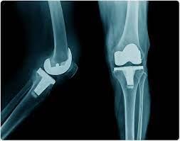
Total Knee Arthroplasty (TKA), or knee replacement surgery, represents a highly effective intervention for end-stage knee arthritis, primarily osteoarthritis (OA). First performed in the 1960s, TKA has evolved significantly in surgical technique, implant design, and perioperative management, transforming into one of the most successful surgical procedures in modern medicine. Its primary objective is to alleviate pain, correct deformity, and restore function in patients with debilitating knee joint disease.
Epidemiologically, OA is the most prevalent form of arthritis, affecting millions globally. Its incidence increases with age, obesity, and prior joint trauma. The projected demand for TKA is substantial and growing rapidly. The Centers for Disease Control and Prevention (CDC) report that OA affects over 32.5 million adults in the United States. Estimates suggest that the annual number of primary TKAs in the U.S. will reach 1.2 million by 2030, representing a significant public health burden and economic impact. This upward trend is attributed to an aging population, rising rates of obesity, and improved patient expectations regarding functional outcomes. While OA remains the predominant indication, inflammatory arthropathies such as rheumatoid arthritis (RA), post-traumatic arthritis, avascular necrosis (and occasionally crystal arthropathy), also contribute to the surgical volume. Revision TKA, addressing failed primary arthroplasties due to aseptic loosening, infection, instability, or polyethylene wear, also constitutes a growing segment of this surgical subspecialty. Understanding the demographic shifts and disease prevalence is crucial for resource allocation, training, and strategic planning within orthopedic surgery.
Surgical Anatomy & Biomechanics
A thorough understanding of the native knee's complex anatomy and biomechanics is fundamental to successful TKA, as the objective is to replicate, as closely as possible, the physiological function of the joint while addressing pathological changes.
Osteology
The knee is formed by the articulation of the distal femur, proximal tibia, and patella.
*
Distal Femur:
Key landmarks include the medial and lateral femoral condyles (which articulate with the tibia), the trochlear groove (for patellar articulation), and the medial and lateral epicondyles (origins for collateral ligaments and gastrocnemius). The surgical epicondylar axis (SEA) and Whiteside's line (perpendicular to the trochlear groove) are critical for femoral component rotation.
*
Proximal Tibia:
Comprises the medial and lateral tibial plateaus, separated by the intercondylar eminence (tibial spines). The posterior slope of the tibia (typically 5-7 degrees) must be respected during resection.
*
Patella:
A sesamoid bone within the quadriceps tendon, its posterior surface has medial and lateral facets articulating with the femoral trochlea. Patellar tracking is paramount for postoperative function.
Ligamentous Anatomy
The knee's stability is largely dependent on its ligamentous structures.
*
Collateral Ligaments:
*
Medial Collateral Ligament (MCL):
Primary restraint to valgus stress, with superficial and deep layers. Understanding its functional length changes through flexion is crucial for gap balancing.
*
Lateral Collateral Ligament (LCL):
Primary restraint to varus stress. Part of the posterolateral corner (PLC) complex, which also includes the popliteus tendon and arcuate complex. Injury or laxity in the PLC requires specific surgical considerations.
*
Cruciate Ligaments:
*
Anterior Cruciate Ligament (ACL):
Resists anterior translation of the tibia relative to the femur and secondary valgus/varus rotation. It is typically sacrificed in most modern TKA designs.
*
Posterior Cruciate Ligament (PCL):
Resists posterior translation of the tibia relative to the femur and secondary valgus/varus rotation. PCL retention (CR designs) versus sacrifice (PS designs) is a significant design philosophy in TKA. PCL-retaining designs aim for more natural kinematics but can be challenging to balance. PCL-substituting designs (PS) incorporate a post-cam mechanism to provide posterior stability.
Musculature and Neurovascular Structures
- Musculature: The quadriceps femoris (rectus femoris, vastus medialis, vastus intermedius, vastus lateralis) is the primary extensor. Hamstrings (semimembranosus, semitendinosus, biceps femoris) are primary flexors. The gastrocnemius contributes to flexion and ankle plantarflexion. Integrity and strength of these muscle groups are vital for rehabilitation.
-
Neurovascular Structures:
Critical for surgical planning and risk mitigation.
- Popliteal Artery and Vein: Positioned immediately posterior to the knee joint capsule, particularly vulnerable during posterior osteophyte removal or aggressive posterior capsular release.
- Common Peroneal Nerve: Courses laterally around the fibular neck, at risk in severe valgus deformities during lateral releases or prolonged tourniquet application.
- Saphenous Nerve: A branch of the femoral nerve, it passes distally on the medial aspect of the knee, often crossing the anterior incision, and is prone to iatrogenic injury leading to paresthesia.
- Femoral Artery/Nerve: Proximal to the operative field but relevant for tourniquet application and regional anesthesia.
Biomechanics of the Native and Arthroplasty Knee
- Native Knee Biomechanics: Involves complex kinematics, including rolling and gliding motions, coupled rotation, and screw-home mechanism. Weight-bearing loads are transmitted through the tibiofemoral joint, with the patellofemoral joint experiencing significant compressive and shear forces. The mechanical axis of the lower limb, passing from the center of the femoral head through the center of the knee to the center of the ankle, is crucial for balanced load distribution.
-
TKA Biomechanics:
TKA aims to restore a neutral mechanical axis, balance soft tissues, and provide a stable, mobile joint.
- Mechanical Alignment (MA): The historical gold standard, aiming for a straight mechanical axis post-TKA (0° varus/valgus) by resecting bone perpendicular to the mechanical axes of the femur and tibia.
- Kinematic Alignment (KA): A more recent concept aiming to restore the native, often subtly varus or valgus, mechanical and kinematic axes of the individual patient, rather than imposing a "neutral" alignment. This involves resecting bone parallel to the native joint lines. Proponents argue for more natural kinematics and potentially better patient satisfaction.
- Gap Balancing: Ensuring equal and rectangular extension and flexion gaps is paramount for stability and range of motion. This is achieved through precise bone cuts and sequential soft tissue releases.
- Joint Line Restoration: Maintaining the anatomical joint line is critical to avoid patella baja/alta and ensure optimal patellofemoral tracking and extensor mechanism function.
Indications & Contraindications
The decision for TKA is multifaceted, requiring careful consideration of patient-specific factors, disease severity, and potential risks versus benefits.
Primary Indications
The primary indication for TKA is severe knee pain and functional limitation caused by end-stage arthropathy, unresponsive to non-operative management.
- Osteoarthritis (OA): Unicompartmental, bicompartmental, or tricompartmental involvement with radiographic evidence of severe joint space narrowing, osteophytes, subchondral sclerosis, and cysts. This remains the most common indication.
- Rheumatoid Arthritis (RA) and Other Inflammatory Arthropathies: Pain, swelling, progressive joint destruction, and deformity secondary to autoimmune or inflammatory processes.
- Post-Traumatic Arthritis: Resulting from intra-articular fractures, meniscectomy, or ligamentous injuries, leading to premature degenerative changes.
- Avascular Necrosis (AVN): Collapse of articular cartilage and subchondral bone due to impaired blood supply.
- Significant Deformity: Fixed varus or valgus deformity (>10-15 degrees), flexion contracture (>10-15 degrees), or gross instability significantly impacting function.
- Failure of Non-Operative Management: A prerequisite for surgical consideration, including weight loss, activity modification, physical therapy, NSAIDs, analgesics, corticosteroid injections, and viscosupplementation.
Contraindications
Contraindications can be absolute or relative, significantly increasing the risk of adverse outcomes or rendering the procedure ineffective.
-
Absolute Contraindications:
- Active Periprosthetic Infection: Current or recent infection in the knee joint or adjacent tissues. Must be treated and resolved prior to TKA.
- Active Infection Elsewhere in the Body: Systemic sepsis or remote foci of infection (e.g., urinary tract infection, dental abscess) increase the risk of hematogenous seeding.
- Extensor Mechanism Insufficiency: Severe quadriceps weakness, rupture of the quadriceps or patellar tendon, or prior failed extensor mechanism repairs that preclude effective rehabilitation and ambulation.
- Recurvatum Deformity: Severe hyperextension that cannot be corrected, indicating significant ligamentous laxity or neuromuscular deficit.
- Neuropathic Arthropathy (Charcot Joint): Characterized by progressive joint destruction due to loss of proprioception and pain sensation, leading to high failure rates and poor outcomes.
- Growing Skeleton: TKA is contraindicated in skeletally immature patients due to potential growth plate disturbance.
- Vascular Insufficiency: Severe peripheral vascular disease that compromises limb viability or healing potential.
- Neurological Deficit: Severe, progressive neurological disorders that prevent rehabilitation or compromise limb control.
-
Relative Contraindications:
- Severe Obesity (BMI > 40-50 kg/m²): Associated with increased surgical time, blood loss, infection risk, DVT/PE, wound complications, and potentially poorer long-term implant survival. Weight loss prior to surgery is strongly advised.
- Poor Skin Condition: Significant dermatitis, psoriasis, or open wounds in the surgical field.
- Unrealistic Patient Expectations: Patients with unrealistic expectations regarding pain relief or functional return are at risk for dissatisfaction.
- Morbid Comorbidities: Uncontrolled diabetes (HbA1c > 8%), severe cardiac or pulmonary disease, or renal failure significantly increase perioperative risks. Medical optimization is crucial.
- Nicotine Use: Smoking is a significant risk factor for wound complications, infection, and impaired bone healing. Cessation is strongly recommended.
- Chronic Regional Pain Syndrome (CRPS): Can be exacerbated by surgery.
- Previous Knee Arthrodesis: While possible, TKA in this setting is technically challenging and often yields poorer outcomes.
Summary of Operative vs. Non-Operative Indications
| Category | Operative Indications | Non-Operative Indications / Relative Contraindications |
|---|---|---|
| Pain | Severe, debilitating knee pain refractory to conservative therapy | Mild to moderate pain, manageable with analgesics or activity modification |
| Function | Significant functional limitation (ADLs), inability to ambulate/work | Minimal functional impairment, able to participate in desired activities |
| Radiographic | Kellgren-Lawrence Grade III-IV OA, joint destruction, severe deformity | Mild OA (K-L Grade I-II), minimal joint space narrowing |
| Etiology | Primary OA, RA, post-traumatic arthritis, AVN | Active infection, neuropathic arthropathy |
| Systemic Factors | Medically optimized for surgery | Uncontrolled diabetes (HbA1c > 8%), severe cardiovascular/pulmonary disease, active infection, severe obesity (BMI > 40) |
| Patient Factors | Realistic expectations, motivated for rehabilitation | Unrealistic expectations, unwillingness to participate in rehab, severe psychosocial issues |
| Local Factors | Intact extensor mechanism, healthy skin envelope | Extensor mechanism insufficiency, poor skin condition in surgical field |
Pre-Operative Planning & Patient Positioning
Meticulous preoperative planning is paramount to optimizing outcomes and minimizing complications in TKA.
Patient Evaluation
A comprehensive patient evaluation extends beyond the knee itself.
*
History:
Detailed medical history focusing on comorbidities (cardiac, pulmonary, renal, endocrine), medication use (especially anticoagulants, immunosuppressants), allergies, prior surgical history, social support, and functional goals. Nutritional status and smoking history are critical.
*
Physical Examination:
*
Lower Extremity Alignment:
Standing varus/valgus deformity, fixed flexion contracture.
*
Range of Motion (ROM):
Active and passive, noting crepitus, pain, or mechanical blocks.
*
Stability:
Assessment of collateral and cruciate ligaments if applicable.
*
Skin and Soft Tissues:
Integrity, scarring, signs of infection or inflammation.
*
Neurovascular Status:
Distal pulses, sensation, motor function.
*
Extensor Mechanism:
Patellar tracking, quadriceps strength.
*
Radiographic Imaging:
*
Weight-Bearing Anteroposterior (AP) View:
Assesses joint space narrowing, osteophytes, and subchondral changes.
*
Lateral View:
Evaluates posterior osteophytes, patellar height (Insall-Salvati, Blackburne-Peel ratios), and flexion contracture.
*
Patellar (Merchant/Skyline) View:
Assesses patellofemoral joint space, tracking, and arthritis.
*
Full-Length Standing AP (Long Leg Alignment) View:
Essential for assessing overall mechanical axis of the limb and planning corrective osteotomies or soft tissue releases. This is crucial for both mechanical and kinematic alignment strategies.
*
Special Imaging (CT/MRI):
Rarely indicated for primary TKA but can be useful for complex deformities, previous hardware, significant bone loss, or tumor evaluation.
*
Pre-Operative Templating:
Using radiographic images and implant templates to estimate component sizes and predict bone resections. This provides a mental roadmap, even with computer-assisted or robotic systems.
Pre-Habilitation & Medical Optimization
- Medical Clearance: From primary care physician and/or specialists for optimization of chronic conditions (e.g., hypertension, diabetes, cardiac disease).
- Glycemic Control: Optimal HbA1c < 7-8% is critical to reduce infection risk.
- Nutritional Optimization: Addressing malnutrition or vitamin deficiencies.
- Smoking Cessation: At least 4-6 weeks preoperatively.
- Physical Therapy: Pre-operative exercises to improve strength and range of motion.
- Patient Education: Comprehensive discussion of the surgical procedure, expected recovery, potential complications, and rehabilitation protocol.
- Blood Management: Anemia correction, consideration of antifibrinolytics (e.g., tranexamic acid).
Infection Prophylaxis
- Antibiotics: Intravenous administration of a first or second-generation cephalosporin (e.g., Cefazolin 1-2g) 30-60 minutes prior to incision. Vancomycin or clindamycin for penicillin-allergic patients or MRSA colonization.
- Skin Preparation: Chlorhexidine-alcohol solution.
Thromboprophylaxis
Risk stratification for Venous Thromboembolism (VTE) is essential.
*
Pharmacological:
Aspirin, low-molecular-weight heparin (LMWH), warfarin, or novel oral anticoagulants (NOACs). Duration varies but typically 2-6 weeks postoperatively.
*
Mechanical:
Intermittent pneumatic compression (IPC) devices.
Patient Positioning
The patient is typically positioned supine on the operating table.
*
Tourniquet:
Applied to the proximal thigh to minimize blood loss and provide a bloodless field. Duration should be monitored (typically < 90-120 minutes).
*
Leg Holder:
A bump or leg holder under the contralateral hip may be used to maintain neutral rotation. A supportive device under the ipsilateral heel allows for full knee flexion.
*
Foot Support:
Ensure the foot is free to allow for full range of motion during intraoperative assessment.
*
Padding:
All bony prominences (heels, fibular head, ulnar nerve at elbow) must be meticulously padded to prevent pressure sores or nerve palsies (e.g., common peroneal nerve).
*
Sterile Preparation and Draping:
Standard sterile technique, often with an impermeable adhesive drape covering the entire limb.
Detailed Surgical Approach / Technique
The standard approach for TKA is the medial parapatellar arthrotomy, which provides excellent exposure of the tibiofemoral and patellofemoral joints. Alternative approaches exist but are less common for primary TKA.
Incision & Dissection
- Skin Incision: A straight longitudinal incision centered over the patella, extending from approximately 5 cm proximal to the patella to 5 cm distal to the tibial tuberosity. The length can vary based on surgeon preference and patient habitus. Curvilinear incisions may be used but increase the risk of skin necrosis at the apex of the curve.
- Subcutaneous Dissection: Dissection through subcutaneous fat to expose the deep fascia. Avoid excessive undermining of skin flaps to preserve vascularity.
-
Arthrotomy (Medial Parapatellar):
- Incise the medial retinaculum, starting proximal to the patella, through the vastus medialis obliquus (VMO) tendon insertion.
- Extend the incision distally along the medial border of the patella, through the medial capsule and synovium, and often just medial to the patellar tendon, into the infrapatellar fat pad.
- This approach is an internervous plane, minimizing muscle damage. The vastus medialis is mobilized laterally, and the rectus femoris superiorly.
- Patellar Eversion: The patella is everted laterally, typically by fully flexing the knee. This provides wide exposure of the distal femur and proximal tibia. Care must be taken to avoid excessive force on the patellar tendon.
Bone Resection
Precise bone resections are critical for achieving balanced gaps and proper alignment. Both intramedullary (IM) and extramedullary (EM) guides are utilized.
-
Femoral Resections:
-
Distal Femoral Cut:
- IM Guide: A rod is inserted into the femoral canal via a small portal in the intercondylar notch. The distal femoral cutting block is then applied, typically set at a valgus angle (5-7 degrees) relative to the femoral mechanical axis to achieve a perpendicular cut to the mechanical axis. The valgus angle is patient-specific and can be determined from preoperative radiographs.
- Resection: The specified amount of distal femur (e.g., 9-10mm) is resected, impacting the extension gap.
- Anterior/Posterior Femoral Sizing: Sizing guides are used to determine the appropriate anteroposterior dimension of the femoral component. This dictates the anterior and posterior femoral cuts. Rotational alignment is crucial here, often referenced to the posterior condylar axis, surgical epicondylar axis (SEA), or Whiteside's line.
- Anterior/Posterior & Chamfer Cuts: Using a 4-in-1 or 5-in-1 cutting block, the anterior, posterior, anterior chamfer, and posterior chamfer cuts are made. These cuts create the shape for the femoral component.
- Notch/Box Cut (for PS designs): If a PCL-substituting implant is used, a box cut is performed in the intercondylar notch to accommodate the femoral cam and tibial post mechanism.
-
Distal Femoral Cut:
-
Tibial Resections:
-
Proximal Tibial Cut:
- EM Guide: Preferred for tibial cuts, aligning the rod with the mechanical axis of the tibia.
- Resection: The proximal tibia is resected, typically 8-10mm from the least affected plateau, aiming for a cut perpendicular to the mechanical axis in the coronal plane, and with a posterior slope (3-7 degrees) in the sagittal plane, replicating the native posterior slope. The depth of resection affects the extension and flexion gaps.
- Osteophytes: Peripheral osteophytes are meticulously removed to ensure proper soft tissue balancing and prevent impingement.
-
Proximal Tibial Cut:
-
Patellar Resection (if resurfacing):
- A freehand or guarded cut removes approximately 8-10mm of bone from the posterior aspect of the patella, ensuring enough bone stock remains (minimum 10-12mm). The aim is to restore patellar thickness to its original dimension with the implant.
- Drill holes are made for patellar button fixation.
Ligamentous Balancing / Gap Balancing
Achieving rectangular and symmetrical flexion and extension gaps is paramount for TKA success, preventing instability or stiffness.
*
Extension Gap Assessment:
With the knee in full extension, trial spacers are inserted. If the gap is tight, posterior osteophytes are removed. If still tight, sequential release of posterior capsule and potentially superficial MCL or lateral structures (for varus/valgus deformities) is performed.
*
Flexion Gap Assessment:
With the knee in 90 degrees of flexion, trial spacers are inserted. If the flexion gap is tighter than the extension gap, this may indicate inadequate posterior femoral resection or a tight PCL (if retained). If looser, it may indicate excessive posterior femoral resection.
*
Soft Tissue Releases:
*
Varus Deformity:
Often requires release of the superficial MCL, posteromedial capsule, and medial osteophytes. Pie-crusting of the superficial MCL may be necessary for significant fixed deformities.
*
Valgus Deformity:
Requires release of the lateral capsule, LCL, popliteus tendon, and potentially iliotibial band (ITB) and posterolateral corner structures. This must be done cautiously to avoid common peroneal nerve injury.
*
Flexion Contracture:
Managed by posterior capsular release, removal of posterior femoral osteophytes, and adequate posterior tibial slope.
Trial Components & Final Implantation
- Trial Insertion: Trial femoral, tibial, and polyethylene components are inserted.
-
Assessment:
- Range of Motion: Full flexion and extension without impingement.
- Stability: Balanced in flexion and extension, checking for mediolateral and anteroposterior stability.
- Patellofemoral Tracking: Observing the patella during knee flexion and extension to ensure smooth tracking in the trochlear groove. Lateral patellar release may be performed if tracking is compromised.
- Joint Line: Ensuring it has been restored to prevent patella baja/alta.
-
Final Implantation:
- The surgical field is thoroughly irrigated with pulsatile lavage.
- Bone surfaces are dried and prepared with hydrogen peroxide to enhance cement-bone interface.
- Cemented Components: Bone cement (PMMA) is mixed and applied in a doughy consistency.
- Tibial Component: Cemented first, pressing firmly onto the resected tibia.
- Femoral Component: Cemented onto the distal femur, ensuring complete coverage.
- Polyethylene Insert: Inserted into the tibial tray.
- Patellar Button (if resurfaced): Cemented onto the resected patella.
- Excess cement is meticulously removed before it cures, especially from the posterior aspect, to prevent impingement.
- The knee is held in extension under compression during cement curing.
-
Wound Closure:
- The medial retinaculum and capsule are repaired using strong absorbable sutures. This restores extensor mechanism integrity.
- Subcutaneous layers are closed.
- Skin is closed with staples or non-absorbable sutures.
- Drains are used selectively and typically removed within 24 hours if used at all (controversial in modern practice).
Complications & Management
Despite high success rates, TKA is not without potential complications, ranging from minor to limb-threatening. Proactive prevention and timely recognition are crucial.
Common Complications & Management Strategies
| Complication | Incidence (%) | Salvage/Management Strategies |
|---|---|---|
| Infection | 0.5 - 2 | Acute (<4 weeks): Debridement and implant retention (DAIR) with exchange of modular components, prolonged IV antibiotics. Chronic (>4 weeks): Two-stage revision (explant, antibiotic spacer, prolonged IV antibiotics, reimplantation) or one-stage revision (less common). Arthrodesis or amputation for intractable cases. |
| Periprosthetic Fracture | 0.3 - 2 | Intraoperative: Depending on location and stability, often managed with internal fixation (wires, plates) or conversion to revision arthroplasty. Postoperative: Vancouver classification guides management; ORIF, revision arthroplasty (stemmed components), or conservative management for stable, non-displaced fractures. |
| Venous Thromboembolism (VTE) | DVT 5-10, PE 1-2 | Prophylaxis: Early mobilization, mechanical compression, pharmacological agents (aspirin, LMWH, NOACs). Treatment: Anticoagulation (LMWH, warfarin, NOACs); IVC filter for recurrent PE or contraindication to anticoagulation. |
| Neurovascular Injury | < 0.5 | Common Peroneal Nerve: Neuropraxia often resolves; observation, bracing, nerve exploration/release in severe cases. Popliteal Vessels: Immediate surgical exploration, vascular repair by a specialist. |
| Arthrofibrosis / Stiffness | 5 - 10 | Aggressive physical therapy. Persistent stiffness (>3 months post-op): Manipulation Under Anesthesia (MUA), arthroscopic or open arthrolysis. Evaluation for underlying infection or malalignment. |
| Instability | 1 - 2 | Acute: Hinged brace, aggressive rehab. Chronic: Polyethylene exchange (for mild laxity), revision TKA with more constrained implant designs (constrained condylar, hinged). |
| Aseptic Loosening / Osteolysis | 0.5 - 1 (per year) | Revision TKA with bone grafting and stemmed components for failed fixation and bone loss. |
| Patellofemoral Complications | 5 - 10 | Maltracking: Lateral retinacular release, revision to more appropriate component. Fracture: ORIF, patellectomy (salvage). Clunk: Arthroscopic debridement. |
| Residual Pain | 10 - 20 | Thorough workup to rule out infection, loosening, instability. Consider conservative management, pain specialist referral, psychiatric evaluation. Revision TKA if a treatable mechanical cause is identified. |
| Wound Complications | 2 - 5 | Debridement, negative pressure wound therapy, primary closure, local muscle flaps in severe cases. |
Detailed Discussion of Key Complications
-
Periprosthetic Joint Infection (PJI):
Remains a devastating complication.
- Diagnosis: Elevated ESR/CRP, positive aspirate culture (cell count, differential, culture), alpha-defensin, synovial fluid leukocyte esterase, periprosthetic membrane histology.
- Management: Varies by chronicity, organism virulence, and host factors. Acute infections may be treated with DAIR, whereas chronic infections typically require a two-stage revision protocol.
-
Periprosthetic Fractures:
Can occur intraoperatively or postoperatively.
- Intraoperative: Often related to vigorous impaction, malpositioning, or stress risers.
- Postoperative: Typically low-energy falls in osteoporotic patients. The Vancouver classification system guides management based on fracture location, stability of implants, and bone stock.
-
Thromboembolic Disease:
TKA patients are at high risk.
- Prevention: Multimodal approach including mechanical (IPC) and pharmacological (aspirin, LMWH) prophylaxis. Early ambulation is key.
- Diagnosis: Ultrasound for DVT, CT pulmonary angiography for PE.
-
Neurovascular Injury:
Rare but limb-threatening.
- Common Peroneal Nerve: Most commonly injured, especially in large valgus deformities requiring extensive lateral release. Typically neuropraxia with potential for recovery.
- Popliteal Vessels: At risk during posterior osteophyte removal or aggressive posterior capsular release. Immediate recognition and vascular surgical consultation are critical.
-
Arthrofibrosis / Stiffness:
Limited ROM post-TKA (e.g., < 90° flexion or > 10° flexion contracture) significantly impacts patient satisfaction and function.
- Causes: Inadequate rehabilitation, PJI, underlying CRPS, surgical technique (e.g., tight gaps, patella baja).
- Management: MUA is effective if performed within 3 months of surgery. Arthroscopic or open arthrolysis for recalcitrant cases.
-
Instability:
Can be due to implant malposition, inadequate soft tissue balancing, or component wear.
- Management: Initial conservative treatment may be attempted. Persistent instability often requires revision surgery, potentially with a more constrained polyethylene insert or a fully constrained revision implant.
-
Aseptic Loosening:
Mechanical failure of the implant-bone interface without infection.
- Causes: Stress shielding, micro-motion, polyethylene wear-induced osteolysis.
- Management: Revision TKA, often requiring stemmed components and bone grafting for associated bone loss.
-
Patellofemoral Complications:
A common source of dissatisfaction.
- Maltracking: Lateral subluxation, tilt, or excessive pressure on facets. Managed by lateral retinacular release, component re-orientation, or revision.
- Patellar Fracture: Often due to direct trauma or osteonecrosis. May require ORIF or patellectomy.
Post-Operative Rehabilitation Protocols
A structured and progressive rehabilitation program is essential for maximizing functional recovery and optimizing TKA outcomes. Protocols vary slightly between institutions and surgeons, but common principles apply.
Immediate Post-Operative Phase (Day 0-3)
-
Goals:
- Pain management (multimodal analgesia: regional blocks, NSAIDs, acetaminophen, opioids).
- Early mobilization and weight-bearing as tolerated (WBAT or PWB depending on fixation).
- Initiate active and passive range of motion.
- Prevent VTE (pharmacological prophylaxis, IPC, ankle pumps).
-
Activities:
- Physiotherapy: Initiate on post-operative day 0 or 1.
- Exercises: Ankle pumps, quadriceps sets, gluteal sets, heel slides (active-assisted), passive knee flexion to 90 degrees.
- Mobility: Bed mobility, sit-to-stand transfers, short-distance ambulation with assistive devices (walker, crutches).
- CPM Machine: Its routine use remains controversial; most evidence suggests no significant benefit over conventional PT, but some surgeons utilize it.
Acute Rehabilitation Phase (Weeks 1-6)
-
Goals:
- Achieve functional ROM (typically 0-90 to 110 degrees).
- Improve quadriceps and hamstring strength.
- Progress weight-bearing and decrease reliance on assistive devices.
- Manage swelling and pain.
-
Activities:
- ROM Exercises: Continued heel slides, wall slides, supine knee flexion, prone hangs (for extension).
- Strengthening: Straight leg raises (SLR) in multiple planes, mini-squats (partial weight-bearing), step-ups (low steps), terminal knee extension (TKE) with resistance, hamstring curls.
- Gait Training: Progression from walker to crutches to cane, focusing on normal gait pattern. Stair climbing training.
- Modalities: Ice, compression, elevation to control edema.
Subacute Rehabilitation Phase (Weeks 6-12)
-
Goals:
- Achieve full functional ROM (>110-120 degrees flexion, full extension).
- Significant improvement in strength and endurance.
- Independent ambulation without assistive devices.
- Return to light household activities.
-
Activities:
- Advanced Strengthening: Lunges, full squats, leg press, calf raises, stationary cycling (low resistance).
- Proprioception and Balance: Single-leg stance, wobble board exercises.
- Functional Training: Return to more challenging ADLs, walking longer distances.
- Low-Impact Aerobics: Swimming, elliptical trainer.
Chronic Rehabilitation / Maintenance Phase (Beyond 12 weeks)
-
Goals:
- Maximize functional independence and return to desired recreational activities.
- Long-term maintenance of strength and ROM.
- Address any residual pain or stiffness.
-
Activities:
- Continuation of strength and conditioning exercises.
- Participation in low-impact sports (e.g., golf, cycling, swimming, doubles tennis). High-impact activities (running, jumping sports) are generally discouraged due to increased risk of implant wear and loosening.
- Patient education on joint protection, activity modification, and long-term care.
Key Considerations in Rehabilitation
- Patient Adherence: Crucial for successful outcomes. Education and motivation are paramount.
- Multidisciplinary Approach: Involving surgeons, nurses, physical therapists, occupational therapists, and pain management specialists.
- Pain Management: Effective multimodal pain control allows for earlier and more aggressive rehabilitation.
- Complication Monitoring: Vigilance for signs of infection, DVT, or neurovascular compromise.
- Individualization: Protocols should be tailored to the patient's individual progress, comorbidities, and goals.
Summary of Key Literature / Guidelines
The field of TKA is supported by extensive research and evidence-based guidelines from major orthopedic societies, aiming to standardize practice and improve outcomes.
Major Society Guidelines
- American Academy of Orthopaedic Surgeons (AAOS): Publishes clinical practice guidelines on various aspects of TKA, including indications, management of DVT, antibiotic prophylaxis, and pain management.
- American Association of Hip and Knee Surgeons (AAHKS): Focuses on arthroplasty, providing educational resources and research.
- National Institute for Health and Care Excellence (NICE - UK): Offers comprehensive guidelines on the management of osteoarthritis and TKA, including implant selection and rehabilitation.
- British Orthopaedic Association (BOA): Provides standards and guidelines for orthopedic practice in the UK.
Evidence-Based Recommendations & Key Literature
- Indications for TKA: Consistently emphasize severe pain and functional limitation refractory to non-operative treatment, coupled with radiographic evidence of end-stage arthritis. Patient age and comorbidities are less strict contraindications than in the past, with increasing evidence supporting TKA in carefully selected older adults and patients with controlled medical conditions.
- Antibiotic Prophylaxis: Strong evidence supports intravenous prophylactic antibiotics (first/second-generation cephalosporin) administered 30-60 minutes pre-incision to reduce the risk of PJI. Prolonged post-operative antibiotics are generally not recommended beyond 24 hours. (e.g., "The Duration of Antibiotic Prophylaxis in Total Joint Arthroplasty" - J Bone Joint Surg Am, 2013 ).
- Thromboprophylaxis: AAOS and ACCP (American College of Chest Physicians) guidelines provide risk stratification and recommendations for pharmacological (e.g., aspirin, LMWH) and mechanical (IPC) prophylaxis for varying durations post-TKA. There is ongoing debate regarding the optimal agent and duration. (e.g., "Prevention of VTE in Orthopedic Surgery Patients: Antithrombotic Therapy and Prevention of Thrombosis, 9th ed" - Chest, 2012 ).
- Patellar Resurfacing: Meta-analyses show no consistent significant difference in patient-reported outcomes between patellar resurfacing and non-resurfacing, but non-resurfacing may be associated with a higher rate of anterior knee pain and re-operation for patellar symptoms. The decision is often surgeon-dependent and based on patellar cartilage quality. (e.g., "Patellar Resurfacing in TKA: A Meta-Analysis" - J Bone Joint Surg Am, 2005 ).
- PCL Retention vs. Sacrifice: Both PCL-retaining (CR) and PCL-substituting (PS) designs yield excellent outcomes. PS designs may offer easier surgical technique and better flexion, while CR designs aim for more natural kinematics. No clear superiority in long-term outcomes for either approach has been definitively established. (e.g., "PCL Retention versus Sacrifice in TKA: A Meta-Analysis" - J Bone Joint Surg Am, 2013 ).
- Continuous Passive Motion (CPM): Most randomized controlled trials and meta-analyses have concluded that routine use of CPM does not provide a significant additional benefit in terms of ROM, pain, or length of hospital stay compared to standard physical therapy. Its use has largely declined. (e.g., "The Efficacy of CPM after TKA: A Meta-Analysis" - J Bone Joint Surg Am, 2004 ).
- Outcomes and Implant Survival: TKA boasts high implant survival rates, with 10-year survival exceeding 90-95% and 20-year survival rates around 80-85% for primary TKA. Patient-reported outcome measures (PROMs) consistently show significant improvements in pain and function. (e.g., "The Long-Term Outcomes of TKA" - J Bone Joint Surg Am, 2009 ).
- Enhanced Recovery After Surgery (ERAS) Protocols: A multidisciplinary, evidence-based approach encompassing pre-operative optimization, intra-operative best practices (e.g., multimodal analgesia, tranexamic acid), and early post-operative mobilization. ERAS pathways have demonstrated reduced hospital stays, lower complication rates, and improved patient satisfaction. (e.g., "Enhanced Recovery after TKA: A Systematic Review and Meta-Analysis" - J Arthroplasty, 2016 ).
- Alignment Strategies: While mechanical alignment has been the gold standard, recent literature explores kinematic alignment (KA) and restricted kinematic alignment (rKA) as alternatives. Studies suggest KA may lead to more patient-reported satisfaction due to more natural joint kinematics, but long-term implant survival data is still maturing. (e.g., "Kinematic Alignment in TKA: A Narrative Review" - J Bone Joint Surg Am, 2020 ).
- Computer-Assisted Navigation and Robotics: These technologies aim to improve accuracy of component placement and alignment, potentially reducing outliers. While improved accuracy is demonstrated, a clear clinical superiority over conventional instrumentation in terms of long-term survival or PROMs is still under active investigation for routine cases. (e.g., "Robotic-Assisted TKA: A Systematic Review and Meta-Analysis" - J Arthroplasty, 2021 ).
The continuous evolution of TKA techniques, implant designs, and perioperative care mandates that orthopedic surgeons remain abreast of current literature and evidence-based guidelines to provide optimal patient care.
You Might Also Like