Pediatric Supracondylar Humerus Fractures: Epidemiology, Anatomy & Management
Key Takeaway
Supracondylar humerus fractures (SCHF) are the most common pediatric elbow fractures, typically occurring in 5-7 year olds from falls onto an outstretched hand. They involve the distal humerus, posing significant risk of neurovascular compromise due to proximity to the brachial artery and nerves. Understanding detailed anatomy, biomechanics (like Gartland classification), and proper research appraisal is crucial for effective management.
Introduction & Epidemiology
Supracondylar humerus fractures (SCHF) are the most common elbow fractures in children, accounting for approximately 60% of pediatric elbow injuries. These injuries primarily occur in children aged 5-7 years, often resulting from a fall onto an outstretched hand with the elbow in hyperextension (extension-type fractures). Less commonly, a direct fall onto the flexed elbow can cause a flexion-type fracture. The clinical significance lies in their propensity for neurovascular compromise and potential for long-term functional deficits or deformities if not managed appropriately.
The continuous evolution of orthopedic practice is underpinned by robust clinical research. When evaluating the existing literature, particularly from diverse global sources, it is crucial to critically appraise study characteristics. For instance, the presented data, while not specific to SCHF nor exclusively from China, illustrates critical parameters for evaluating research quality:
*
Sample Size:
Studies range from 100 to 3,456 participants. Adequate sample size is paramount for statistical power and generalizability of findings, particularly in pediatric populations where incidence rates may vary.
*
Demographics:
The mean age in these examples, predominantly 4-5 years with a standard deviation of approximately 2 years, aligns well with the typical age range for SCHF. The male predominance (53-62%) is also a common demographic pattern observed in pediatric trauma research.
*
Follow-up Duration:
A 12-week follow-up, while sufficient for assessing immediate post-operative complications and early union, may be insufficient for evaluating long-term outcomes such as cubitus varus deformity or chronic stiffness, which can manifest later.
*
Geographic Distribution:
The studies cited originate from various countries including the UK, USA, Germany, Canada, Spain, and Italy. This highlights the global collaborative nature of orthopedic research. The increasing contribution of research from China, characterized by large populations and diverse clinical settings, is a significant development in the global orthopedic landscape. Analyzing such research requires careful consideration of methodology, patient populations, and healthcare systems, often demanding similar scrutiny of sample demographics, outcome measures, and follow-up periods.
Understanding the epidemiology and the critical appraisal of research characteristics allows for evidence-based decision-making in the management of SCHF.
Surgical Anatomy & Biomechanics
A thorough understanding of the pediatric distal humerus anatomy and the biomechanics of SCHF is crucial for successful management.
Distal Humerus Anatomy
The distal humerus transitions from a cylindrical shaft to a flattened, triangular structure culminating in the humeral condyles. Key anatomical landmarks include:
*
Capitellum:
Lateral articular surface, ossifies around 1 year. Articulates with the radial head.
*
Trochlea:
Medial articular surface, ossifies around 3 years. Articulates with the ulna.
*
Medial Epicondyle:
Prominent subcutaneous bony landmark, origin for wrist flexors, ossifies around 5-7 years. The ulnar nerve courses posterior to this epicondyle in the cubital tunnel.
*
Lateral Epicondyle:
Less prominent, origin for wrist extensors, ossifies around 10-12 years.
*
Olecranon Fossa:
Posterior depression accommodating the olecranon during elbow extension.
*
Coronoid Fossa:
Anterior depression accommodating the coronoid process during elbow flexion.
The growth plate (physis) for the distal humerus is located just proximal to the articular surface. The fracture line in SCHF typically occurs through the metaphysis, proximal to the physeal plate, but distal to the olecranon and coronoid fossae. This particular location makes the fragment susceptible to displacement and neurovascular injury.
Neurovascular Structures
The intricate relationship between the fracture site and vital neurovascular structures is paramount:
*
Brachial Artery:
Courses anteromedially, anterior to the distal humerus, often tethered by the lacertus fibrosus and straddling the fracture site. It is at high risk of injury or entrapment, particularly in displaced extension-type fractures.
*
Median Nerve:
Travels alongside the brachial artery, anterior to the distal humerus.
*
Radial Nerve:
Located more laterally, anterior to the lateral epicondyle, and divides into superficial and deep branches distal to the elbow.
*
Ulnar Nerve:
Courses posteriorly and medially within the cubital tunnel, behind the medial epicondyle. It is especially vulnerable to injury during medial pin placement or if the fracture fragment displaces posteromedially.
Biomechanics of Injury
-
Extension-type (95%):
Occurs with a fall onto an outstretched hand with the elbow in hyperextension. The olecranon impinges on the olecranon fossa, acting as a fulcrum, causing the distal fragment to displace posteriorly and proximally relative to the humeral shaft. This type is further classified by Gartland:
- Gartland Type I: Nondisplaced.
- Gartland Type II: Displaced posteriorly, intact posterior cortex.
- Gartland Type III: Completely displaced, no cortical contact.
- Gartland Type IV (Wilkins modification): Multidirectionally unstable in both flexion and extension, periosteum torn circumferentially.
- Flexion-type (5%): Occurs with a direct fall onto a flexed elbow. The distal fragment displaces anteriorly.
Understanding these anatomical and biomechanical principles guides surgical approach, reduction maneuvers, and fixation strategies to minimize complications.
Indications & Contraindications
The management of supracondylar humerus fractures in children varies significantly based on the degree of displacement and the presence of neurovascular compromise.
Operative vs. Non-Operative Indications
| Indication Category | Non-Operative Management | Operative Management |
|---|---|---|
| Gartland Classification | Type I: Nondisplaced or minimally displaced fracture (fat pad sign only or <2mm displacement). | Type II: Displaced with intact posterior cortex, but significant angulation/rotation. |
| Type III: Completely displaced with no cortical contact. | ||
| Type IV (Wilkins): Multidirectionally unstable. | ||
| Neurovascular Status | Intact neurovascular status. | Any neurovascular compromise: Absent or diminished pulses, pallor, paresthesias, paralysis, or signs of compartment syndrome (pain out of proportion, pain with passive stretch, paresthesias, tense compartment, paralysis, pulselessness - the 6 P's, though early signs are more reliable). |
| Fracture Type | Stable, closed fracture. | Open fractures: Any skin breach communicating with the fracture site. |
| Polytrauma: Associated injuries requiring surgical intervention, where SCHF fixation facilitates patient care. | ||
| Reduction Status | Achieved and maintained acceptable reduction with immobilization. | Unreducible fractures: Failed closed reduction attempts (e.g., due to soft tissue interposition, severe comminution). |
| Unstable fractures: Reduction achieved but cannot be maintained with casting. |
Contraindications
Absolute contraindications to operative fixation of a displaced SCHF are rare and generally relate to the child's overall physiological stability rather than the fracture itself.
*
Patient Instability:
Severe systemic injury or medical comorbidities that preclude safe anesthesia and surgery. In such cases, stabilization and transfer to a higher level of care are paramount.
*
Non-Salvageable Limb:
In extreme polytrauma scenarios, an unsalvageable limb may necessitate different management strategies, though this is exceedingly rare for SCHF.
*
Minor Displacement (Gartland Type I):
Operative intervention is generally contraindicated for truly nondisplaced fractures, as the risks outweigh the benefits.
Pre-Operative Planning & Patient Positioning
Meticulous pre-operative planning and appropriate patient positioning are critical for successful SCHF fixation and minimization of complications.
Pre-Operative Assessment
- Clinical History: Ascertain mechanism of injury, time of injury, prior medical history, and allergies.
-
Physical Examination:
- Neurovascular Status: Thoroughly document baseline neurological function (median, ulnar, radial nerve integrity – motor and sensation) and vascular status (radial and ulnar pulses, capillary refill, warmth, color of the hand). Any deficits must be documented pre-operatively.
- Skin Integrity: Inspect for open wounds or impending skin compromise.
- Compartment Syndrome Check: Assess for signs of compartment syndrome, particularly in significantly swollen or tense forearms.
-
Imaging:
-
Standard X-rays:
AP and true lateral views of the elbow are essential.
- The anterior humeral line (drawn along the anterior cortex of the humerus) should normally pass through the middle third of the capitellum on a true lateral view. In SCHF, it typically passes anterior to the capitellum.
- Coronoid-capitellar line: On a true lateral view, a line drawn through the center of the capitellum should pass through the coronoid fossa in flexion.
- Fat pad signs: Anterior and posterior fat pad signs indicate joint effusions, common with intra-articular fractures or significant trauma.
- Oblique views: May be helpful for complex fractures or to better visualize displacement.
- Comparison views: Less commonly needed, but can aid in evaluating ossification centers in younger children if epiphyseal injury is suspected.
-
Standard X-rays:
AP and true lateral views of the elbow are essential.
Anesthesia Considerations
General anesthesia is typically required. The anesthesiologist should be aware of potential blood loss, nerve injury, and the need for tourniquet application. Regional blocks (e.g., supraclavicular, axillary) can provide post-operative analgesia but should be carefully considered due to potential masking of compartment syndrome symptoms if administered prior to definitive fixation.
Patient Positioning
- Supine on a Radiographs-Compatible Operating Table: The patient is placed supine.
-
Arm Positioning:
- The injured arm is often positioned on a hand table or arm board to allow free manipulation and unimpeded access for fluoroscopy.
- Alternatively, a chest roll placed beneath the patient's ipsilateral chest can allow the arm to hang freely off the side of the table, facilitating reduction maneuvers, especially for highly displaced fractures.
- C-arm Access: Ensure the C-arm can be brought in from the opposite side of the table (typically from the patient's head or foot) for true lateral and AP views without repositioning the patient or the arm. This requires sufficient space and sterile draping to allow full C-arm rotation.
- Tourniquet: A pneumatic tourniquet is routinely applied to the upper arm proximally to the anticipated surgical field to facilitate a bloodless field, crucial for visualization during reduction and pin placement.
- Sterile Preparation and Draping: The entire upper extremity, from shoulder to fingertips, is prepped with an antiseptic solution and draped to allow sterile manipulation of the arm and access for the C-arm. The axilla should be included in the prep.
Detailed Surgical Approach / Technique
The primary goal of surgical management for displaced SCHF is to achieve anatomical reduction and stable fixation, protecting neurovascular structures. Closed reduction percutaneous pinning (CRPP) is the gold standard. Open reduction is reserved for specific indications.
General Principles
- Gentle Tissue Handling: Avoid excessive traction or rough manipulation to minimize soft tissue injury and neurovascular compromise.
- Minimize Soft Tissue Stripping: Preserve periosteal attachments as much as possible to maintain blood supply and promote healing.
- Achieve Anatomical Reduction: Restore the distal humerus to its normal alignment in all planes (AP, lateral, rotation).
- Stable Fixation: Use K-wires to maintain reduction until healing occurs.
- Fluoroscopic Control: Constant use of C-arm for real-time visualization of reduction and pin placement.
Closed Reduction Percutaneous Pinning (CRPP)
This is the preferred method for most displaced SCHF (Gartland Types II, III, IV) without open wounds or failed closed reduction.
-
Reduction Maneuvers (Sequential Technique):
- Traction: Gentle, continuous longitudinal traction is applied to the forearm, counteracted by an assistant stabilizing the upper arm. This disengages the fracture fragments and restores length.
- Correct Medial/Lateral Displacement: While maintaining traction, apply direct pressure to align the fragments in the coronal plane.
- Correct Rotation: The key maneuver for extension-type fractures: The forearm is pronated to untwist the distal fragment. Pronation tightens the medial collateral ligament, which is usually intact, allowing it to act as a hinge to derotate the distal fragment. It also moves the ulnar nerve away from the medial epicondyle, theoretically reducing its risk during medial pin placement.
- Correct AP Displacement (Flexion): While maintaining traction and pronation, flex the elbow to approximately 100-120 degrees. This brings the olecranon into the olecranon fossa, pushing the distal fragment anteriorly and reducing the posterior displacement.
-
Verify Reduction:
Immediately obtain true lateral and AP fluoroscopic views.
- On lateral: Anterior humeral line should pass through the middle third of the capitellum. Check for absence of posterior displacement.
- On AP: Check for proper carrying angle and absence of medial/lateral shift or rotation. Ensure the "figure-of-8" configuration of the medial and lateral columns is restored.
-
Pinning Techniques (K-wires):
- K-wires (typically 1.6mm or 2.0mm, 0.062" or 0.045") are used. Smooth wires are preferred to minimize physeal damage.
-
Lateral Entry Pinning (Preferred):
This technique minimizes iatrogenic ulnar nerve injury.
-
Two Divergent Lateral Pins:
- Insert the first pin just proximal to the lateral epicondyle, aiming superomedial into the medial column. Advance under fluoroscopy to engage both cortices of the proximal humerus.
- Insert the second pin also from the lateral side, proximal to the lateral epicondyle, diverging significantly from the first, aiming superoanteromedial into the medial column. The goal is to maximize the spread of the pins in the distal fragment to create a stable construct and achieve adequate purchase in the proximal fragment.
- Advantages: Lower risk of ulnar nerve injury.
- Disadvantages: Potentially less stable biomechanically than cross-pinning in highly unstable fractures, but clinical outcomes are generally equivalent if performed correctly.
- Three Lateral Pins: Can be used for increased stability, forming a triangular construct. One pin aimed medially, one anteriorly, one posteriorly.
-
Two Divergent Lateral Pins:
-
Cross-Pinning (Medial and Lateral Pins):
- The lateral pin is placed first as described above.
-
Medial Pin Placement (Controversial due to Ulnar Nerve):
- Risk: The ulnar nerve is at high risk of iatrogenic injury (incidence up to 5-10% in some series) when a medial pin is inserted blindly.
-
Minimizing Risk:
- Maximal elbow flexion (≥110 degrees) can pull the ulnar nerve anteriorly, away from the medial epicondyle.
- Percutaneous "Mini-Open" or Direct Visualization: A small incision (1-2 cm) over the medial epicondyle can be made to directly visualize and protect the ulnar nerve, retracting it anteriorly, before inserting the medial K-wire. This significantly reduces nerve injury risk and is increasingly advocated, especially for Type III/IV fractures.
- The medial pin is inserted through the medial epicondyle, aiming superolaterally into the lateral column of the proximal humerus, crossing the lateral pin above the fracture site.
- Advantages: Biomechanically superior stability for highly unstable fractures.
- Disadvantages: Risk of ulnar nerve injury if not protected.
-
Post-Fixation Checks:
- Verify reduction and pin placement with AP and lateral fluoroscopy.
- Check for stability by gently stressing the elbow.
- Re-assess neurovascular status of the hand.
- Bend and cut the K-wires outside the skin, leaving enough wire to facilitate removal. Place sterile caps or dressings over the pin sites.
- Apply a long-arm plaster cast in approximately 70-90 degrees of flexion with the forearm in pronation. Avoid excessive flexion to prevent compartment syndrome.
Open Reduction
Indications for open reduction include:
*
Failed Closed Reduction:
Due to soft tissue interposition (e.g., periosteum, triceps muscle, median nerve, brachial artery), which prevents anatomical reduction.
*
Open Fractures:
Requires debridement and irrigation, followed by reduction and fixation.
*
Vascular Injury Requiring Exploration:
If vascular compromise persists after reduction, open exploration is necessary.
*
Significant Nerve Injury Requiring Exploration:
Rare, but can occur.
Common Surgical Approaches for Open Reduction:
*
Anterior (Henry) Approach:
Often used for vascular exploration. Allows access to the brachial artery and median nerve. Dissection is between the brachioradialis laterally and pronator teres/biceps medially.
*
Medial Approach (Epitrochlear):
Allows direct visualization of the ulnar nerve and medial column. Dissection is between the triceps and brachialis.
*
Lateral Approach (Kaplan):
Less common for primary reduction, but can be used for persistent entrapment or for pinning in flexion-type fractures. Dissection between the brachioradialis and brachialis.
After open reduction, internal fixation is typically achieved with K-wires, similar to CRPP. Occasionally, small plates may be considered in older, larger children or adolescents, but this is uncommon.
Complications & Management
Complications following SCHF, while relatively uncommon, can significantly impact long-term function. Prompt recognition and appropriate management are crucial.
Common Complications, Incidence, and Salvage Strategies
| Complication | Incidence | Description & Etiology | Salvage Strategy / Management |
|---|---|---|---|
| Neurovascular Injury | Pre-op: 10-20% (nerve), 5-10% (vascular); Post-op (iatrogenic): 0-5% (nerve) | Pre-existing: Due to initial trauma (traction, contusion, entrapment). Most common: median nerve, then radial, ulnar. Brachial artery injury leading to pulselessness. Iatrogenic: Nerve injury during pin placement (ulnar nerve with medial pins) or excessive manipulation. | Pre-existing Pulseless Hand: Evaluate for perfusion. Pulseless but Perfused Hand: Often managed by urgent closed reduction and pinning. Re-assess pulse post-reduction. If pulse doesn't return, observe (often resolves) or consider angiography/exploration if signs of ischemia. Pulseless and Ischemic Hand: Immediate exploration of brachial artery. Consider fasciotomy if compartment syndrome. Nerve Palsy (Pre-existing): Observe. Most (>90%) resolve spontaneously within 3-6 months. Electromyography (EMG) and nerve conduction studies (NCS) if no recovery. Iatrogenic Nerve Injury: If intra-operative injury suspected, remove offending pin. If post-operative, observe. Surgical exploration if no improvement or severe deficit. |
| Compartment Syndrome | 0.1-0.5% | Increased pressure within a closed fascial compartment, compromising blood flow. Can result from severe swelling post-trauma/surgery, or excessive cast flexion. Critical signs: pain out of proportion, pain with passive stretch, paresthesias, tense compartment. | Immediate recognition is key. Loosen cast/dressing. If symptoms persist or worsen, emergent forearm fasciotomy is indicated. Elevate limb, monitor renal function. |
| Pin Site Infection | 1-5% | Superficial or deep infection at K-wire entry sites. | Superficial: Oral antibiotics, local wound care, daily pin site cleaning. Deep: IV antibiotics, surgical debridement, and pin removal (if fracture stable). |
| Loss of Reduction | 1-5% | Inadequate fixation, post-operative displacement, or premature pin removal. | Early detection on follow-up X-rays. If significant displacement and union has not occurred: Re-reduction and re-pinning . If minimal displacement and early healing, may manage conservatively with watchful waiting. |
| Cubitus Varus (Gunstock Deformity) | 10-30% | Most common long-term cosmetic deformity. Not a functional deficit typically. Results from malunion in extension, internal rotation, and varus. Usually due to inadequate initial reduction, rotational malalignment, or settling. | No treatment needed for mild cosmetic deformity or functionally asymptomatic cases. For severe cosmetic deformity or rare functional issues (e.g., triceps impingement), a corrective osteotomy (lateral closing wedge or dome osteotomy) is performed, typically after skeletal maturity. |
| Elbow Stiffness/Contracture | 5-10% | Restricted range of motion (ROM) due to capsular scarring, heterotopic ossification, or prolonged immobilization. Avoid aggressive passive ROM post-injury. | Gentle active range of motion (AROM) exercises, physical therapy. Avoid passive stretching , as it can exacerbate stiffness or lead to heterotopic ossification. Most stiffness resolves with time and gentle AROM. Surgical release is rarely indicated. |
| Prominent Hardware | Common (pins) | K-wires can irritate skin or be uncomfortable. | Once fracture is healed (typically 3-4 weeks), K-wire removal in the clinic or operating room is performed. |
| Other (Rare) | <1% | Nonunion, Growth arrest, Heterotopic ossification, Complex regional pain syndrome (CRPS). | Management is specific to the complication: Nonunion (rare) may require open reduction and bone grafting. Growth arrest (rare) may require osteotomy. Heterotopic ossification (rare) treated with gentle ROM and NSAIDs; excision if severe. CRPS requires multidisciplinary pain management. |
Post-Operative Rehabilitation Protocols
Post-operative rehabilitation following SCHF fixation aims to restore full elbow function while protecting the healing fracture and avoiding complications like stiffness or re-displacement. The protocol is typically phased, progressing from immobilization to gradual mobilization and activity resumption.
Phase 1: Immobilization and Acute Protection (Weeks 0-3/4)
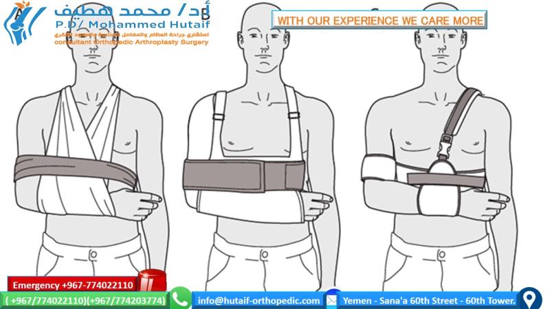
- Immobilization: A long-arm plaster cast or splint is applied in 70-90 degrees of elbow flexion with the forearm in pronation (for extension-type fractures) to maintain reduction and protect pin sites. Excessive flexion should be avoided to prevent compartment syndrome.
- Pin Site Care: If pins are exposed, daily cleaning with antiseptic solution (e.g., chlorhexidine) and sterile dressing changes are performed. Parents are instructed on signs of infection.
- Neurovascular Monitoring: Continue monitoring for signs of neurovascular compromise (e.g., increasing pain, numbness, pallor, decreased capillary refill) for the first 24-48 hours.
- Edema Control: Elevate the limb above heart level to reduce swelling.
- Shoulder and Hand Exercises: Encourage gentle active range of motion of the shoulder and hand/fingers to prevent stiffness in adjacent joints.
- Weight Bearing: No weight bearing or strenuous activity with the affected arm.
- Pain Management: As prescribed by the orthopedic surgeon.
Phase 2: Pin Removal and Initiation of Mobilization (Weeks 3/4 - 6/8)
- Pin Removal: K-wires are typically removed in the clinic or minor operating room after 3-4 weeks, once radiographic signs of early union are visible (e.g., bridging callus).
-
Gentle Active Range of Motion (AROM):
- Following pin removal, a removable splint may be applied for comfort and protection, especially during sleep.
- Initiate gentle, active, gravity-assisted elbow flexion and extension exercises.
- Crucially, avoid passive stretching or forced manipulation. Aggressive passive range of motion can induce pain, muscle guarding, and lead to heterotopic ossification or increased stiffness.
- The child should be encouraged to use the arm for light activities of daily living within comfortable limits.
- Forearm Rotation: Begin gentle active pronation and supination exercises.
- Scar Management (if open reduction): If an open reduction was performed, begin gentle scar massage once the wound is well-healed.
- Weight Bearing: Continue to restrict heavy lifting or pushing activities.
Phase 3: Progressive Strengthening and Functional Restoration (Weeks 6/8 - 12+)
- Physical Therapy (as needed): Formal physical therapy may be initiated if the child is not progressing well with home exercises, exhibits significant stiffness, or has persistent functional limitations. The focus remains on gentle, active-assisted and active ROM.
- Gradual Strengthening: Introduce light strengthening exercises using body weight or very light resistance bands, progressively increasing as tolerated.
-
Return to Activity:
- Gradual return to non-contact sports and recreational activities.
- Avoid high-impact or contact sports until full pain-free range of motion is achieved and strength is adequate, typically 3-6 months post-injury.
- Parental Education: Emphasize the importance of continued home exercises and caution against overdoing activities or allowing others to force the elbow into positions of pain. Reassure parents that full range of motion may take several months to achieve and that some minor loss of terminal extension is not uncommon but rarely functionally significant.
Long-Term Monitoring
- Follow-up X-rays: Routinely obtained at 6-12 weeks post-injury to confirm union and assess for any early signs of malunion (e.g., cubitus varus).
- Clinical Evaluation: Periodic clinical assessment of elbow range of motion, strength, and function. Long-term follow-up (e.g., 6 months, 1 year) is recommended to identify late-onset complications like cubitus varus.
Summary of Key Literature / Guidelines
The management of pediatric supracondylar humerus fractures is well-established, guided by extensive literature and supported by consensus from major orthopedic societies. The core principles emphasize accurate diagnosis, prompt intervention for displaced fractures, meticulous surgical technique, and a structured rehabilitation protocol.
-
Closed Reduction Percutaneous Pinning (CRPP) as Standard of Care: Numerous studies, systematic reviews, and meta-analyses consistently demonstrate that CRPP is the most effective and safest treatment for displaced (Gartland Types II, III, IV) extension-type SCHF. It provides stable fixation, minimizes soft tissue dissection, and allows for rapid healing with low complication rates. The current evidence supports a preference for lateral-entry pinning over cross-pinning to reduce the risk of iatrogenic ulnar nerve injury, without significant compromise in biomechanical stability for most fractures. When cross-pinning is deemed necessary for highly unstable fractures, a mini-open technique with direct visualization of the ulnar nerve is strongly recommended to mitigate nerve injury risk.
- Example studies (though not in the seed data): Many comparative studies from the early 2000s onwards have compared lateral-only vs. cross-pinning, contributing to the shift in practice patterns.
-
Importance of Neurovascular Assessment: Guidelines from organizations such as the American Academy of Orthopaedic Surgeons (AAOS) and the Pediatric Orthopaedic Society of North America (POSNA) highlight the critical importance of serial neurovascular assessment both pre- and post-operatively. Pulseless but perfused hands often recover after reduction and pinning alone, but careful observation for signs of evolving ischemia or compartment syndrome is paramount. Pulseless and ischemic hands necessitate immediate operative exploration.
-
Risk of Cubitus Varus: Malunion leading to cubitus varus remains the most common long-term complication, primarily cosmetic. Literature consistently links inadequate reduction, rotational malalignment, and settling to its development. While not typically functionally limiting, severe cases may warrant corrective osteotomy, usually after skeletal maturity. The goal of initial fixation should be anatomical reduction in all planes to minimize this risk.
-
Avoidance of Aggressive Rehabilitation: The literature strongly advises against passive stretching or aggressive physical therapy in the early post-operative period. Overly aggressive rehabilitation can exacerbate pain, lead to re-displacement, or promote heterotopic ossification, ultimately prolonging recovery or worsening stiffness. Gentle, active-assisted and active range of motion is the cornerstone of successful rehabilitation.
-
Global Contributions to Evidence Base: The global nature of orthopedic research, as highlighted by the diverse countries in the initial data, is essential. While the provided seed data doesn't explicitly contain studies from China, it is imperative to acknowledge the burgeoning volume and increasing quality of orthopedic literature emanating from Chinese research institutions. These contributions, often involving large patient cohorts, are critical for refining diagnostic criteria, surgical techniques, and rehabilitation protocols. As with all research, critical appraisal of study design, methodology, sample characteristics (e.g., age, sex, follow-up duration as presented in the initial data), and reporting standards is essential to integrate findings from all global sources, including China, into best clinical practice. Continued research, including comparative effectiveness studies and long-term outcome analyses, is crucial for further optimizing the management of SCHF and other pediatric orthopedic conditions.
You Might Also Like