Anterior Cruciate Ligament (ACL) Rupture: Epidemiology, Anatomy & Surgical Management
Key Takeaway
ACL reconstruction is often indicated for symptomatic instability in active individuals, high-level athletes aiming to return to pivoting sports, and cases with concomitant knee injuries like meniscal tears. The goal is to restore knee stability, prevent further damage, and enable a safe return to desired physical activities after rehabilitation.
Introduction & Epidemiology
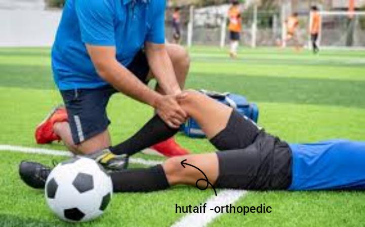
Sports injuries represent a significant burden on athletic populations, ranging from professional athletes to recreational participants. While preventive strategies are paramount, a subset of these injuries necessitates surgical intervention. This review focuses on the current understanding and surgical management of a common, often debilitating, sports injury: anterior cruciate ligament (ACL) rupture. ACL tears are a leading cause of time lost from sport and a significant contributor to long-term knee morbidity, including the development of post-traumatic osteoarthritis.
The incidence of ACL injuries is estimated at 30-78 per 100,000 person-years in the general population, with higher rates observed in specific athletic cohorts. Females demonstrate a 2-8 times higher incidence than males in certain sports, particularly soccer and basketball, a disparity attributed to a complex interplay of anatomical, biomechanical, hormonal, and neuromuscular factors. The majority of ACL injuries (approximately 70-80%) are non-contact in nature, typically occurring during sudden deceleration, cutting, pivoting, or landing maneuvers. Contact injuries, while less common, often involve direct valgus stress to the knee. Understanding the epidemiology of ACL tears underscores the need for robust surgical approaches and comprehensive rehabilitation strategies to restore knee stability and facilitate a safe return to sport.
Surgical Anatomy & Biomechanics
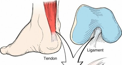
The anterior cruciate ligament is a critical intra-articular stabilizer of the knee, originating from the posteromedial aspect of the lateral femoral condyle and inserting onto a broad area in the anteromedial intercondylar eminence of the tibia. It is an extra-synovial, intra-capsular structure. The ACL is comprised of two functional bundles: the anteromedial (AM) bundle, which is taut in flexion, and the posterolateral (PL) bundle, which is taut in extension.
The femoral attachment spans approximately 18 mm in the long axis, with its anterior-most fibers positioned anterior to the cartilage margin and its posterior-most fibers located superior to the posterior articular cartilage. The tibial footprint is C-shaped, measuring approximately 11x17 mm, located between the medial and lateral tibial spines, anterior to the transverse meniscal ligament. The AM bundle inserts anteromedially on the tibia, while the PL bundle inserts posterolaterally.
Histologically, the ACL is composed primarily of type I collagen fibers (90-95%), with smaller amounts of type III collagen, elastin, and proteoglycans. Mechanoreceptors (Ruffini, Pacinian, Golgi-like endings) and free nerve endings are present, contributing to proprioception and kinesthetic awareness. The ligament’s blood supply is primarily from the middle genicular artery, with contributions from the inferior genicular arteries.
Biomechanically, the ACL is the primary restraint to anterior translation of the tibia relative to the femur, resisting approximately 85% of this anterior shear force, particularly at full extension. It also acts as a secondary restraint to internal rotation of the tibia, valgus/varus rotation (especially in extension), and hyperextension. The AM bundle is more significant in resisting anterior translation in flexion, while the PL bundle provides greater rotational stability in extension. An intact ACL is crucial for dynamic knee stability during pivoting and cutting maneuvers, preventing pathological rotatory laxity (pivot shift phenomenon). Rupture of the ACL results in altered knee kinematics, leading to tibiofemoral subluxation, increased stress on secondary stabilizers (menisci, articular cartilage), and a heightened risk of meniscal tears and articular cartilage damage over time.
Indications & Contraindications
The decision to proceed with surgical reconstruction of the ACL is complex and individualized, balancing patient factors, activity level, associated injuries, and shared decision-making. The primary goal of ACL reconstruction is to restore knee stability, prevent recurrent episodes of instability, reduce the risk of secondary meniscal and chondral damage, and enable a safe return to desired activities.
Indications for Operative Management (ACL Reconstruction)
- Functional Instability: Symptomatic instability ("giving way") during activities of daily living or sport, particularly in young, active individuals involved in pivoting or cutting sports.
- High Activity Level/Athletic Goals: Patients who desire to return to sports requiring agility, rapid deceleration, and pivoting movements (e.g., soccer, basketball, football, skiing).
-
Concomitant Injuries:
- Meniscal tears (especially reparable tears of the medial or lateral meniscus) that would benefit from a stable knee environment for healing.
- Associated collateral ligament injuries (e.g., MCL Grade III or PLC injuries) that require concurrent or staged reconstruction.
- Articular cartilage lesions that may progress with ongoing instability.
- Skeletally Immature Patients: While historically a relative contraindication, physeal-sparing or transphyseal techniques are now performed in skeletally immature patients to prevent growth disturbances, particularly in those with significant instability limiting activity.
- Chronic Instability with Progressive Symptoms: Patients who have failed an adequate trial of non-operative management and continue to experience instability or develop secondary pathology.
Indications for Non-Operative Management
- Low Demand Individuals: Sedentary individuals, those who do not participate in pivoting sports, or older patients whose activity levels do not necessitate a stable knee for high-impact activities.
- Partial ACL Tears: Tears without symptomatic instability or significant functional limitation, confirmed by physical examination and MRI.
- Successful Rehabilitation with No Instability: Patients who can achieve functional stability through comprehensive neuromuscular rehabilitation, strengthening, and activity modification.
- Significant Medical Comorbidities: Patients with medical conditions that preclude safe surgical intervention or rehabilitation compliance.
- Unwillingness to Undergo Surgery or Rehabilitation: Patient preference, after thorough counseling regarding the risks of ongoing instability and potential long-term degenerative changes.
Contraindications for ACL Reconstruction
-
Absolute Contraindications:
- Active infection in or around the knee joint.
- Severe, rapidly progressing degenerative joint disease (though reconstruction can be considered in select cases to address instability prior to arthroplasty).
- Non-ambulatory patient or those with severe neurological deficits precluding meaningful rehabilitation.
-
Relative Contraindications:
- Extreme skeletal immaturity (consider physeal-sparing techniques or delayed reconstruction).
- Significant medical comorbidities posing an unacceptable surgical risk.
- Poor patient compliance or psychological readiness for demanding post-operative rehabilitation.
- Severe generalized ligamentous laxity, which may affect graft tensioning and stability.
Table 1: Operative vs. Non-Operative Indications for ACL Rupture
| Factor | Operative Management | Non-Operative Management |
|---|---|---|
| Activity Level | High-demand athlete, desire to return to pivoting/cutting sports, physically active lifestyle | Low-demand individual, sedentary lifestyle, willingness to modify activities |
| Symptomology | Recurrent functional instability ("giving way"), episodes of knee buckling | No symptomatic instability, well-controlled through bracing/rehabilitation |
| Age | Young (adolescent to middle-aged adult), skeletally mature (or appropriate physeal-sparing for immature) | Older patient, low functional expectations, significant degenerative changes |
| Associated Injuries | Meniscal tears (especially repairable), collateral ligament injuries (e.g., MCL Grade III), articular cartilage lesions | Isolated ACL tear without significant associated injuries that require surgical stabilization |
| Functional Outcome | Desire for high-level athletic function, prevention of recurrent instability and secondary pathology | Acceptance of potential activity limitations, willingness to manage symptoms conservatively |
| Rehabilitation Adherence | High motivation and expected compliance with intensive post-operative rehabilitation program | Unwillingness or inability to commit to rigorous rehabilitation, poor compliance history |
| Medical Comorbidities | Acceptable surgical risk profile | Significant medical comorbidities precluding safe surgery or anesthetic risks |
| Previous Treatment | Failed conservative management with persistent instability or progressive secondary damage | Successful trial of conservative management, stable knee with activity modification |
Pre-Operative Planning & Patient Positioning
Thorough pre-operative planning is essential for a successful ACL reconstruction. This involves comprehensive patient evaluation, detailed imaging review, graft selection, and meticulous surgical setup.
Pre-Operative Planning
-
Clinical Evaluation:
- History: Detailed account of injury mechanism, symptoms (pain, swelling, instability), previous knee injuries, and activity level.
- Physical Examination: Assessment of effusion, range of motion, and ligamentous laxity (Lachman test, anterior drawer test, pivot shift test). A positive pivot shift test is highly specific for ACL insufficiency and indicates rotatory instability. Evaluation of concomitant injuries (menisci, collateral ligaments, posterolateral corner).
-
Imaging:
- Radiographs: AP, lateral, Merchant (patellofemoral) views to rule out fractures, assess growth plates in skeletally immature patients, and identify signs of early osteoarthritis or osteophytes. Rosenberg view (posteroanterior weight-bearing flexion) can reveal joint space narrowing. Stress radiographs may be used in specific cases.
- Magnetic Resonance Imaging (MRI): Confirms ACL tear, identifies associated injuries (meniscal tears, chondral lesions, collateral ligament sprains, bone bruises), and assesses ligamentous integrity. Pre-operative MRI also aids in identifying the native ACL footprint remnants, which can guide anatomical tunnel placement.
-
Graft Selection:
The choice of graft is critical and depends on surgeon preference, patient factors (age, activity level, prior surgeries), and anticipated graft harvest morbidity.
-
Autografts (patient's own tissue):
- Bone-Patellar Tendon-Bone (BPTB): Gold standard for high-demand athletes, excellent primary stability, bone-to-bone healing. Disadvantages: anterior knee pain, patellar fracture risk, patellar tendon shortening.
- Hamstring Tendons (Semitendinosus and Gracilis - STG): Lower harvest site morbidity, cosmetically more appealing. Disadvantages: potentially slower bone-to-tendon healing, hamstring weakness.
- Quadriceps Tendon (QT): Increasingly popular, especially with a bone block. Offers robust tissue and low harvest site morbidity, similar biomechanical properties to BPTB. Disadvantages: anterior knee pain, quadriceps weakness.
-
Allografts (donor tissue):
- Less harvest site morbidity, shorter operative time. Disadvantages: potential for disease transmission (though rare with current processing), slower incorporation, higher re-rupture rates in younger, active patients, higher cost.
- Synthetic Grafts: Primarily used for augmentation or revision cases, not recommended for primary reconstruction due to high failure rates and synovitis.
-
Autografts (patient's own tissue):
- Surgical Strategy: Determine type of reconstruction (single vs. double bundle - single bundle is standard), tunnel placement technique (transtibial, anteromedial portal, outside-in), and fixation methods.
- Pre-operative Education: Inform the patient about the surgical procedure, potential risks, expected post-operative course, and the demanding rehabilitation protocol.
Patient Positioning
- Supine Position: The patient is placed supine on the operating table.
- Leg Holder/Side Support: A padded leg holder or lateral side support is applied to the contralateral thigh to stabilize the pelvis and prevent rotation. The ipsilateral leg is positioned to allow full range of motion, particularly hyperextension and flexion.
- Tourniquet: A pneumatic tourniquet is typically applied to the proximal thigh to ensure a bloodless field, crucial for optimal visualization during arthroscopy. The tourniquet is inflated after exsanguination of the limb.
- Foot Drape/Distraction: The foot may be left free or placed in a foot holder/distraction boot, which can be connected to an adjustable support to facilitate valgus/varus stress and flexion/extension maneuvers, especially during meniscal repair or graft tensioning.
- Sterile Preparation and Draping: The entire leg, from mid-thigh to the foot, is meticulously prepped with an antiseptic solution and sterilely draped to isolate the operative field. A sterile stockinette or impervious drape may be used over the foot to allow manipulation by the assistant.
- Pillow/Bump: A small bump or pillow may be placed under the ipsilateral gluteal region to rotate the hip internally, facilitating better access to the medial aspect of the knee for hamstring graft harvest. A bump under the distal thigh/proximal calf can aid in achieving optimal knee flexion.
Detailed Surgical Approach / Technique (ACL Reconstruction with Hamstring Autograft)
The following describes a common arthroscopic single-bundle ACL reconstruction using a quadrupled semitendinosus and gracilis autograft.
1. Diagnostic Arthroscopy & Debridement
- Standard Portals: Anterolateral (AL) and Anteromedial (AM) portals are established. The AL portal, typically 1 cm lateral to the patellar tendon at the level of the inferior pole of the patella, is used for the arthroscope. The AM portal, 1 cm medial to the patellar tendon, serves as the working portal. An accessory AM portal may be used for specific instruments or sutures.
- Systematic Evaluation: A comprehensive arthroscopic survey is performed to confirm the ACL rupture, assess meniscal integrity (repair or partial meniscectomy as needed), evaluate articular cartilage surfaces, and inspect other ligaments.
- Debridement: The remnant torn ACL tissue is debrided using a shaver or motorized resector to expose the native femoral and tibial footprints. Care is taken to preserve any viable ACL remnant tissue that may aid in graft healing and vascularization, provided it does not impede tunnel placement or graft passage.
2. Graft Harvest (Semitendinosus and Gracilis Autograft)
- Incision: A 2-3 cm oblique longitudinal incision is made over the anteromedial aspect of the proximal tibia, approximately 2-3 cm distal and medial to the tibial tubercle, parallel to the sartorius fascia.
- Dissection: The subcutaneous tissue is incised, and the sartorius fascia is identified. The fascia is incised longitudinally, revealing the semitendinosus and gracilis tendons lying deep to it. The pes anserinus is identified.
- Tendon Release: The semitendinosus tendon, typically superior and larger, and the gracilis tendon, inferior and smaller, are identified. A tendon stripper (open or closed-end) is passed along the length of each tendon, carefully detaching them from their muscular bellies while minimizing collateral tissue damage.
- Graft Preparation: The harvested tendons are cleared of muscle and fascia. The ends are whip-stitched with strong non-absorbable suture. The tendons are then folded to create a quadrupled graft of appropriate length and diameter (typically 8-10 mm). The graft is pre-tensioned on a graft board to minimize creep.
3. Femoral Tunnel Creation (Anteromedial Portal Technique)
- Femoral Footprint Identification: With the knee hyperflexed (110-130 degrees), the native femoral ACL footprint is identified on the posteromedial aspect of the lateral femoral condyle. The goal is an anatomical tunnel position.
- Guide Pin Placement: An ACL guide pin (e.g., 2.4 mm) is introduced through the AM portal. The tip of the guide pin is placed at the center of the desired femoral footprint, typically at the 10 o'clock position (right knee) or 2 o'clock position (left knee), ensuring it is posterior to the posterior femoral cortex to avoid a "killer turn" and posterior wall blow-out.
- Tunnel Reaming: A cannulated femoral reamer, matched to the prepared graft diameter, is advanced over the guide pin. The reamer is stopped once the desired tunnel depth (typically 20-25 mm) is achieved. The depth is measured from the articular surface. The reamer is then reversed and removed. The tunnel edges are chamfered to prevent graft abrasion.
4. Tibial Tunnel Creation
- Tibial Footprint Identification: Using the AM portal, the native tibial ACL footprint is identified between the tibial spines, anterior to the posterior cruciate ligament (PCL).
- Tibial Guide Placement: An offset tibial guide (e.g., 55 degrees) is used. The tip of the guide is placed at the center of the desired tibial footprint, aiming for an anatomical position that avoids impingement on the roof or notch in full extension.
- Guide Pin Placement: The guide pin is drilled from the anteromedial tibia, approximately 3-4 cm distal to the joint line and medial to the tibial tubercle, exiting at the desired intra-articular footprint.
- Tunnel Reaming: A cannulated tibial reamer, matched to the graft diameter, is advanced over the guide pin. The reamer is advanced until it breaches the joint space, creating a full-length tunnel. The tunnel should be smooth, without sharp edges. The position is checked arthroscopically throughout the range of motion for impingement.
5. Graft Passage and Fixation
- Suture Passage: A passing suture (e.g., shuttle wire) is retrieved from the femoral tunnel, through the notch, and out the tibial tunnel. The sutures from the graft are attached to this passing suture.
- Graft Passage: The graft is carefully pulled through the tibial tunnel, across the joint, and into the femoral tunnel using the passing suture. Care is taken to avoid graft impingement, fraying, or kinking. A notchplasty may be performed if impingement is anticipated.
-
Femoral Fixation:
- Suspensory Fixation (e.g., Endobutton, adjustable loop device): This is a common method for hamstring grafts. The device is passed through the femoral tunnel and flipped on the lateral femoral cortex, allowing the graft to be suspended from it.
- Interference Screw: A metallic or bioabsorbable interference screw can be placed between the graft and tunnel wall, compressing the graft within the tunnel.
-
Tibial Fixation:
- Interference Screw: After tensioning the graft (typically at 20-30 degrees of flexion to reduce posterior tibial sag and allow for an isometric position), an interference screw is carefully inserted between the graft and the tibial tunnel wall. The length and diameter of the screw are selected to provide adequate fixation without damaging the graft or widening the tunnel.
- Additional Fixation: A post screw and washer, a staple, or a hybrid fixation technique may be used to augment tibial fixation, especially with hamstring grafts, to prevent slippage.
- Tensioning: The graft is tensioned with continuous cycling through a full range of motion (0-90 degrees) multiple times to ensure proper seating and reduce viscoelastic creep. Final tensioning is typically performed at 20-30 degrees of knee flexion, and then fixation is applied.
- Final Assessment: After fixation, arthroscopic visualization confirms secure graft position, absence of impingement, and appropriate tension. The Lachman and pivot shift tests are performed to confirm stability.
6. Closure
- The arthroscopic portals are closed with a simple suture or sterile strips.
- The hamstring harvest incision is closed in layers.
- A sterile dressing is applied, and the knee is often placed in a post-operative brace set to limit range of motion.
Complications & Management
Despite advancements in surgical technique and rehabilitation, complications following ACL reconstruction can occur, impacting patient outcomes. Recognition and prompt management are crucial.
Intra-operative Complications
-
Nerve/Vascular Injury (Incidence <1%):
Popliteal artery, peroneal nerve, saphenous nerve.
- Management: Meticulous dissection, careful portal placement, clear visualization, tourniquet control. If recognized, immediate repair (vascular) or nerve grafting/neurolysis.
-
Chondral Damage (Incidence 1-5%):
Iatrogenic damage to articular cartilage during arthroscopy or tunnel reaming.
- Management: Careful instrument handling, precise portal placement. Microfracture or debridement if significant; consider cartilage repair strategies in large defects.
-
Fracture (Femoral/Tibial, Patellar with BTB harvest - Incidence <1%):
- Management: Careful reaming technique, avoiding over-reaming. Open reduction internal fixation (ORIF) if significant, or conservative management for stable, non-displaced fractures.
-
Graft Malpositioning (Incidence 5-10%):
Femoral or tibial tunnel malposition (e.g., too anterior/posterior, too vertical/horizontal).
- Management: Prevention through careful anatomical landmark identification and precise guide placement. If recognized intraoperatively, new tunnels may be drilled and the graft repositioned; otherwise, revision surgery may be required for symptomatic instability.
-
Posterior Wall Blow-out (Femoral Tunnel) (Incidence <1%):
Reaming too far posteriorly, compromising femoral fixation.
- Management: Prevention by confirming adequate posterior wall thickness. May require alternative fixation methods (e.g., larger suspensory device, additional screw) or revision tunnel creation.
-
Graft Impingement:
Graft impinging on the intercondylar notch or femoral roof in full extension.
- Management: Prevention through proper tunnel placement and adequate notchplasty if indicated. If persistent, revision notchplasty may be required.
Early Post-operative Complications
-
Infection (Incidence 0.1-1%):
Septic arthritis or superficial wound infection.
- Management: Prophylactic antibiotics. For septic arthritis, urgent arthroscopic irrigation and debridement, appropriate antibiotic therapy. Superficial infections managed with oral antibiotics and local wound care.
-
Deep Vein Thrombosis (DVT) / Pulmonary Embolism (PE) (Incidence DVT 0.5-2%, PE <0.1%):
- Management: Early mobilization, mechanical prophylaxis (compression stockings, intermittent pneumatic compression devices). Pharmacological prophylaxis for high-risk patients. Treatment with anticoagulation.
-
Hemarthrosis (Incidence 5-10%):
Excessive bleeding into the joint.
- Management: Aspiration of the knee if significant, rest, ice, compression, elevation. Occasionally, arthroscopic washout.
-
Arthrofibrosis / Stiffness (Incidence 2-10%):
Loss of range of motion, particularly extension, due to scar tissue formation.
- Management: Aggressive rehabilitation, dynamic splinting. If conservative measures fail, manipulation under anesthesia (MUA) or arthroscopic lysis of adhesions (arthrolysis) may be necessary, typically not before 3 months post-op.
-
Patellar Fracture (BTB harvest - Incidence <0.5%):
Stress riser from bone block harvest.
- Management: Non-operative for non-displaced fractures. ORIF for displaced fractures.
-
Anterior Knee Pain (BTB harvest - Incidence 10-30%):
Pain around the patella and patellar tendon.
- Management: Rehabilitation focus on quadriceps strengthening, patellar mobilization, physical therapy modalities. May persist despite treatment.
-
Flexion Contracture:
Inability to achieve full extension.
- Management: Aggressive physiotherapy, static progressive stretching, dynamic splinting. MUA or arthroscopic lysis if refractory.
Late Post-operative Complications
-
Graft Failure / Re-rupture (Incidence 2-10%):
Recurrent instability due to graft elongation, mechanical failure, or new traumatic event. Higher rates in young athletes returning to high-risk sports.
- Management: Revision ACL reconstruction, addressing specific causes of failure (e.g., malposition, tunnel widening, unrecognized instability).
-
Persistent Instability:
Despite an intact graft, functional instability may persist due to residual laxity or unrecognized associated ligamentous injury.
- Management: Intensive rehabilitation, bracing. Revision surgery if severe and symptomatic, possibly with lateral extra-articular tenodesis.
-
Post-Traumatic Osteoarthritis (PTOA) (Incidence 10-50% long-term):
Degenerative changes of articular cartilage, regardless of successful reconstruction.
- Management: Prevention through early stabilization, managing meniscal tears effectively, and appropriate rehabilitation. Symptomatic management with NSAIDs, physical therapy, injections. Arthroplasty in severe cases.
-
Hardware Complications (Incidence 1-5%):
Prominent or painful screws/buttons, hardware loosening.
- Management: Removal of hardware if symptomatic, typically after graft incorporation (6-12 months).
-
Tunnel Widening (Incidence 10-20%):
Expansion of femoral or tibial tunnels, especially with suspensory fixation or bioabsorbable screws.
- Management: Usually asymptomatic. May complicate revision surgery. Bone grafting for significant widening.
Table 2: Common Complications, Incidence, and Salvage Strategies
| Complication | Incidence (Approx.) | Salvage Strategies / Management |
|---|---|---|
| Intra-operative | ||
| Nerve/Vascular Injury | <1% | Immediate repair, careful technique, clear visualization |
| Chondral Damage | 1-5% | Microfracture, debridement, cartilage repair for larger defects |
| Graft Malposition | 5-10% | Immediate revision of tunnel/graft placement; if recognized late, revision surgery |
| Posterior Wall Blow-out | <1% | Alternative fixation (e.g., larger suspensory), revision tunnel if needed |
| Early Post-operative | ||
| Infection (Septic Arthritis) | 0.1-1% | Urgent arthroscopic I&D, targeted antibiotics |
| DVT/PE | DVT 0.5-2%, PE <0.1% | Early mobilization, mechanical/pharmacological prophylaxis, anticoagulation |
| Arthrofibrosis / Stiffness | 2-10% | Aggressive physical therapy, MUA, arthroscopic lysis of adhesions |
| Anterior Knee Pain (BPTB) | 10-30% | PT, activity modification, NSAIDs; rarely hardware removal |
| Late Post-operative | ||
| Graft Failure / Re-rupture | 2-10% | Revision ACL reconstruction (addressing cause), potentially with lateral augmentation |
| Persistent Instability | 1-5% | Intensive PT, bracing, revision surgery (possibly with extra-articular tenodesis) |
| Post-Traumatic Osteoarthritis | 10-50% (long-term) | Symptomatic management (PT, NSAIDs, injections), activity modification; arthroplasty in severe cases |
| Hardware Complications | 1-5% | Removal of symptomatic hardware after graft incorporation |
| Tunnel Widening | 10-20% | Typically asymptomatic; may require bone grafting for revision surgery |
Post-Operative Rehabilitation Protocols
Post-operative rehabilitation is as critical as the surgery itself for a successful outcome in ACL reconstruction. Protocols are typically structured into phases, emphasizing progressive loading, restoration of range of motion (ROM), strength, neuromuscular control, and gradual return to sport. These protocols are usually criterion-based rather than purely time-based, allowing for individualized progression.
Phase I: Protection and Early Motion (Weeks 0-4)
- Goals: Protect the healing graft, minimize pain and swelling, achieve full knee extension, restore quadriceps activation, and initiate early flexion.
- Weight-Bearing (WB): Typically partial weight-bearing (PWB) with crutches for 1-2 weeks, progressing to full weight-bearing (FWB) as tolerated. Brace locked in extension for ambulation initially.
- Bracing: Post-operative brace worn for ambulation for 4-6 weeks, allowing controlled ROM.
-
Exercises:
- ROM: Immediate passive knee extension exercises (e.g., heel props), active-assisted flexion up to 90 degrees by 2 weeks.
- Quadriceps Activation: Quadriceps setting exercises, straight leg raises (SLR) in various planes with brace locked in extension.
- Other: Ankle pumps (DVT prophylaxis), gentle hamstring curls (if BPTB graft), gluteal sets, core strengthening.
- Key Consideration: Avoid open-kinetic chain (OKC) knee extension exercises from 0-45 degrees to protect the healing graft from anterior shear forces.
Phase II: Strength and Neuromuscular Control (Weeks 4-12)
- Goals: Restore full knee ROM, normalize gait, regain lower extremity strength, improve proprioception and neuromuscular control.
- WB/Bracing: Discontinue crutches and often the brace once stable gait is achieved and quadriceps control is sufficient.
-
Exercises:
- ROM: Progress full ROM (0-135 degrees) gradually.
-
Strengthening:
- Closed Kinetic Chain (CKC): Wall squats, mini-squats, leg press, step-ups/downs, lunges. Emphasize proper mechanics.
- Open Kinetic Chain (OKC): Hamstring curls, calf raises. Gentle OKC knee extension (e.g., leg extension machine) may be introduced beyond 45-90 degrees flexion, cautiously progressing range as appropriate for graft type (later for hamstring).
- Neuromuscular Control: Balance exercises (single leg stance, wobble board, foam pad), perturbation training.
- Core Strengthening: Plank variations, bridging.
- Key Consideration: Continue to monitor for effusion and pain. Avoid activities that induce pivoting or excessive valgus stress.
Phase III: Return to Activity (Weeks 12-24)
- Goals: Enhance strength, power, agility, and endurance; introduce sport-specific movements.
-
Exercises:
- Progressive Strengthening: Increase resistance and intensity of CKC and OKC exercises. Plyometrics (box jumps, hopping, jumping).
- Agility Drills: Ladder drills, cone drills, lateral shuffling, cutting maneuvers (gradually progressing from controlled to unpredictable).
- Sport-Specific Training: Introduce light sport-specific drills, gradually increasing intensity and complexity.
- Cardiovascular: Treadmill running, elliptical, cycling, swimming.
- Key Consideration: Functional testing (e.g., single leg hop tests, Y-balance test) becomes increasingly important for guiding progression. Graft protection remains a priority.
Phase IV: Return to Sport (Weeks 24+)
- Goals: Full return to sport-specific training and competitive play, minimizing risk of re-injury.
-
Criteria for Return to Sport:
- Time: Generally 9-12 months post-surgery, with growing evidence suggesting waiting longer (e.g., >9 months) reduces re-rupture risk, especially in younger athletes.
- Strength: Quadriceps and hamstring strength 90% or more compared to the uninjured leg (isokinetic testing).
- Functional Testing: Hop test symmetry >90% (single hop, triple hop, crossover hop, 6-meter timed hop). Y-balance test symmetry.
- Neuromuscular Control: Excellent dynamic stability and balance.
- Psychological Readiness: Patient confidence and lack of kinesiophobia (fear of movement/re-injury).
- Clinical Evaluation: Absence of pain, swelling, or instability on examination.
- Key Consideration: A multidisciplinary team approach (surgeon, physical therapist, athletic trainer, sports psychologist) is beneficial. Gradual integration back into sport is crucial. The risk of re-rupture is highest in the first 1-2 years post-op, particularly in adolescents and young adults.
Summary of Key Literature / Guidelines
The landscape of ACL reconstruction has been shaped by extensive research, leading to evolving guidelines and best practices. Key areas of ongoing study include graft choice, tunnel positioning, fixation techniques, and rehabilitation strategies.
-
Graft Choice:
- Autografts vs. Allografts: Multiple systematic reviews and meta-analyses, including those by the MOON (Multicenter Orthopaedic Outcomes Network) group , consistently demonstrate superior outcomes and lower re-rupture rates with autografts, particularly in younger, active individuals. Allografts show higher failure rates, slower incorporation, and higher costs, generally reserved for revision surgery, older patients, or those unwilling to accept autograft harvest morbidity.
- BPTB vs. Hamstring: The choice between BPTB and hamstring autografts remains a topic of debate. While BPTB historically had slightly lower re-rupture rates in some high-level athletes, it is associated with higher rates of anterior knee pain and patellofemoral issues. Hamstring grafts generally have comparable clinical outcomes but may demonstrate slightly greater residual laxity in some studies. Quadriceps tendon autograft is gaining popularity, showing robust biomechanical properties and potentially lower anterior knee pain compared to BPTB, with good graft volume.
- Level of Evidence: Predominantly Level I and II evidence supports the use of autografts over allografts for primary ACL reconstruction in active populations. Comparisons between BPTB, hamstring, and quadriceps autografts often show similar functional outcomes but differing patterns of donor site morbidity.
-
Surgical Technique - Anatomical Reconstruction:
- The concept of anatomical ACL reconstruction has largely replaced earlier isometric placement theories. Anatomical reconstruction aims to reproduce the native ACL's femoral and tibial footprints, often through the use of the anteromedial (AM) portal drilling technique for the femoral tunnel, which allows for more horizontal graft placement and potentially better rotational stability compared to the transtibial technique.
- Single-Bundle vs. Double-Bundle: While double-bundle reconstruction anatomically mimics the AM and PL bundles, systematic reviews have not consistently shown superior clinical outcomes (e.g., preventing PTOA, improving return to sport rates) compared to well-performed single-bundle anatomical reconstructions. Double-bundle techniques are technically more demanding, require larger tunnels, and may have increased risks of complications (e.g., tunnel confluence, stiffness). Current consensus often favors robust single-bundle anatomical reconstruction as the standard.
- Lateral Extra-articular Tenodesis (LET): The role of concomitant LET or anterolateral ligament (ALL) reconstruction, particularly in revision cases or high-risk primary cases (young athletes with high-grade pivot shift, generalized laxity), is gaining attention. Emerging evidence, including randomized controlled trials, suggests that adding an LET may reduce re-rupture rates in specific high-risk cohorts, although its routine use in all primary cases remains debated due to potential for stiffness and over-constraint.
-
Fixation Methods:
- Various fixation methods (interference screws, suspensory devices, staples, cross-pins) are available. Current literature supports that effective fixation is achieved by multiple techniques, with no single method consistently demonstrating clear superiority in terms of graft incorporation or long-term outcomes. The choice often depends on graft type and surgeon preference.
- Bioabsorbable vs. Metallic Screws: Both are effective. Bioabsorbable screws have the advantage of being incorporated over time, potentially avoiding hardware complications, but may be associated with tunnel widening in some cases.
-
Rehabilitation Guidelines:
- Current rehabilitation protocols emphasize early range of motion, early weight-bearing, and progressive strengthening , moving away from prolonged immobilization.
- Criteria-based progression is strongly advocated over purely time-based protocols, as it allows for individualized advancement based on specific milestones (e.g., strength, hop tests, neuromuscular control).
- Delayed Return to Sport: Increasing evidence suggests that delaying return to pivoting/cutting sports beyond 9 months, and ideally closer to 12 months, particularly in adolescent athletes, significantly reduces the risk of re-rupture. Functional testing and psychological readiness are critical components.
- Focus on Neuromuscular Control: Emphasis on balance, proprioception, and landing mechanics is paramount to address neuromuscular deficits post-injury and post-surgery.
-
Future Directions:
- Biologic Augmentation: Research into the use of biologics (platelet-rich plasma, stem cells) to enhance graft healing and reduce failure rates is ongoing, though strong clinical evidence for routine use is still lacking.
- Internal Bracing: Augmentation of primary ACL repair with an internal brace (suture tape) is an emerging technique, potentially enabling earlier return to activity with certain graft-sparing benefits.
- Predictors of Outcome: Identifying genetic, biomechanical, and psychological factors that predict graft failure or the development of PTOA is a critical area of research.
In conclusion, ACL reconstruction is a highly effective procedure for restoring knee stability in appropriately selected patients. Success hinges on meticulous surgical technique, careful graft selection, and a rigorous, individualized rehabilitation program. Ongoing research continues to refine these aspects, aiming to further optimize patient outcomes and minimize long-term morbidity.
You Might Also Like