Achieve Your Comeback: Orthopedic Rehab to Get Back to Your Best
Key Takeaway
Looking for accurate information on Achieve Your Comeback: Orthopedic Rehab to Get Back to Your Best? Orthopedic rehabilitation is a specialized therapy aiding recovery from musculoskeletal injuries or surgeries affecting bones, muscles, tendons, and ligaments. It uses exercise, manual therapy, and modalities to improve range of motion, strength, and flexibility. This therapy is crucial for reducing pain, restoring functional ability, and getting you back to your best, enhancing overall quality of life.
Introduction & Epidemiology
Orthopedic rehabilitation represents a critical continuum of care extending from pre-operative optimization through acute post-operative recovery and long-term functional restoration. Within the rigorous framework of musculoskeletal surgery, rehabilitation is not merely an adjunctive therapy but an integral component determining the ultimate success of operative intervention and the patient's return to maximal functional capacity. This perspective emphasizes rehabilitation as a scientific discipline, grounded in biomechanics, physiology, and evidence-based practice, essential for surgical trainees and practicing surgeons.
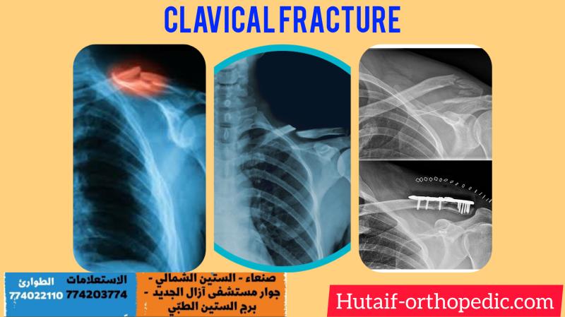
Epidemiologically, musculoskeletal conditions account for a substantial global health burden, impacting quality of life and imposing significant economic costs. Injuries, degenerative diseases, and congenital deformities frequently necessitate surgical intervention. Fractures, for instance, affect millions annually worldwide, with osteoporotic fractures representing a growing challenge in an aging population. Osteoarthritis and rheumatoid arthritis drive millions of joint replacement procedures each year. Sports-related injuries, ranging from ligamentous tears to complex articular fractures, exhibit high incidence rates, particularly in athletic cohorts. The successful management of these conditions, from surgical technique to structured post-operative rehabilitation, is paramount to mitigating disability and facilitating a robust "comeback" for patients. Effective rehabilitation minimizes complications, accelerates recovery, and optimizes long-term outcomes, thereby influencing healthcare resource utilization and patient satisfaction metrics.
Surgical Anatomy & Biomechanics
A profound understanding of surgical anatomy and biomechanics is foundational for any orthopedic surgeon. Surgical approaches are meticulously planned to minimize iatrogenic damage, exploit internervous planes, and optimize visualization of critical structures. Precise anatomical knowledge of bones, articular surfaces, capsuloligamentous complexes, musculotendinous units, and the neurovascular network is indispensable for safe and effective surgery.
- Bones: Understanding bone morphology, density, vascular supply, and healing potential is crucial for fracture fixation, osteotomy planning, and arthroplasty. Cortical versus cancellous bone properties dictate screw purchase and implant selection.
- Joints: Detailed knowledge of articular congruity, meniscal/labral structures, and capsuloligamentous stabilizers is vital for joint preservation, reconstruction, and arthroplasty. The specific kinematic patterns of each joint (e.g., knee screw-home mechanism, shoulder rotation) inform operative technique and guide post-operative rehabilitation.
- Muscles & Tendons: Understanding muscle origins, insertions, bellies, and their synergistic/antagonistic actions is critical for soft tissue releases, repairs, and transfers. Tendon tensile strength, elasticity, and healing biology dictate repair strategies and early mobilization protocols.
- Neurovascular Structures: Meticulous identification and protection of nerves and vessels within surgical corridors are paramount to prevent devastating complications. Familiarity with common variations and danger zones is essential. For example, the common peroneal nerve is vulnerable posterolateral to the knee, and the axillary nerve is at risk during deltoid-splitting approaches to the shoulder.
Biomechanics dictates the etiology of injury, principles of surgical repair, and the progression of rehabilitation.
- Injury Biomechanics: Understanding load application, stress risers, and failure modes helps surgeons anticipate injury patterns and plan reconstructive strategies (e.g., tensile failure of ligaments, compressive failure of vertebrae, shear forces in complex fractures).
- Fixation Biomechanics: Principles such as load sharing, stress shielding, interfragmentary compression, and relative stability govern implant selection and application in fracture management. For instance, absolute stability for articular fractures versus relative stability for diaphyseal fractures.
- Rehabilitation Biomechanics: Controlled loading of healing tissues, progressive resistance training, and functional movement patterns are designed to optimize tissue regeneration and functional adaptation while protecting repairs. Wolff's Law (bone remodeling in response to stress) and Davis's Law (soft tissue adaptation) are fundamental concepts. Understanding lever arms, muscle force vectors, and joint reaction forces guides exercise prescription and activity progression.
Indications & Contraindications
The decision to proceed with orthopedic surgical intervention is a complex process, weighing potential benefits against risks, patient factors, and the natural history of the condition. It necessitates a thorough evaluation of operative versus non-operative indications.
General Operative Indications:
- Failure of Non-Operative Management: Persistent pain, functional limitation, or progressive deformity despite adequate conservative treatment (e.g., physical therapy, medication, injections, bracing).
- Mechanical Instability: Gross joint instability, recurrent dislocations, or pathological motion compromising function or risking further damage (e.g., ACL rupture in an active individual, recurrent patellar dislocation).
- Structural Derangement: Displaced or unstable fractures requiring anatomical reduction and stable fixation, severe articular incongruity, large irreparable soft tissue defects, or mechanical blocks to motion.
- Neurological Compromise: Acute or progressive neurological deficit related to spinal or peripheral nerve compression requiring decompression (e.g., cauda equina syndrome, acute carpal tunnel syndrome).
- Tumor/Infection: Biopsy for diagnosis, débridement, or resection of malignant or aggressive benign tumors, or management of septic arthritis/osteomyelitis unresponsive to antibiotics.
- Life/Limb Threatening Conditions: Compartment syndrome, open fractures, vascular injury requiring surgical exploration/repair.
General Contraindications:
-
Absolute Contraindications:
- Active systemic infection or local infection at the surgical site.
- Severe uncorrectable medical comorbidities precluding safe anesthesia or surgical stress (e.g., uncontrolled cardiac disease, severe coagulopathy).
- Lack of critical tissue for repair or reconstruction (e.g., insufficient bone stock, severely compromised soft tissue envelope).
-
Relative Contraindications:
- Unrealistic patient expectations or poor patient compliance with post-operative rehabilitation protocols.
- Significant psychological comorbidities unaddressed.
- Poor nutritional status.
- Active substance abuse.
- Advanced age or frailty, where surgical risks may outweigh potential benefits, necessitating a careful risk-benefit analysis.
- Obesity, which can increase surgical complexity and complication rates, often warrants pre-operative weight loss.
- Smoking, a known deterrent to bone and soft tissue healing, often requires cessation prior to elective procedures.
Operative vs. Non-Operative Indications
| Indication Category | Operative Criteria | Non-Operative Criteria | Examples |
|---|---|---|---|
| Fractures | Displaced, unstable, open, intra-articular step-off > 2mm, poly-trauma | Non-displaced, stable, closed, minimal articular involvement, low demand patient | Tibia shaft fracture (displaced) vs. Non-displaced distal radius fracture |
| Ligamentous Injuries | Complete rupture (Grade III) with instability, failed conservative therapy, young active patient | Partial tears (Grade I/II), stable joint, older/low demand patient, excellent conservative response | ACL rupture (active) vs. Grade I MCL sprain |
| Degenerative Conditions | Severe pain/functional impairment despite conservative care, radiographic evidence of advanced arthritis, mechanical symptoms | Mild-moderate symptoms, early radiographic changes, good response to conservative management | End-stage hip osteoarthritis vs. Mild knee OA treated with PT/injections |
| Tendon Injuries | Complete rupture (e.g., Achilles tendon, rotator cuff, quadriceps tendon) in functional patient | Partial tears, tendinopathy, good response to conservative measures | Full-thickness rotator cuff tear vs. Supraspinatus tendinopathy |
| Nerve Compression | Progressive neurological deficit, intractable pain, failure of conservative therapy, motor weakness | Sensory symptoms only, intermittent pain, good response to conservative measures | Carpal Tunnel Syndrome with thenar atrophy vs. mild CTS with nocturnal paresthesia |
Pre-Operative Planning & Patient Positioning
Meticulous pre-operative planning is a cornerstone of successful orthopedic surgery, reducing operative time, mitigating complications, and optimizing outcomes.
Pre-Operative Planning
-
Clinical Assessment:
- History & Physical Examination: Comprehensive medical history, current medications, allergies, social history (smoking, alcohol), functional status, and detailed examination of the affected region, including neurovascular assessment.
- Comorbidity Management: Optimization of chronic medical conditions (e.g., diabetes, hypertension, cardiac disease) with primary care or specialist consultations. Assessment of anesthetic risk (ASA classification).
- Nutritional Status: Evaluation and optimization, as malnutrition can impair wound healing and increase infection risk.
- Psychosocial Factors: Assessment of patient expectations, motivation for rehabilitation, social support, and potential psychological barriers to recovery.
-
Imaging Review & Templating:
- Radiographs: Standard views are essential. Weight-bearing views for lower extremity joint pathology.
- Advanced Imaging: CT for complex fractures, articular involvement, or malunion assessment; MRI for soft tissue injuries (ligaments, tendons, menisci, labrum) and occult fractures.
- Surgical Templating: Critical for arthroplasty (implant size, alignment, offset), osteotomies (correction angles), and complex fracture fixation (plate length, screw trajectories). Digital templating has become standard, improving accuracy and reducing revision rates.
- Informed Consent: Detailed discussion of the proposed procedure, alternative treatments (including non-operative), potential risks (general and specific to the procedure), expected benefits, and the rehabilitation pathway. Clear communication regarding realistic outcomes is vital.
- Blood Management: Pre-operative anemia screening, consideration of blood conservation strategies (e.g., antifibrinolytics, cell savers), and blood product availability.
- Infection Prophylaxis: Administration of prophylactic antibiotics (typically first- or second-generation cephalosporins) within 60 minutes of incision. Skin preparation protocols.
- Pre-habilitation: Engaging patients in pre-operative physical therapy can improve strength, range of motion, and functional status, potentially leading to faster recovery post-surgery. Education on post-operative expectations and exercises.
Patient Positioning
Correct patient positioning is critical for surgical exposure, patient safety, and optimal outcomes.
-
Principles:
- Exposure: Adequate visualization of the surgical field while protecting surrounding tissues.
- Safety: Prevention of iatrogenic nerve compression, vascular compromise, skin breakdown, and compartment syndrome.
- Anesthesia Access: Ensuring unhindered access for airway management and monitoring.
- Stability: Secure positioning to prevent unwanted movement during the procedure.
-
Common Positions & Considerations:
- Supine: Most common for anterior approaches to the hip, knee, and shoulder. Careful padding of heels, sacrum, and elbows. Head in neutral position.
- Prone: Used for posterior spinal approaches, posterior hip approaches (rarely), and posterior lower extremity surgery. Chest rolls to allow diaphragmatic excursion, careful neck alignment, meticulous padding of knees, shins, and feet.
- Lateral Decubitus: Common for shoulder arthroscopy/arthroplasty (beach chair variant), lateral hip approaches, or lateral lower extremity procedures. Axillary roll to protect brachial plexus, careful padding of bony prominences (greater trochanter, fibular head).
- Beach Chair: A variant of semi-sitting supine, primarily for shoulder surgery. Requires careful head and neck positioning to prevent cerebral hypoperfusion and ensure airway patency. Neuromonitoring (e.g., transcranial Doppler) may be considered.
- Pressure Point Management: Use of gel pads, foam, and careful draping to distribute pressure and prevent nerve palsies (e.g., ulnar nerve at elbow, common peroneal nerve at fibular head), skin necrosis, or rhabdomyolysis from prolonged compression.
- Neurovascular Assessment: Baseline assessment and vigilant monitoring throughout the procedure, especially in prolonged cases or those requiring significant traction.
Detailed Surgical Approach / Technique
While specific surgical techniques vary widely, fundamental principles underpin all orthopedic interventions, ensuring anatomical restoration, stable fixation, and optimal biological healing. This section outlines general principles applicable across common orthopedic surgical categories.
1. Incision and Dissection
-
Incision Planning:
- Length & Location: Adequate for exposure without excessive soft tissue stripping. Typically longitudinal or curvilinear, respecting Langer's lines where possible for cosmetic and healing outcomes. Placement for future potential reoperations.
- Anatomical Landmarks: Utilize palpable bony prominences and muscular intervals.
- Neurovascular Protection: Incisions are planned to avoid direct injury to superficial nerves and vessels.
-
Superficial Dissection:
- Skin & Subcutaneous Tissue: Sharp dissection, precise hemostasis using electrocautery.
- Deep Fascia: Incised sharply, usually in line with muscle fibers or to release compartments.
-
Internervous Planes:
The optimal surgical approach utilizes internervous planes, which are intervals between muscles supplied by different nerves. Dissection through these planes allows direct access to the target anatomy with minimal muscle fiber disruption and nerve injury, facilitating faster recovery and reduced post-operative pain.
- Example: Deltopectoral interval for anterior shoulder approaches (cephalic vein, pectoralis major supplied by medial/lateral pectoral nerves, deltoid by axillary nerve).
- Example: Paratricipital approach to the distal humerus (interval between triceps medial head and lateral head, preserving radial nerve).
2. Exposure and Pathology Management
- Retraction: Judicious use of retractors to achieve adequate visualization while minimizing soft tissue trauma. Avoid excessive pressure or prolonged retraction on neurovascular structures.
- Capsuloligamentous Structures: Careful capsulotomy or ligamentous release for joint access, aiming for anatomical repair or reconstruction if necessary. Preserve structures vital for joint stability where possible.
-
Pathology Addressing:
-
Fracture Management:
- Debridement: Removal of devitalized tissue, hematoma, and contaminants, especially in open fractures.
- Reduction: Restoration of anatomical alignment. Can be direct (open reduction) or indirect (closed reduction with external manipulation and imaging guidance). Key principles include restoring length, alignment, and rotation, and achieving articular congruity.
-
Fixation:
Application of implants to maintain reduction.
- Internal Fixation: Plates, screws, intramedullary nails, wires. Aims for absolute stability (compression) for articular fractures or relative stability (bridging) for diaphyseal fractures, respecting the biology of healing.
- External Fixation: Used for temporary stabilization in trauma, limb lengthening, or severe open fractures.
-
Arthroplasty (Joint Replacement):
- Resection: Precise bony cuts guided by jigs and navigation to remove arthritic surfaces.
- Component Implantation: Sequential preparation and insertion of prosthetic components (femoral, tibial, patellar for knee; femoral, acetabular for hip), ensuring proper alignment, stability, and soft tissue balancing.
-
Ligament/Tendon Reconstruction/Repair:
- Direct Repair: Re-apposing torn ends, often augmented with sutures or anchors.
- Reconstruction: Using autograft (patient's own tissue), allograft (cadaveric tissue), or synthetic grafts to recreate torn structures. Requires precise tunnel placement and tensioning.
-
Fracture Management:
3. Closure
- Hemostasis: Thorough hemostasis of all layers to prevent hematoma formation, which can increase infection risk and cause pain.
- Deep Fascia/Capsule Closure: Anatomical repair of deep layers to restore muscle function and joint stability.
- Subcutaneous Closure: Approximation of subcutaneous tissues to eliminate dead space.
- Skin Closure: Fine sutures, staples, or adhesive strips for aesthetic results and to minimize tension on the incision.
- Dressings: Sterile dressing application, often with compression to minimize edema.
Complications & Management
Orthopedic surgery, despite advancements, carries inherent risks. A comprehensive understanding of potential complications, their incidence, and salvage strategies is crucial for patient safety and optimal outcomes.
| Complication Category | Description & Incidence (Approximate) | Salvage Strategies / Management Principles |
|---|---|---|
| Infection |
Superficial:
2-5%. Cellulitis, wound dehiscence.
Deep (SSI): 0.5-2% (elective arthroplasty), higher in trauma/open fractures (up to 20-30%). Periprosthetic joint infection (PJI) is devastating. |
Superficial:
Oral antibiotics, local wound care.
Deep: Surgical débridement, irrigation, retention vs. exchange of components, long-term IV antibiotics, potentially Girdlestone resection arthroplasty or amputation (PJI). |
| Neurovascular Injury |
Nerve:
0.1-5% (traction, direct transection, compression). E.g., peroneal nerve in knee surgery, radial nerve in humerus fractures.
Vascular: <1% (direct injury, thrombosis, spasm). E.g., femoral artery in hip surgery. |
Nerve:
Observation (neurapraxia), neurolysis, direct repair (neurotmesis), nerve graft, tendon transfer.
Vascular: Prompt recognition, vascular repair/graft, thrombectomy, fasciotomy for compartment syndrome. |
| Thromboembolic Events |
DVT:
5-20% (clinical), higher (subclinical).
PE: 0.5-2%. Higher risk in lower extremity trauma, arthroplasty. Fatal PE <0.1-0.5%. |
Prophylaxis:
Chemical (LMWH, DOACs) and mechanical (IPC, early mobilization).
Treatment: Anticoagulation (therapeutic), IVC filter (contraindications to anticoagulation). |
| Nonunion/Malunion |
Nonunion:
5-10% (fractures). Failure of bone healing after 6-9 months.
Malunion: >5% (fractures). Healing in an unacceptable anatomical position. |
Nonunion:
Revision surgery (debridement, stable fixation, bone grafting, biological augmentation).
Malunion: Corrective osteotomy and fixation, sometimes observation if asymptomatic. |
| Implant-Related Failure |
Breakage/Loosening:
1-5% (e.g., plate fracture, screw pullout, arthroplasty loosening).
Periprosthetic Fracture: 1-5% in arthroplasty. |
Revision surgery with stronger implants, different fixation principles, or bone grafting. Periprosthetic fracture fixation or revision arthroplasty. |
| Stiffness / Loss of Motion | 5-20% depending on joint/procedure (e.g., arthrofibrosis, heterotopic ossification, frozen shoulder). | Intensive physical therapy, manipulation under anesthesia, arthroscopic or open lysis of adhesions, NSAIDs/radiation prophylaxis for HO. |
| Pain Syndromes |
Chronic Post-Surgical Pain:
Variable, often multifactorial.
CRPS (Complex Regional Pain Syndrome): 1-5% (after extremity trauma/surgery). |
Multidisciplinary pain management (medications, nerve blocks, psychological support, PT/OT). For CRPS: early diagnosis, stellate ganglion blocks, gabapentinoids, PT, desensitization. |
| Hematoma/Seroma | 5-10%. Collection of blood or serous fluid. | Aspiration, drain insertion, sometimes surgical evacuation, compression dressings. |
| Hardware Irritation | 5-15%. Symptoms due to prominent or irritating implants (e.g., screw head under skin). | Hardware removal after adequate healing, typically 12-18 months post-op. |
Post-Operative Rehabilitation Protocols
Post-operative rehabilitation is the orchestrated continuum of care designed to restore function, minimize disability, and facilitate the patient's return to activities of daily living, work, and sport. It is a highly individualized process, dictated by the surgical procedure, tissue healing biology, patient comorbidities, and functional goals.
Core Principles of Rehabilitation
-
Phased Progression:
Rehabilitation typically follows a structured, progressive approach through distinct phases:
- Phase I: Protection and Early Motion (Inflammatory Phase): Focus on pain and edema control, protection of the surgical repair, gradual restoration of passive and controlled active range of motion (ROM) within safe limits, and isometric strengthening.
- Phase II: Controlled Motion and Early Strengthening (Proliferative Phase): Progress to active ROM, light resistance exercises, neuromuscular re-education, and gradual weight-bearing (if applicable). Emphasis on restoring joint kinematics and early muscle activation.
- Phase III: Progressive Strengthening and Proprioception (Remodeling Phase): Advanced strengthening, proprioceptive training, balance exercises, and sport-specific or activity-specific drills. Focus on power, endurance, and functional integration.
- Phase IV: Return to Activity/Sport (Maturation Phase): Gradual reintroduction to full activity, often with continued advanced conditioning and maintenance exercises. Criteria for return to sport are objective and functional-based.
- Tissue Healing Biology: All protocols are designed to respect the biological timeline of tissue healing (inflammation, proliferation, remodeling). Overstressing healing tissues can lead to re-injury or delayed healing, while under-stressing can lead to stiffness or muscle atrophy.
- Individualization: Protocols serve as guidelines; actual progression is tailored to patient response, pain levels, edema, and achievement of objective milestones.
- Multidisciplinary Approach: Requires close collaboration between the surgeon, physical therapist, occupational therapist, and potentially pain management specialists and psychologists. The surgeon sets the parameters and precautions; the therapist guides the daily progression.
Key Components of Rehabilitation
- Pain and Edema Management: Cryotherapy, compression, elevation, pharmacological interventions (NSAIDs, analgesics).
-
Range of Motion (ROM):
- Passive ROM: Movement performed by an external force (e.g., therapist, CPM machine) to prevent stiffness.
- Active-Assistive ROM: Patient contributes some effort with external assistance.
- Active ROM: Patient performs movement independently.
-
Strengthening:
- Isometric: Muscle contraction without joint movement.
- Isotonic: Muscle contraction with joint movement against constant resistance (e.g., free weights).
- Isokinetic: Muscle contraction at a constant velocity against variable resistance.
- Eccentric: Lengthening contraction (e.g., lowering a weight slowly) – crucial for tendon healing and power.
- Neuromuscular Re-education & Proprioception: Exercises to improve balance, coordination, and the body's awareness of joint position (e.g., balance boards, single-leg stance).
- Functional Training: Progression from basic movements to complex activities mimicking daily living, work, or sport.
- Patient Education: Crucial for compliance, understanding limitations, home exercise program adherence, and recognizing warning signs.
Condition-Specific Considerations
-
Total Joint Arthroplasty (Hip/Knee):
- Goals: Rapid mobilization, pain control, restoration of functional ROM, ambulation, and independence.
- Protocol: Early out-of-bed activity (Day 0/1), progressive weight-bearing (often full weight-bearing as tolerated for cemented/ingrowth prostheses), continuous passive motion (CPM) for knees (controversial benefits), strengthening of quadriceps/gluteal muscles. Precautions for hip dislocation depending on approach.
-
Anterior Cruciate Ligament (ACL) Reconstruction:
- Goals: Graft protection, quadriceps control, full knee extension, gradual return to sport.
- Protocol: Initial bracing, controlled ROM (avoiding excessive anterior tibial translation), early quadriceps activation (isometric, closed-chain exercises), progressive open-chain hamstring strengthening (if patellar tendon graft), progressive plyometrics and agility drills. Return to sport typically 9-12 months, based on functional criteria (isokinetic strength, hop tests).
-
Rotator Cuff Repair:
- Goals: Protect repair, prevent stiffness, restore shoulder mechanics and strength.
- Protocol: Initial immobilization (sling, abduction pillow) for 4-6 weeks to protect healing. Gradual passive/active-assistive ROM, followed by active ROM. Isometric and light isotonic strengthening after 8-12 weeks. Scapular stabilization exercises. Full return to overhead activity typically 6-12 months.
-
Fracture Fixation:
- Goals: Allow bone healing, maintain adjacent joint ROM, restore strength.
- Protocol: Dictated by fracture stability and fixation type. Non-weight-bearing, touch-down weight-bearing, or protected weight-bearing. Early adjacent joint motion. Progressive strengthening once radiological signs of healing are evident.
Summary of Key Literature / Guidelines
The field of orthopedic surgery and rehabilitation is continually evolving, driven by evidence-based medicine. Staying abreast of key literature and clinical practice guidelines is imperative for academic orthopedic surgeons and trainees.
- Evidence-Based Practice (EBP): The overarching principle is to integrate the best available research evidence with clinical expertise and patient values. High-quality randomized controlled trials (RCTs) and systematic reviews form the pinnacle of evidence. Examples include the debate on accelerated vs. conventional rehabilitation protocols for ACL reconstruction, the utility of CPM after total knee arthroplasty, and optimal timing for weight-bearing in specific fracture types.
- Clinical Practice Guidelines (CPGs): Professional organizations, such as the American Academy of Orthopaedic Surgeons (AAOS), British Orthopaedic Association (BOA), and American Orthopaedic Society for Sports Medicine (AOSSM), regularly publish CPGs. These guidelines synthesize current evidence to provide recommendations for diagnosis, treatment (surgical and non-surgical), and rehabilitation for various orthopedic conditions (e.g., management of hip fractures, shoulder instability, low back pain). Adherence to CPGs is associated with improved patient outcomes and reduced variability in care.
-
Landmark Studies & Meta-Analyses:
- Early Mobilization: Numerous studies have demonstrated the benefits of early post-operative mobilization across various procedures (e.g., total joint arthroplasty, spinal surgery), leading to reduced complication rates (DVT, PE, pneumonia) and faster functional recovery.
- Multimodal Pain Management: Evidence supports combining pharmacological agents (opioids, NSAIDs, acetaminophen) with regional anesthesia (nerve blocks) and non-pharmacological interventions (PT, education) to optimize post-operative pain control and reduce opioid reliance.
- Pre-habilitation: Growing literature suggests that pre-operative exercise and education can significantly improve post-operative strength, function, and reduce length of hospital stay, particularly for elective arthroplasty.
- Personalized Rehabilitation: Recognition that "one-size-fits-all" protocols may not be optimal, leading to research into patient-specific factors (genetics, psychological profile, baseline function) influencing rehabilitation response and guiding more tailored approaches.
-
Technological Advancements:
- Advanced Imaging: Continued refinement of MRI, CT, and motion analysis systems allows for more precise pre-operative planning and post-operative assessment.
- Smart Implants & Wearable Technology: Emerging research on "smart" implants that monitor load or healing, and wearable sensors that track activity and adherence to rehab protocols, hold promise for future personalized care.
- Robotics & Navigation: Enhancing surgical precision and consistency, potentially impacting rehabilitation requirements due to minimized tissue trauma and optimized alignment.
Continuous professional development, critical appraisal of new research, and participation in collaborative learning are essential for orthopedic surgeons to deliver the highest standard of care, ensuring patients achieve their maximal functional comeback.
You Might Also Like