Comprehensive Review of Knee Pathologies: Surgical Anatomy, Biomechanics & Management
Key Takeaway
A comprehensive academic review of knee pathologies details the epidemiology, surgical anatomy (osseous, ligamentous, meniscal structures), and biomechanics of the tibiofemoral and patellofemoral joints. It provides a high-yield reference for orthopedic surgeons, residents, and medical students on the diagnosis and surgical management of significant knee injuries, including ACL tears and meniscal lesions.
Introduction & Epidemiology
Knee injuries encompass a broad spectrum of pathologies affecting the osseous, ligamentous, meniscal, and chondral structures of the tibiofemoral and patellofemoral joints. These injuries represent a significant burden on healthcare systems globally, impacting individuals across all age groups and activity levels. From an epidemiological standpoint, knee injuries are highly prevalent, particularly in athletic populations and the elderly.
Ligamentous injuries, such as anterior cruciate ligament (ACL) tears, are common in sports involving pivoting, jumping, and cutting, with an estimated incidence of 100,000 to 200,000 cases annually in the United States. Meniscal tears are even more frequent, often occurring in conjunction with ligamentous injuries or as degenerative phenomena in older populations. Articular cartilage injuries can range from focal chondral defects to osteochondral lesions, frequently resulting from acute trauma or chronic overload. Osseous injuries, including tibial plateau fractures, patellar fractures, and distal femoral fractures, typically result from high-energy trauma but can also occur with low-energy mechanisms in osteoporotic patients. Tibial plateau fractures, for instance, account for approximately 1% of all fractures, with a bimodal distribution peaking in young males and elderly females.
The economic implications of knee injuries are substantial, involving direct costs related to surgical intervention, hospitalization, rehabilitation, and indirect costs associated with lost productivity and long-term disability. Understanding the specific injury patterns, demographics, and mechanisms is paramount for accurate diagnosis, appropriate management, and effective prevention strategies. This academic review will delve into the surgical management of significant knee pathologies, providing a high-yield reference for orthopedic surgeons, residents, and medical students.
Surgical Anatomy & Biomechanics
A thorough understanding of the knee's intricate surgical anatomy and biomechanics is fundamental for effective diagnosis and successful operative intervention.
Osseous Structures:
*
Distal Femur:
Comprises medial and lateral condyles, trochlear groove, intercondylar notch. The condyles articulate with the tibial plateau, while the trochlear groove engages the patella. The epicondyles serve as origins for collateral ligaments.
*
Proximal Tibia:
Features medial and lateral tibial plateaus, separated by the intercondylar eminence (tibial spines). The plateaus are subtly concave, with the medial plateau being larger and more weight-bearing. The tibial tuberosity serves as the insertion for the patellar tendon.
*
Patella:
The largest sesamoid bone, embedded within the quadriceps tendon. It articulates with the femoral trochlear groove, enhancing the lever arm of the quadriceps mechanism and protecting the joint.
Ligamentous Structures:
*
Cruciate Ligaments:
*
Anterior Cruciate Ligament (ACL):
Originates from the posterior aspect of the lateral femoral condyle (posteromedial aspect of the lateral femoral condylar notch) and inserts onto the anteromedial aspect of the tibial intercondylar eminence. It resists anterior tibial translation and internal rotation. Functionally divided into anteromedial (AM) and posterolateral (PL) bundles.
*
Posterior Cruciate Ligament (PCL):
Originates from the anterolateral aspect of the medial femoral condyle and inserts into the posterior intercondylar fossa of the tibia. It resists posterior tibial translation and external rotation. Stronger and broader than the ACL.
*
Collateral Ligaments:
*
Medial Collateral Ligament (MCL):
Comprises superficial (sMCL) and deep (dMCL) layers. The sMCL originates from the medial femoral epicondyle and inserts distal to the joint line on the tibia. The dMCL is a capsular thickening, blending with the medial meniscus. It is the primary restraint to valgus stress.
*
Lateral Collateral Ligament (LCL):
A cord-like structure originating from the lateral femoral epicondyle and inserting onto the fibular head. It is the primary restraint to varus stress and is part of the posterolateral corner (PLC).
*
Posterolateral Corner (PLC):
A complex group of structures including the LCL, popliteofibular ligament, popliteus tendon, arcuate ligament complex, and posterior capsule. It provides critical restraint against varus angulation, external rotation, and posterior translation of the tibia.
*
Posteromedial Corner (PMC):
Less defined than the PLC, comprising the sMCL, dMCL, posterior oblique ligament (POL), semimembranosus tendon, and posterior capsule. It contributes to medial stability and limits external rotation.
Meniscal Structures:
*
Medial Meniscus:
C-shaped, broader posteriorly. Less mobile than the lateral meniscus due to strong attachments to the MCL and tibia (coronary ligaments).
*
Lateral Meniscus:
O-shaped, more mobile due to fewer capsular attachments and lack of direct connection to the LCL.
* Both menisci are fibrocartilaginous structures, acting as shock absorbers, load distributors, and secondary stabilizers. Their peripheral 10-30% (red-red zone) is vascularized by genicular arteries, supporting repair, while the inner white-white zone is avascular.
Extensor Mechanism:
Comprises the quadriceps femoris muscle, quadriceps tendon, patella, and patellar tendon (ligament). This system extends the knee, and its integrity is critical for ambulation.
Innervation and Vascularity:
The knee joint is innervated primarily by branches of the femoral, obturator, common peroneal, and tibial nerves. The major vascular supply is from the popliteal artery and its genicular branches, forming an anastomotic network around the joint. Surgical approaches must meticulously protect these neurovascular structures.
Biomechanics:
*
Kinematics:
The knee exhibits a combination of rolling and gliding movements during flexion and extension, coupled with axial rotation. The "screw-home mechanism" involves external rotation of the tibia during terminal extension, creating maximal congruency.
*
Stability:
Provided by a combination of static restraints (ligaments, capsule, menisci, osseous congruity) and dynamic restraints (muscles). ACL and PCL are primary sagittal plane stabilizers. MCL and LCL are primary coronal plane stabilizers.
*
Load Transmission:
Menisci significantly increase the contact area between the femur and tibia, reducing peak stresses on articular cartilage by up to 50%. Meniscectomy leads to increased contact pressure and accelerates degenerative changes.
Indications & Contraindications
The decision-making process for operative versus non-operative management of knee injuries is complex, requiring careful consideration of patient factors, injury characteristics, and functional demands.
General Principles:
*
Operative Indications:
Typically involve mechanical instability, failed non-operative management, significant articular disruption, loss of extensor mechanism function, symptomatic meniscal tears in vascular zones, and combined ligamentous injuries. The goal is to restore stability, preserve joint function, and mitigate the risk of post-traumatic arthritis.
*
Non-Operative Indications:
Often include low-grade ligamentous sprains, stable non-displaced fractures, asymptomatic meniscal tears, or patients with significant comorbidities precluding surgery. Rehabilitation and symptom management are central.
Specific Injury Indications:
-
Anterior Cruciate Ligament (ACL) Tear:
- Operative: Complete tears in active individuals requiring return to pivoting or cutting sports; symptomatic instability with activities of daily living (ADLs); young patients with open physes (physeal-sparing techniques); associated meniscal tears amenable to repair (particularly lateral meniscal tears and bucket-handle tears); concomitant ligamentous injuries.
- Non-Operative: Partial tears without instability; low-demand individuals not participating in pivoting sports; stable knee despite tear; inability to comply with rehabilitation protocol; significant medical comorbidities.
-
Meniscal Tears:
- Operative (Repair): Longitudinal tears >1 cm in vascularized (red-red or red-white) zones; radial tears with a stable rim; bucket-handle tears (displaced); meniscocapsular separations; tears associated with ACL reconstruction. Goals are to preserve meniscal tissue and restore function.
- Operative (Partial Meniscectomy): Degenerative tears causing mechanical symptoms (locking, catching) and not amenable to repair; complex tears in avascular zones; tears with significant maceration; irreparable tears.
- Non-Operative: Asymptomatic tears; stable tears without mechanical symptoms; small tears in avascular zones; degenerative tears with mild symptoms in older patients.
-
Medial Collateral Ligament (MCL) Tear:
- Operative: Grade III (complete) tears with significant valgus instability, especially with concomitant ACL or PCL injury; tibial avulsion fractures of the MCL.
- Non-Operative: Isolated Grade I and II tears; Grade III tears without concomitant ligamentous injury, managed with bracing and protected weight-bearing often heal well.
-
Lateral Collateral Ligament (LCL) / Posterolateral Corner (PLC) Injury:
- Operative: Acute Grade III LCL tears with significant varus instability; acute and chronic multi-ligamentous PLC injuries (e.g., LCL, popliteofibular ligament, popliteus tendon); irreducible knee dislocations. Primary repair or reconstruction is often indicated due to poor healing potential.
- Non-Operative: Isolated Grade I and II LCL injuries; chronic, low-grade instability in low-demand patients.
-
Tibial Plateau Fractures (Schatzker Classification):
- Operative: Articular step-off >2-3 mm; condylar widening >5 mm; significant articular depression; open fractures; compartment syndrome; irreducible dislocations; associated vascular/nerve injury; bicondylar involvement (Schatzker V/VI).
- Non-Operative: Non-displaced, stable Schatzker I fractures; minimally displaced, stable Schatzker I, II, or III fractures without significant articular depression or condylar widening; patients with prohibitive medical comorbidities.
-
Patellar Fractures:
- Operative: Displaced transverse fractures >2-3 mm; articular step-off >2 mm; loss of extensor mechanism integrity; comminuted fractures with significant displacement; open fractures. Tension band wiring, lag screws, or partial patellectomy.
- Non-Operative: Non-displaced stable fractures with intact extensor mechanism; vertical fractures without displacement.
-
Osteochondral Lesions:
- Operative: Symptomatic lesions (>1-2 cm²) causing mechanical symptoms; lesions involving critical weight-bearing areas; failed non-operative management. Techniques include microfracture, osteochondral autograft transfer system (OATS), autologous chondrocyte implantation (ACI), matrix-associated autologous chondrocyte implantation (MACI).
- Non-Operative: Small, asymptomatic lesions; stable lesions without mechanical symptoms; lesions in low-demand areas.
Contraindications (General):
*
Absolute:
Active systemic or local infection; severe medical comorbidities precluding safe anesthesia and surgery; non-viable soft tissue envelope preventing wound closure.
*
Relative:
Unrealistic patient expectations; non-compliance with post-operative rehabilitation; severe osteoporosis precluding stable fixation; uncontrolled psychiatric disorders.
Operative vs. Non-Operative Indications
| Injury Type | Operative Indications | Non-Operative Indications |
|---|---|---|
| ACL Tear | Symptomatic instability in active individuals; complete tears with desire for pivoting sports; associated meniscal tear requiring repair; multi-ligamentous injury. | Partial tears without instability; low-demand individuals; stable knee despite complete tear; significant medical comorbidities; inability to comply with rehabilitation. |
| Meniscal Tear | Symptomatic tears in vascular zones (red-red, red-white) >1cm (repair); displaced bucket-handle tears (repair); mechanical symptoms (locking/catching) from irreparable tears (partial meniscectomy); associated with ACL reconstruction. | Asymptomatic tears; stable tears without mechanical symptoms; small tears in avascular zones; degenerative tears with mild symptoms in older patients. |
| MCL Tear | Isolated Grade III tears with significant valgus instability and/or tibial avulsion; multi-ligamentous injuries involving MCL (e.g., combined ACL/MCL). | Isolated Grade I and II tears; isolated Grade III tears without other ligamentous injury (often heal with bracing); stable knee despite tear. |
| LCL/PLC Injury | Acute Grade III LCL tears with significant varus instability; acute/chronic multi-ligamentous PLC injuries (often requires reconstruction); irreducible knee dislocations. | Isolated Grade I and II LCL injuries; chronic, low-grade instability in low-demand patients. |
| Tibial Plateau Fractures | Articular step-off >2-3mm; condylar widening >5mm; significant articular depression; open fractures; compartment syndrome; associated neurovascular injury; bicondylar fractures (Schatzker V/VI). | Non-displaced, stable Schatzker I fractures; minimally displaced, stable fractures without significant articular depression or widening; prohibitive medical comorbidities. |
| Patellar Fractures | Displaced transverse fracture >2-3mm; articular step-off >2mm; loss of extensor mechanism integrity; comminuted displaced fractures; open fractures. | Non-displaced stable fractures with intact extensor mechanism; vertical fractures without displacement. |
| Osteochondral Lesions | Symptomatic lesions >1-2cm²; lesions in critical weight-bearing areas; failed non-operative management. | Small, asymptomatic lesions; stable lesions without mechanical symptoms; lesions in low-demand areas. |
Pre-Operative Planning & Patient Positioning
Meticulous pre-operative planning and precise patient positioning are critical for optimizing surgical outcomes and minimizing complications in knee surgery.
Pre-Operative Planning:
1.
History & Physical Examination:
*
History:
Ascertain mechanism of injury, symptom duration, previous knee surgeries, functional limitations, activity level, and patient expectations. Review medical comorbidities (cardiac, pulmonary, metabolic) and current medications (anticoagulants, immunosuppressants).
*
Physical Exam:
Assess range of motion (ROM), stability (Lachman, anterior/posterior drawer, pivot shift, varus/valgus stress tests at 0° and 30°), meniscal signs (McMurray, Apley), extensor mechanism integrity, neurovascular status, and presence of effusion or tenderness. Document any pre-existing angular deformities or laxity.
2.
Imaging Review:
*
Plain Radiographs:
AP, lateral, Merchant (patellofemoral), and oblique views. Stress radiographs may be indicated for ligamentous injuries (e.g., varus/valgus stress views). Full-length standing radiographs are essential for assessing mechanical axis and planning for osteotomies or total knee arthroplasty in chronic conditions. For fractures, they help determine fracture pattern, displacement, and comminution.
*
Magnetic Resonance Imaging (MRI):
Gold standard for evaluating soft tissue injuries (ligaments, menisci, articular cartilage). Crucial for assessing tear patterns, extent of injury, and associated pathologies. Provides detailed information for graft selection in ligament reconstruction.
*
Computed Tomography (CT) Scan:
Indicated for complex intra-articular fractures (e.g., tibial plateau, distal femur) to delineate articular depression, condylar widening, and fracture comminution, particularly for surgical templating and approach planning. 3D reconstructions can be invaluable.
3.
Surgical Templating & Graft Planning:
* For fractures, template plate length, screw trajectories, and fixation strategy using radiographs and CT.
* For ligament reconstructions (e.g., ACL), determine graft choice (autograft vs. allograft) and size requirements. Autograft harvest site planning (e.g., semitendinosus/gracilis, patellar tendon, quadriceps tendon).
4.
Informed Consent:
Thoroughly discuss diagnosis, proposed surgical procedure, risks (infection, neurovascular injury, DVT/PE, stiffness, pain, failure, hardware issues), benefits, and alternative treatment options.
5.
Medical Optimization:
Pre-operative medical clearance, cardiopulmonary risk assessment, glycemic control, nutritional optimization. DVT prophylaxis protocol initiation.
6.
Anesthesia Consultation:
Discuss regional (femoral nerve block, adductor canal block, sciatic nerve block) vs. general anesthesia, and post-operative pain management.
Patient Positioning:
1.
Operating Room Setup:
* Position all necessary equipment (arthroscopy tower, instrument tables, C-arm) to avoid conflict with the sterile field and surgical team.
2.
Patient Position:
* Most knee procedures are performed with the patient in a
supine position
.
* A
well-padded leg holder
(e.g., pneumatic tourniquet holder) or a sandbag/bolster can be used to support the contralateral limb and allow the operative leg to hang free, facilitating flexion and extension.
* Ensure proper padding of all pressure points (heels, sacrum, ulnar nerves, brachial plexus) to prevent neuropraxia.
* A
thigh tourniquet
is typically applied high on the operative thigh for hemostasis, ensuring adequate padding and pressure limits (e.g., 250-300 mmHg, or 100 mmHg above systolic BP). The skin under the tourniquet is not prepped.
3.
Draping & Sterile Field:
* Standard sterile prep from hip to ankle, circumferentially.
* Sterile stockinette applied over the foot and ankle, often cut to expose the operative field.
* Arthroscopic procedures require specific draping to create fluid-containment pouches.
4.
C-arm Access:
* Ensure unobstructed access for the C-arm during fracture cases, potentially requiring a radiolucent table extension or positioning of the leg in a fracture table boot.
5.
Surgical Team Positioning:
* The primary surgeon typically stands on the ipsilateral side of the knee for arthroscopy or on the side from which the primary exposure will be made for open procedures. Assistant and scrub nurse positions are critical for ergonomic efficiency.
Detailed Surgical Approach / Technique
Given the extensive scope of knee injuries, a detailed approach for every pathology is beyond this review. Instead, we will provide exemplar techniques for common, high-yield surgical procedures: Arthroscopic ACL Reconstruction and Open Reduction Internal Fixation (ORIF) of a Tibial Plateau Fracture.
1. Arthroscopic Anterior Cruciate Ligament (ACL) Reconstruction (Hamstring Autograft)
I. Graft Harvest (Semitendinosus and Gracilis Autografts):
*
Incision:
Small (2-3 cm) vertical or transverse incision 2-3 cm medial to the tibial tuberosity, approximately 1 cm below the joint line. Identify the sartorial fascia.
*
Dissection:
Incise the sartorial fascia. Identify the semitendinosus and gracilis tendons, which lie superficially. The semitendinosus is typically more robust.
*
Harvest:
Use a specialized tendon stripper (e.g., closed-ended or open-ended) to detach the tendons proximally. Ensure complete harvest by maintaining tension and advancing the stripper smoothly.
*
Preparation:
Clean muscle tissue from tendons. Prepare a quadruple-stranded graft by folding the harvested tendons. Measure graft length and diameter, ensuring appropriate sizing for tunnels (typically 8-10 mm diameter). Whipstitch the ends for cortical button fixation and ensure adequate strength.
II. Arthroscopic Portal Placement:
*
Standard Anteromedial (AM) Portal:
Approximately 1 cm medial to the patellar tendon, at or just above the joint line. Used for instrument insertion and visualization.
*
Standard Anterolateral (AL) Portal:
Approximately 1 cm lateral to the patellar tendon, at or just above the joint line. Primarily for scope insertion and visualization.
*
Accessory Anteromedial Portal:
(If needed for femoral tunnel drilling via AM portal) Higher and more medial than the standard AM portal.
III. Diagnostic Arthroscopy:
*
Systematic Evaluation:
Thoroughly inspect all compartments (patellofemoral, medial, lateral) using the arthroscope from the AL portal.
*
Assess Pathology:
Evaluate the ACL remnant, meniscal tears, articular cartilage defects, and any other intra-articular pathologies. Address meniscal tears (repair or partial meniscectomy) at this stage.
IV. ACL Remnant Debridement and Notchplasty (if indicated):
*
Debridement:
Use a shaver to remove enough of the ACL remnant to visualize the native ACL footprints on the femur and tibia. Preserve healthy tissue where possible.
*
Notchplasty:
Rarely required with modern techniques but can be performed with a burr if intercondylar notch impingement is anticipated, especially for smaller notches or larger grafts.
V. Femoral Tunnel Creation:
*
Anteromedial Portal Technique (Preferred):
Place a femoral aiming guide through the AM portal, positioning the tip on the desired femoral footprint (deep in the posterior aspect of the lateral femoral condyle). Aim for the desired tunnel length (e.g., 25-30 mm). Drill a guide wire, then ream to the appropriate graft diameter (e.g., 8-10 mm).
*
Transtibial Technique:
Pass a femoral aimer through the tibial tunnel (after it is created) to guide the femoral tunnel drilling. This technique can lead to a more vertical femoral tunnel, potentially compromising rotational stability.
*
Outside-In Technique:
Rarely used, involves drilling from the lateral femoral cortex inwards.
VI. Tibial Tunnel Creation:
*
Tibial Aiming Guide:
Place a tibial aiming guide (typically at 55-60 degrees in the sagittal plane) to locate the tibial footprint. The goal is to avoid impingement on the PCL and roof of the notch, while maintaining an anatomical position.
*
Drilling:
Drill a guide wire, then ream to the appropriate graft diameter. Irrigate thoroughly to remove debris.
VII. Graft Passage and Fixation:
*
Femoral Fixation:
Pass the prepared graft through the tibial tunnel, into the femoral tunnel. For cortical button fixation (e.g., Endobutton), the button is flipped on the lateral femoral cortex, pulling the graft snug into the femoral tunnel.
*
Tibial Fixation:
*
Interference Screw:
With the knee in 20-30 degrees of flexion and firm anterior drawer force, insert a bioabsorbable or titanium interference screw alongside the graft in the tibial tunnel.
*
Other options:
Post-tie, staple, adjustable loop devices.
*
Tensioning:
Cycle the knee through a full ROM several times to tension the graft and ensure no impingement. Verify stable fixation.
VIII. Closure:
* Close arthroscopic portals with non-absorbable sutures or Steri-Strips. Close graft harvest site in layers. Apply sterile dressings and a knee brace.
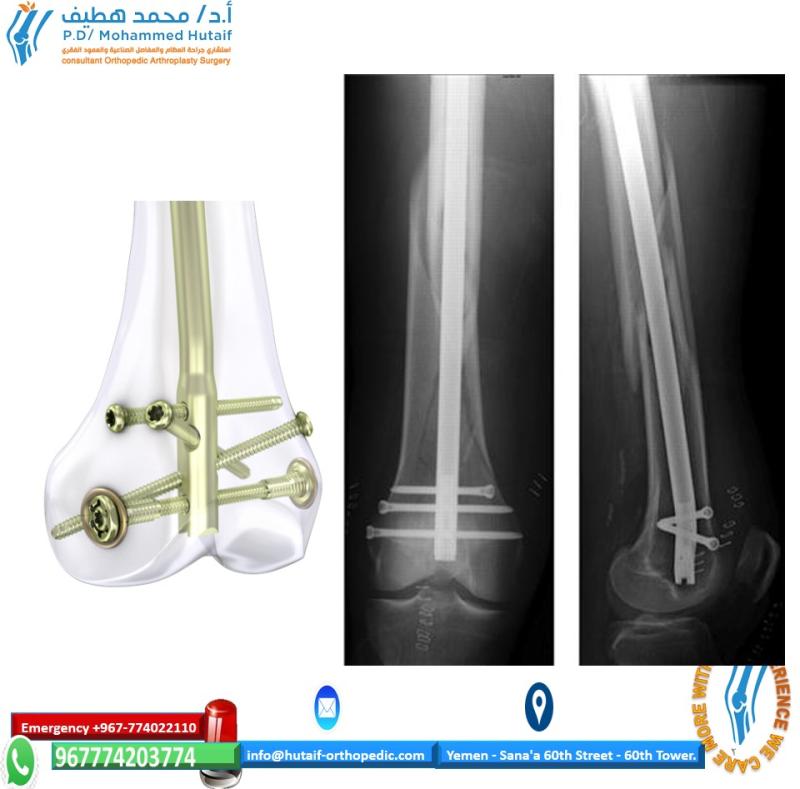
2. Open Reduction Internal Fixation (ORIF) of a Tibial Plateau Fracture (Schatzker II - Lateral Condylar Split-Depression)
I. Patient Positioning and Approach:
*
Position:
Supine on a radiolucent table. Place a bump under the ipsilateral hip to internally rotate the leg and expose the lateral aspect of the knee. Tourniquet application.
*
Incision:
Standard anterolateral approach. A slightly curvilinear incision extending from the lateral femoral epicondyle distally to the fibular head, curving anteriorly around the fibular head to end just distal to the Gerdy's tubercle. Alternatively, a straight anterolateral incision.
*
Internervous Plane:
The critical internervous plane for an anterolateral approach is between the tibialis anterior (anterior compartment) and the fibularis longus (lateral compartment). Care must be taken to protect the superficial peroneal nerve, which runs superficially in the distal aspect of this plane, and the common peroneal nerve, which wraps around the fibular neck.
II. Exposure and Soft Tissue Management:
*
Deep Dissection:
Incise the fascia lata. Develop the interval between the anterior border of the fibularis longus and the posterior border of the tibialis anterior.
*
Gerdy's Tubercle Osteotomy (Optional):
If additional anterior exposure of the lateral plateau is needed, a small osteotomy of Gerdy's tubercle (insertion of IT band) can be performed and later reattached.
*
Submeniscal Arthrotomy:
Perform a submeniscal arthrotomy (typically via incision along the meniscotibial ligament) to visualize the articular surface directly. Carefully reflect the lateral meniscus superiorly to expose the entire lateral plateau. Avoid detachment of the meniscus peripherally if possible.
III. Reduction of Articular Depression and Fracture Fragments:
*
Indirect Reduction:
Apply longitudinal traction to the extremity to help distract the fracture and restore length.
*
Direct Reduction:
Use a pointed reduction clamp or laminar spreaders to reduce displaced main fracture fragments.
*
Elevation of Depressed Segments:
Use a periosteal elevator or a custom tamp (e.g., bone graft impactor) to elevate the depressed articular segments. This is typically done through a cortical window created in the lateral metaphysis, approximately 1-2 cm distal to the fracture line. Elevate the fragments under direct vision via the submeniscal arthrotomy, ensuring an anatomical reduction of the articular surface.
*
Void Filling:
Once the articular surface is reduced and supported, fill the metaphyseal void created by the elevation with structural bone graft (autograft from iliac crest, allograft, or synthetic bone substitute) to prevent re-collapse.
IV. Provisional and Definitive Fixation:
*
Provisional Fixation:
Use K-wires to temporarily hold the reduced articular surface and main fragments. Confirm reduction and alignment with fluoroscopy (AP, lateral, oblique views) and direct visualization.
*
Definitive Fixation:
*
Buttress Plating:
Apply a pre-contoured locking plate (e.g., variable angle lateral locking plate for tibial plateau) to the lateral aspect of the tibia, ensuring adequate distal fixation and proximal buttressing of the articular segment.
*
Lag Screws:
Insert lag screws across the fracture planes to achieve interfragmentary compression, particularly for split components. Subchondral lag screws can be used to stabilize elevated articular fragments, ensuring they do not penetrate the joint surface.
*
Screw Length and Trajectory:
Use fluoroscopy to confirm appropriate screw length and trajectory, avoiding articular penetration and neurovascular structures. The screws should capture the subchondral bone effectively.
V. Closure:
*
Meniscal Reattachment:
If a submeniscal arthrotomy was performed, reattach the meniscus to the capsule using absorbable sutures (e.g., 2-0 Vicryl).
*
Layered Closure:
Close the fascia lata. Subcutaneous closure with absorbable sutures. Skin closure with staples or non-absorbable sutures. Apply sterile dressing.
Complications & Management
Complications following knee surgery, while generally low, can significantly impact patient outcomes. A proactive approach to identification, prevention, and management is essential.
General Complications:
*
Infection (Surgical Site Infection - SSI):
*
Incidence:
0.5-2% for arthroscopic procedures; 2-5% for open fracture fixation. Higher in revision surgery, open fractures, and immunocompromised patients.
*
Management:
Superficial infections: local wound care, oral antibiotics. Deep infections (periprosthetic or hardware-related): aggressive surgical debridement (DAIR - debridement, antibiotics, implant retention), irrigation, intravenous antibiotics. May require hardware removal and staged reconstruction in severe cases.
*
Deep Vein Thrombosis (DVT) / Pulmonary Embolism (PE):
*
Incidence:
DVT 1-5% symptomatic; PE <1%. Higher risk for lower extremity trauma and prolonged immobilization.
*
Management:
Prophylaxis (mechanical and/or chemical anticoagulation). Diagnosis: Duplex ultrasound (DVT), CT pulmonary angiography (PE). Treatment: Therapeutic anticoagulation, Greenfield filter for recurrent PE or contraindication to anticoagulation.
*
Neurovascular Injury:
*
Incidence:
<1%. Common peroneal nerve (most vulnerable for lateral approaches), saphenous nerve (medial approaches, hamstring harvest), popliteal artery/vein.
*
Management:
Immediate recognition, intra-operative repair of vascular injuries. Nerve injury: observation for neuropraxia, exploration and repair for transection.
*
Arthrofibrosis (Stiffness):
*
Incidence:
5-15% (higher in ACL reconstruction with early aggressive therapy or delayed motion after trauma/fracture).
*
Management:
Aggressive physical therapy, dynamic splinting. If conservative measures fail, arthroscopic or open arthrolysis with manipulation under anesthesia. Prevention: early controlled motion.
*
Compartment Syndrome:
*
Incidence:
Rare but limb-threatening, particularly after high-energy trauma (tibial plateau fractures), prolonged tourniquet use, or significant soft tissue swelling.
*
Management:
Immediate diagnosis (5 P's: pain, pallor, pulselessness, paresthesia, paralysis), urgent fasciotomy.
*
Hardware Failure / Prominence:
*
Incidence:
Varies with implant and fracture stability.
*
Management:
Symptomatic hardware removal (usually after fracture union), revision fixation if failure leads to loss of reduction or nonunion.
Specific Complications:
-
ACL Reconstruction:
-
Graft Failure / Re-tear:
- Incidence: 5-10%. Causes: re-injury, malpositioning, insufficient tension, tunnel widening, aggressive rehabilitation.
- Management: Revision ACL reconstruction with different graft type, tunnel placement, or staged procedure.
-
Cyclops Lesion:
- Incidence: 2-5%. Anterior proliferative fibrous tissue impeding terminal extension.
- Management: Arthroscopic debridement.
-
Tunnel Widening:
- Incidence: Common radiographically, but often asymptomatic. Symptomatic widening can lead to graft laxity.
- Management: Conservative for asymptomatic. Revision surgery with bone grafting for symptomatic tunnel widening prior to re-reconstruction.
-
Extensor Mechanism Injury (Graft Harvest):
Patellar fracture (BTB harvest), quadriceps tendon rupture, patellar tendon rupture.
- Incidence: Rare.
- Management: ORIF for patellar/quads/patellar tendon rupture.
-
Graft Failure / Re-tear:
-
Meniscal Surgery:
-
Re-tear / Persistent Symptoms:
- Incidence: Meniscal repair 15-30% failure rate; meniscectomy can lead to pain from cartilage degeneration.
- Management: Revision repair, partial meniscectomy, or observation. For pain post-meniscectomy: consider conservative management, biologics, or osteotomy/arthroplasty for degenerative changes.
-
Iatrogenic Chondral Damage:
- Incidence: Rare, but possible during instrument manipulation.
- Management: Microfracture if lesion small and contained, otherwise observe or staged cartilage procedure.
-
Re-tear / Persistent Symptoms:
-
Tibial Plateau Fracture ORIF:
-
Post-traumatic Arthritis:
- Incidence: Up to 50% long-term, especially with severe articular damage, malreduction, or infection.
- Management: Conservative initially, ultimately requires osteotomy (for angular deformity) or arthroplasty (uni- or total knee).
-
Malunion / Nonunion:
- Incidence: 5-10%. Can lead to deformity, pain, and arthritis.
- Management: Corrective osteotomy and revision fixation, bone grafting.
-
Loss of Reduction:
- Incidence: 2-5%. Early collapse of articular fragments or delayed union.
- Management: Revision surgery if significant.
-
Soft Tissue Complications:
- Incidence: 5-10%. Wound dehiscence, skin necrosis, particularly with large incisions or significant swelling.
- Management: Local wound care, serial debridement, skin grafting, rotational flaps.
-
Post-traumatic Arthritis:
Common Complications & Management
| Complication | Incidence (Approx.) | Salvage Strategies |
|---|---|---|
| Infection (SSI) | 0.5-5% | Debridement & irrigation (DAIR), IV antibiotics, hardware removal (if deep/persistent), staged reconstruction. |
| DVT / PE | 1-5% / <1% | Therapeutic anticoagulation, IVC filter (for contraindication to anticoagulation or recurrent PE). |
| Neurovascular Injury | <1% | Immediate recognition, vascular repair, nerve exploration/repair or observation (for neuropraxia). |
| Arthrofibrosis (Stiffness) | 5-15% | Aggressive physical therapy, dynamic splinting, manipulation under anesthesia (MUA), arthroscopic or open arthrolysis. |
| Compartment Syndrome | Rare | Urgent fasciotomy. |
| Hardware Failure/Prominence | Varies | Symptomatic hardware removal (after union), revision fixation (for loss of reduction/nonunion). |
| ACL Graft Failure | 5-10% | Revision ACL reconstruction (different graft, tunnel revision, staged if tunnel widening severe). |
| ACL Cyclops Lesion | 2-5% | Arthroscopic debridement. |
| Meniscal Re-tear | 15-30% (repair) | Revision repair, partial meniscectomy, or conservative management. |
| Post-traumatic Arthritis | Up to 50% | Conservative management, osteotomy (for malalignment), uni- or total knee arthroplasty. |
| Malunion / Nonunion | 5-10% | Corrective osteotomy, revision internal fixation with bone grafting, external fixation. |
| Loss of Fracture Reduction | 2-5% | Revision ORIF if symptomatic or severe. |
| Soft Tissue Complications | 5-10% | Local wound care, serial debridement, negative pressure wound therapy (NPWT), skin grafting, rotational flaps. |
Post-Operative Rehabilitation Protocols
Post-operative rehabilitation is a crucial, integrated component of surgical management, critical for achieving optimal functional recovery and preventing long-term complications. Protocols are tailored to the specific injury, surgical procedure, and patient's individual needs.
General Principles:
1.
Pain and Edema Control:
Early focus on pain management (multimodal analgesia) and edema reduction (RICE: rest, ice, compression, elevation).
2.
Protected Weight-Bearing:
Gradual progression of weight-bearing according to fracture stability, fixation strength, and soft tissue healing. Non-weight bearing (NWB), toe-touch weight-bearing (TTWB), partial weight-bearing (PWB), full weight-bearing (FWB).
3.
Controlled Range of Motion (ROM):
Balance between early mobilization to prevent stiffness and protection of healing structures. Passive, active-assistive, and active ROM exercises.
4.
Gradual Strengthening:
Progressive resistance exercises to restore muscle strength and endurance, starting with isometric, then isotonic, and finally isokinetic exercises.
5.
Neuromuscular Control and Proprioception:
Essential for dynamic stability and return to activity. Balance exercises, proprioceptive training.
6.
Functional Progression:
Sport-specific or activity-specific training for return to previous levels of function.
Phases of Rehabilitation (Example: ACL Reconstruction):
Phase I: Acute Protective Phase (Weeks 0-2/3)
*
Goals:
Control pain and swelling, achieve full knee extension (0 degrees), protect healing graft, quadriceps activation, establish independent ambulation.
*
Precautions:
Limited knee flexion (e.g., 0-90° initially), protected weight-bearing (TTWB to PWB with crutches), avoid open-chain hamstring exercises (if hamstring autograft).
*
Exercises:
*
RICE:
Continuous cryotherapy, compression.
*
ROM:
Passive knee extension (heel props), active-assistive flexion, patellar mobilizations.
*
Strengthening:
Quadriceps setting (quadriceps activation), straight leg raises (SLRs) in all planes, ankle pumps.
*
Gait:
Initiate gait training with crutches, focusing on quadriceps activation during weight-bearing.
Phase II: Intermediate Controlled Motion Phase (Weeks 3-12)
*
Goals:
Full pain-free ROM, restore normal gait pattern, normalize quadriceps strength, improve neuromuscular control.
*
Precautions:
Avoid premature return to high-impact activities. Progressive increase in resistance.
*
Exercises:
*
ROM:
Continue stretching and active ROM to achieve full flexion.
*
Strengthening:
Closed-chain exercises (mini-squats, leg press, wall slides, step-ups), hamstring curls (if quadriceps or BTB graft), gluteal strengthening.
*
Proprioception:
Balance board exercises, single-leg stance.
*
Cardiovascular:
Stationary bike, elliptical.
Phase III: Advanced Strength and Power Phase (Months 3-6)
*
Goals:
Achieve near-normal strength, power, and endurance. Initiate sport-specific activities.
*
Precautions:
Gradual return to plyometrics and cutting activities. Adherence to a structured progression.
*
Exercises:
*
Strengthening:
Progressive resistance training, plyometrics (box jumps, hopping), lunges.
*
Agility Drills:
Ladder drills, cone drills, change-of-direction exercises.
*
Sport-Specific Training:
Mimic movements from desired sport or activity.
*
Running:
Initiate straight-line running, progress to curvilinear.
Phase IV: Return to Sport/Activity Phase (Months 6-12+)
*
Goals:
Safe and effective return to full activity/sport. Prevent re-injury.
*
Criteria for Return to Sport:
* Full, pain-free ROM.
* Quadriceps strength symmetry >90% compared to contralateral limb (isokinetic testing).
* Functional hop testing symmetry >90%.
* Excellent dynamic stability and neuromuscular control.
* Psychological readiness.
* No effusion.
*
Exercises:
High-level plyometrics, agility, and sport-specific drills, emphasizing controlled landings and cutting mechanics. Maintenance strength and conditioning program.
Rehabilitation for Other Injuries:
*
Tibial Plateau Fractures:
*
NWB (6-12 weeks):
Crucial to protect fracture healing. Initial ROM often limited to prevent stress on fixation.
*
Gradual Weight-Bearing:
Initiated based on radiographic evidence of healing and fracture stability, often with a brace.
*
ROM:
Progressive, but avoid stressing fracture site. Potential for arthrofibrosis is high, so gentle, consistent motion is key.
*
Meniscal Repair:
*
Protected Weight-Bearing:
NWB to PWB for 4-6 weeks, especially for posterior horn repairs.
*
Limited Flexion:
Often restricted to 0-90° for 4-6 weeks to prevent excessive stress on the repair.
*
LCL/PLC Reconstruction:
*
Limited Weight-Bearing/Bracing:
Often NWB or TTWB for 6 weeks, with a hinged knee brace restricting extension (e.g., 0-90°).
*
Slow ROM Progression:
Due to the complexity of the reconstruction, ROM progression is typically slower and more protected.
The close collaboration between the surgeon, physical therapist, and patient is paramount for successful rehabilitation. Regular assessments and protocol adjustments are necessary to address individual patient responses and optimize outcomes.
Summary of Key Literature / Guidelines
The management of knee injuries is constantly evolving, guided by robust clinical research and expert consensus. Adherence to evidence-based medicine is crucial for providing high-quality care.
1. Anterior Cruciate Ligament (ACL) Reconstruction:
*
Autograft vs. Allograft:
Meta-analyses consistently demonstrate superior outcomes and lower re-rupture rates with autografts (hamstring, patellar tendon, quadriceps tendon) compared to allografts, especially in younger, active patients (Piedmont et al.,
AJSM
2013; Kaeding et al.,
AAOS JAAOS
2011). Hamstring autografts are favored for their lower donor site morbidity, while BTB autografts may offer slightly faster bone-to-bone healing and return to sport in some contexts. Quadriceps tendon autografts are gaining popularity due to their robust nature and versatility.
*
Tunnel Placement:
Anatomical ACL reconstruction, focusing on recreating the native femoral and tibial footprints, has superseded transtibial techniques for improving rotational stability and reducing pivot shift phenomenon (Zarins et al.,
Sports Med Arthrosc Rev
2010; Lubowitz et al.,
Arthroscopy
2010). The anteromedial portal technique for femoral tunnel drilling is widely accepted.
*
Rehabilitation:
Accelerated rehabilitation protocols emphasizing early weight-bearing and controlled motion have become standard, demonstrating improved outcomes without increased graft failure rates (Shelbourne et al.,
AJSM
1999). However, return-to-sport criteria, including strength and hop test symmetry (>90%), are critical and often delay return to cutting sports for 9-12 months (Grindem et al.,
BJSM
2016).
2. Meniscal Repair vs. Meniscectomy:
*
Meniscal Preservation:
The long-term deleterious effects of meniscectomy on articular cartilage are well-established, leading to accelerated osteoarthritis (Fairbank,
J Bone Joint Surg Br
1948). Current guidelines strongly advocate for meniscal repair whenever biologically feasible, particularly in younger patients and tears in the vascularized red-red or red-white zones (Mariani et al.,
Knee Surg Sports Traumatol Arthrosc
2019).
*
Repair Techniques:
Inside-out, outside-in, and all-inside repair devices have comparable clinical outcomes when indicated, with choice often dictated by tear location and surgeon preference (Paxton et al.,
Arthroscopy
2015).
*
Concomitant ACL Reconstruction:
Repairing a meniscal tear concurrently with ACL reconstruction generally yields higher success rates for meniscal healing due to the biological stimulation from the ACL drilling and improved knee stability (Shelbourne et al.,
AJSM
2010).
3. Tibial Plateau Fractures:
*
Classification:
The Schatzker classification remains the most widely used system for describing tibial plateau fractures, guiding treatment decisions (Schatzker et al.,
Clin Orthop Relat Res
1979). The AO/OTA classification provides more comprehensive detail for research purposes.
*
Surgical Fixation:
Open Reduction Internal Fixation (ORIF) with buttress plating and lag screws is the gold standard for displaced intra-articular fractures (Schatzker II-VI) to restore articular congruity and mechanical axis (Marmol et al.,
Injury
2015). Minimally invasive techniques and external fixation may be utilized in specific high-energy or compromised soft-tissue scenarios.
*
Bone Grafting:
Autograft or allograft is frequently employed to support elevated articular segments and fill metaphyseal voids, aiming to prevent secondary collapse and promote healing (Pratibha et al.,
J Orthop Sci
2017).
*
Outcomes:
While ORIF aims to prevent post-traumatic arthritis, it remains a significant long-term complication, especially with malreduction, infection, or severe initial cartilage damage. Functional outcomes are influenced by initial injury severity, quality of reduction, and rehabilitation adherence.
4. Multi-Ligamentous Knee Injuries:
*
Reconstruction vs. Repair:
High-grade (Grade III) tears of the LCL and PLC often require surgical repair or reconstruction due to their poor intrinsic healing potential and critical role in knee stability. Acute surgical intervention within 3 weeks is often recommended to improve outcomes (LaPrade et al.,
AJSM
2010).
*
Surgical Timing:
Acute reconstruction (within 2-3 weeks) vs. delayed reconstruction (after soft tissue quiescence) remains debated, with evidence supporting both approaches depending on patient factors and associated injuries.
5. American Academy of Orthopaedic Surgeons (AAOS) Guidelines:
* The AAOS regularly publishes clinical practice guidelines for various knee conditions (e.g., "Management of Osteoarthritis of the Knee," "Management of ACL Injuries"), providing evidence-based recommendations for diagnosis, non-operative treatment, and surgical indications. These guidelines are invaluable resources for practitioners.
Areas of Ongoing Research:
*
Biologics:
Role of platelet-rich plasma (PRP), bone marrow aspirate concentrate (BMAC), and stem cells in augmenting meniscal repair, cartilage regeneration, and graft healing in ACL reconstruction.
*
Cartilage Restoration:
Advancements in matrix-associated chondrocyte implantation (MACI), allograft osteochondral transplantation, and synthetic cartilage implants.
*
Robotics & Navigation:
Increasing adoption of robotic assistance and computer navigation for precision in total knee arthroplasty and potentially for ligament reconstruction.
*
Personalized Medicine:
Tailoring treatment protocols based on individual patient biomechanics, genetics, and activity demands.
In conclusion, the surgical management of knee injuries necessitates a profound understanding of anatomy, biomechanics, and pathology, coupled with a commitment to evidence-based practice and continuous learning. Integrating current literature and guidelines ensures optimal patient care and outcomes.
You Might Also Like