Hip Replacement Surgery: Costs, Recovery, and Is It Right for You?
Key Takeaway
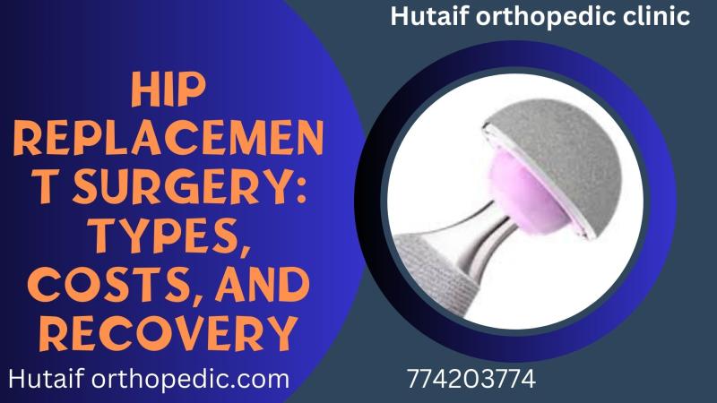
In this comprehensive guide, we discuss everything you need to know about Hip Replacement Surgery: Costs, Recovery, and Is It Right for You?. Hip replacement surgery is a procedure that replaces a damaged or diseased hip joint with an artificial one to relieve pain and improve function, commonly due to arthritis. The two main types are total hip replacement, which replaces both the femoral head and socket, and hemiarthroplasty, which replaces only the femoral head.
Introduction & Epidemiology
Hip replacement surgery, or arthroplasty, represents a highly effective intervention for advanced hip joint pathology, primarily aimed at alleviating pain and restoring function. The procedure involves the surgical excision of damaged articular cartilage and subchondral bone, followed by reconstruction with artificial prosthetic components. While the seed content broadly defines total hip replacement (THR) and hemiarthroplasty, a nuanced understanding is critical for the orthopedic surgeon.
THR, or total hip arthroplasty, involves the replacement of both the femoral head and the acetabular articulating surface with prosthetic components. This typically includes a femoral stem, a femoral head, an acetabular cup, and a liner (articulating bearing surface). Hemiarthroplasty, conversely, replaces only the femoral head, articulating with the native acetabular cartilage. While historically performed for degenerative conditions, its role is now largely confined to specific femoral neck fractures in elderly, low-demand patients, or in situations where acetabular reconstruction is contraindicated or impractical due to patient comorbidity or severe acetabular bone stock deficiency.
The evolution of hip arthroplasty from early attempts by Themistocles Gluck and Robert Jones, through the groundbreaking work of Sir John Charnley with low-friction arthroplasty and acrylic bone cement in the 1960s, to contemporary cementless designs and advanced bearing surfaces, underscores its continuous refinement. This progression has significantly improved implant longevity and patient outcomes.
Epidemiologically, THR is one of the most frequently performed and successful orthopedic procedures globally. The incidence of THR has been steadily increasing, driven by an aging population, rising rates of osteoarthritis, and expanding indications. Data from national registries indicate that primary THR procedures in the United States alone exceed 450,000 annually, with projections suggesting continued growth, potentially doubling by 2030. The primary indication remains osteoarthritis, accounting for over 90% of cases, followed by avascular necrosis, rheumatoid arthritis, and post-traumatic arthritis. While the average costs cited ($40,000 for THR, $25,000 for hemiarthroplasty) represent typical charges in the US healthcare system, these figures are subject to considerable variation based on geographic region, facility type (e.g., academic medical center vs. ambulatory surgery center), implant choice, and the prevalent reimbursement models, such as bundled payments or DRG-based systems. These economic considerations significantly influence healthcare resource allocation and system sustainability.
Surgical Anatomy & Biomechanics
A thorough understanding of hip joint anatomy and biomechanics is paramount for successful hip arthroplasty, influencing surgical approach selection, implant positioning, and complication avoidance.
Surgical Anatomy
-
Bony Anatomy
:
- Pelvis : The acetabulum is formed by the fusion of the ilium, ischium, and pubis. Key features include the acetabular rim, the lunate surface (the primary weight-bearing articular surface), the acetabular fossa (non-articular, containing the ligamentum teres and fat pad), and the transverse acetabular ligament. Acetabular orientation, including anteversion and inclination, significantly impacts stability and wear.
- Proximal Femur : Comprises the femoral head, neck, greater trochanter, lesser trochanter, and the intertrochanteric line. The femoral head is typically spherical, covered by articular cartilage. The femoral neck connects the head to the shaft, with the angle of inclination and anteversion influencing hip mechanics. The greater trochanter serves as the insertion site for the gluteus medius and minimus, critical abductor muscles. The lesser trochanter is the insertion for the iliopsoas.
-
Capsule and Ligaments
: The fibrous joint capsule is reinforced by three strong intrinsic ligaments:
- Iliofemoral ligament (Y-ligament of Bigelow) : Anterior, strongest, prevents hyperextension.
- Pubofemoral ligament : Anteromedial, prevents hyperabduction and external rotation.
- Ischiofemoral ligament : Posterior, prevents hyperextension and internal rotation.
- Ligamentum Teres : Intracapsular, provides minor blood supply to the femoral head in childhood, but minimal mechanical stability in adults.
-
Musculature
: Muscles surrounding the hip are crucial for function and define surgical approaches:
- Gluteal muscles (maximus, medius, minimus) : Primary abductors and extensors. The gluteus medius and minimus insert onto the greater trochanter and are vital for gait stability. The superior gluteal nerve innervates medius and minimus.
- Short External Rotators : Piriformis, superior gemellus, obturator internus, inferior gemellus, quadratus femoris. These muscles attach from the pelvis to the greater trochanter/posterior femur and are frequently encountered in posterior approaches.
- Iliopsoas : Primary hip flexor, inserts on the lesser trochanter.
- Adductors : Pectineus, adductor longus, brevis, magnus, gracilis.
- Quadriceps Femoris : Rectus femoris originates from the anterior inferior iliac spine (AIIS) and is a key landmark/retractor in direct anterior approaches.
-
Neurovascular Structures
:
- Sciatic Nerve : Courses inferior to the piriformis muscle, posterior to the short external rotators and quadratus femoris. At significant risk in posterior approaches, especially with excessive limb lengthening or cement extrusion.
- Femoral Nerve : Lies within the femoral triangle, lateral to the femoral artery and vein, anterior to the hip joint capsule. At risk during anterior approaches, particularly during medial retraction or cement extrusion.
- Lateral Femoral Cutaneous Nerve (LFCN) : Exits the pelvis near the anterior superior iliac spine (ASIS) and crosses the sartorius muscle. Highly variable course and susceptible to traction or direct injury in anterior approaches, leading to meralgia paresthetica.
- Superior Gluteal Nerve/Artery : Emerge superior to piriformis and supply the gluteus medius and minimus. At risk in direct lateral approaches (transgluteal split).
- Vascular structures : Femoral artery and vein (anterior), superior/inferior gluteal arteries (posterior). Branches of the lateral femoral circumflex artery (ascending branch) are encountered in the direct anterior approach.
Biomechanics
Optimal biomechanical reconstruction is fundamental to long-term THR success.
*
Hip Center of Rotation
: Re-establishing the anatomical hip center of rotation minimizes joint reaction forces and optimizes abductor muscle efficiency. Medializing the center of rotation decreases the abductor lever arm but can also reduce joint reaction forces.
*
Femoral Offset
: The horizontal distance from the center of the femoral head to the axis of the femoral shaft. Restoring femoral offset is critical for abductor muscle tension, moment arm, and gait mechanics. Loss of offset leads to abductor weakness, Trendelenburg gait, and increased joint loads.
*
Leg Length Discrepancy (LLD)
: Accurate restoration of leg length prevents gait abnormalities, back pain, and patient dissatisfaction. Intraoperative assessment through various techniques (e.g., specific landmarks, fluoroscopy, surgical navigation) is crucial.
*
Bearing Surfaces
: The selection of articulating surfaces influences wear, friction, and long-term implant survival.
*
Metal-on-polyethylene (MoP)
: Most common. Polyethylene has evolved from conventional to highly cross-linked polyethylene (HXLPE) to reduce wear and osteolysis, improving implant longevity.
*
Ceramic-on-polyethylene (CoP)
: Offers extremely low wear rates due to the hardness and wettability of ceramic. Risk of ceramic fracture (rare) and squeaking (also rare but reported).
*
Ceramic-on-ceramic (CoC)
: Lowest wear rates, theoretically infinite implant life in terms of wear. Primary concerns include ceramic fracture (rare), squeaking, and material cost.
*
Metal-on-metal (MoM)
: Historically used for low wear and large head options, but largely abandoned due to concerns over metal ion release, pseudotumor formation, and systemic effects.
*
Femoral Stem Design
: Cemented stems rely on a mantle of polymethylmethacrylate (PMMA) for fixation. Cementless stems achieve biological fixation through bony ingrowth into porous-coated surfaces, relying on initial press-fit stability. Hybrid constructs (cementless cup, cemented stem) are also utilized. Stem morphology (tapered, anatomical, straight) and metaphyseal/diaphyseal fit are considered.
*
Acetabular Cup Design
: Primarily press-fit, often augmented with screws for initial stability, allowing for biological ingrowth. Cemented cups are used in specific scenarios, such as poor bone quality or certain revision settings.
Indications & Contraindications
The decision for hip arthroplasty is a complex balance of pain, functional limitation, radiographic findings, and patient-specific factors.
Operative Indications for Hip Replacement Surgery
-
Degenerative Arthritis
:
- Primary Osteoarthritis : Most common indication, characterized by progressive cartilage loss, osteophyte formation, and subchondral sclerosis, leading to pain and stiffness.
- Secondary Osteoarthritis : Resulting from pre-existing conditions such as hip dysplasia, femoroacetabular impingement (FAI), Legg-Calve-Perthes disease, slipped capital femoral epiphysis (SCFE), or post-traumatic injury (e.g., acetabular fracture, femoral neck fracture malunion).
-
Inflammatory Arthropathy
:
- Rheumatoid Arthritis : Systemic autoimmune disease causing symmetrical joint inflammation, cartilage destruction, and bone erosion.
- Ankylosing Spondylitis : Chronic inflammatory disease primarily affecting the axial skeleton, but can lead to severe hip arthritis.
- Psoriatic Arthritis, Lupus Arthritis : Other less common inflammatory conditions.
- Avascular Necrosis (AVN) of the Femoral Head : Ischemic death of osteocytes and bone marrow, leading to collapse of the femoral head and subsequent degenerative changes. Etiologies include steroid use, alcoholism, trauma, sickle cell disease, and idiopathic causes.
- Certain Femoral Neck Fractures : Displaced femoral neck fractures in elderly patients (often >65-70 years) are typically treated with hemiarthroplasty or THR, depending on patient mobility, pre-existing arthritis, and life expectancy. THR is favored in active, healthy elderly patients with displaced fractures or those with pre-existing hip arthritis.
- Failed Prior Hip Surgery : Including failed internal fixation of femoral neck or acetabular fractures, failed osteotomies, or failed arthroplasty (e.g., hemiarthroplasty converting to THR).
- Benign or Malignant Tumors : Resection of tumors involving the proximal femur or acetabulum that require reconstruction with endoprosthesis.
- Severe Pain and Functional Limitation : Despite adequate trials of non-operative management (pharmacological, physical therapy, activity modification, injections). Radiographic evidence of end-stage arthritis is essential.
Relative and Absolute Contraindications to Hip Arthroplasty
| Category | Operative Indications (THA/Hemi) | Relative/Absolute Contraindications (THA/Hemi) |
|---|---|---|
| Arthritis/Diagnosis | Osteoarthritis (primary/secondary), Rheumatoid Arthritis, Avascular Necrosis, Post-traumatic Arthritis, Hip Dysplasia with secondary OA, Femoral neck fractures (displaced in elderly), Failed previous hip surgery, Tumors of proximal femur/acetabulum. |
Absolute
: Active systemic infection, active local infection (e.g., cellulitis near hip), Charcot arthropathy (high risk of early loosening), Significant medical instability precluding anesthesia (e.g., unstable angina, recent MI, severe uncontrolled diabetes).
Relative : Mild symptoms responsive to conservative treatment, rapidly progressive neurological disease, inability to participate in rehab. |
| Patient Health/Status | Medically stable patients with good overall health and reasonable life expectancy, ability to participate in rehabilitation. |
Absolute
: Inadequate abductor musculature (severe paralysis), skeletal immaturity.
Relative : Morbid obesity (BMI >40) due to increased complication rates (infection, dislocation, VTE), severe peripheral vascular disease, significant cognitive impairment precluding informed consent or rehabilitation compliance, uncontrolled psychiatric illness, chronic uncontrolled infection (e.g., dental, urinary tract). |
| Local Factors | Sufficient bone stock for implant fixation, good soft tissue envelope. | Relative : Prior skin incision directly over planned surgical approach (potential for vascular compromise), severe osteopenia (compromised fixation, but often manageable), poor skin integrity at surgical site. |
| Patient Expectation | Realistic expectations regarding pain relief, functional improvement, activity limitations, and recovery timeline. | Relative : Unrealistic patient expectations regarding activity level or complete absence of pain, patient non-compliance with post-operative instructions. |
Pre-Operative Planning & Patient Positioning
Meticulous pre-operative planning and appropriate patient positioning are critical determinants of surgical efficacy, minimizing complications and optimizing outcomes.
Pre-Operative Evaluation
-
Clinical Assessment
:
- History : Detailed medical history focusing on comorbidities (cardiac, pulmonary, renal, neurological, endocrine), current medications (anticoagulants, antiplatelets, NSAIDs, immunomodulators), allergies, and prior surgical history. Crucially, assess the patient's primary complaint, pain characteristics, functional limitations (e.g., walking distance, sleep disturbance, ADL independence), and prior non-operative treatments and their efficacy.
- Physical Examination : Comprehensive examination including gait analysis (Trendelenburg sign), range of motion (ROM), limb length discrepancy (true and apparent), neurovascular status of the lower extremities, abductor muscle strength, and inspection of skin integrity around the hip. Identification of any pre-existing infection foci (e.g., dental, urinary, skin) is mandatory.
-
Radiographic Assessment
:
- Standard Radiographs : Antero-posterior (AP) pelvis with both hips, AP view of the affected hip, and a true lateral view of the affected hip (cross-table lateral or frog-leg lateral). These images allow for assessment of joint degeneration, bone stock quality, femoral head and neck morphology, acetabular orientation, and identification of any pre-existing hardware.
-
Templating
: Essential for pre-operative planning of implant size, type (cemented/cementless), and position. Digital templating with calibrated radiographs is standard practice. Goals include:
- Restoration of the hip center of rotation.
- Accurate reproduction of femoral offset.
- Equalization of limb length.
- Estimation of prosthetic component sizes (femoral stem, head, acetabular cup).
- Identification of potential challenges (e.g., severe osteophytes, complex acetabular morphology, previous hardware).
-
Advanced Imaging
:
- CT Scan : Indicated for complex cases such as severe dysplasia, post-traumatic deformity, previous acetabular fracture with malunion, or revision surgery to assess bone defects and component orientation.
- MRI : Useful for diagnosing avascular necrosis, occult fractures, or evaluating soft tissue pathology, though rarely indicated for routine primary THR planning.
-
Medical Optimization
:
- Anesthesia Consultation : Pre-operative cardiac risk stratification, pulmonary function assessment, and optimization of chronic medical conditions (e.g., diabetes, hypertension).
- Infection Prophylaxis : Pre-operative screening for Methicillin-resistant Staphylococcus aureus (MRSA) and decolonization if positive. Dental clearance for active dental infections. Pre-operative antibiotic protocol.
- Blood Management : Evaluation for anemia and optimization with iron supplementation or erythropoietin. Consideration of anti-fibrinolytic agents (e.g., tranexamic acid) to reduce intraoperative blood loss. Autologous blood donation is rarely used now.
- Medication Management : Protocol for perioperative management of anticoagulants, antiplatelets, and other crucial medications.
- Patient Education and Rehabilitation Assessment : Pre-operative physical therapy assessment provides an opportunity for patient education regarding the surgical procedure, post-operative precautions, and initial rehabilitation expectations.
Patient Positioning
The choice of patient positioning is dictated by the selected surgical approach. Careful attention to padding and patient security is crucial to prevent iatrogenic injury.
-
Lateral Decubitus Position (for Posterior and Direct Lateral Approaches)
:
- Setup : The patient is placed on their unaffected side. A beanbag or specialized hip positioner (e.g., pegboard) is used to stabilize the torso and pelvis. The anterior iliac spine is typically positioned slightly anterior to the mid-axillary line, and the posterior iliac spine slightly posterior, ensuring the pelvis is not rotated.
- Padding : All pressure points must be meticulously padded: axillary roll (to protect brachial plexus), between the knees, ankles, and along the dependent arm.
- Extremity Draping : The operative leg is typically draped free to allow for full range of motion and manipulation during component implantation and stability testing. The non-operative leg is flexed at the hip and knee to provide a stable base.
- Considerations : This position provides excellent exposure to the posterior and lateral aspects of the hip. It requires precise patient stabilization to prevent intraoperative rotation.
-
Supine Position (for Direct Anterior and Anterolateral Approaches)
:
- Setup : The patient is positioned supine on the operating table. For the direct anterior approach, often a specialized fracture table (e.g., Hana table) is utilized to facilitate hyperextension and external rotation of the operative leg for femoral preparation. Alternatively, a standard operating table with leg holders or a perineal post may be used, requiring skilled assistant maneuvering for femoral exposure.
- Padding : Pressure points (heels, sacrum, occiput, elbows) are padded. The contralateral leg is often slightly abducted and flexed.
- Fluoroscopy : Supine positioning is conducive to intraoperative fluoroscopic guidance, which is frequently used in direct anterior approaches to verify component position, limb length, and offset.
- Considerations : This position typically offers a true anterior perspective and allows for precise control of limb length and offset through fluoroscopy. Nerve injuries (LFCN, femoral nerve) and vascular injuries (femoral vessels) are specific risks.
-
Draping and Sterilization
:
- Standard surgical skin preparation (e.g., chlorhexidine-alcohol or povidone-iodine solutions) from the umbilical region to the knee circumferentially.
- Application of sterile drapes to establish a sterile field, ensuring the operative limb is isolated and remains freely mobile for surgical maneuvers.
Detailed Surgical Approach / Technique
The choice of surgical approach significantly influences surgical exposure, potential soft tissue damage, and immediate postoperative precautions. Three primary approaches dominate contemporary practice: posterior, direct anterior, and direct lateral. Each involves distinct internervous planes or muscle-splitting techniques, leading to varying risk profiles and recovery characteristics.
1. Posterior Approach (Kocher-Langenbeck / Modified Moore)
This is the most common approach globally, offering excellent exposure of both the acetabulum and femur.
- Patient Positioning : Lateral decubitus.
- Skin Incision : A curvilinear or straight incision is made centered over the greater trochanter, extending proximally along the posterior aspect of the iliac crest and distally along the line of the femoral shaft.
-
Fascial Dissection
:
- Incision of the fascia lata in line with the skin incision.
- The gluteus maximus is identified. Its fibers are split bluntly in line with their orientation, or the anterior border is elevated from the gluteus medius.
-
Internervous Plane & Muscle Reflection
: There is no true internervous plane in the initial superficial dissection. The deep dissection involves:
- Identification and release of the short external rotator muscles (piriformis, gemelli superior and inferior, obturator internus) from their insertions on the greater trochanter. The piriformis is typically released first, providing access to the obturator internus and gemelli. The quadratus femoris may also be released. These muscles are tagged for later repair.
- The capsule is then incised, typically in a T-shape or complete capsulectomy.
- Neurovascular Structures at Risk : The sciatic nerve lies deep and posterior to the short external rotators. It is imperative to protect this nerve throughout the procedure, especially during retraction and disarticulation. Excessive traction or lengthening can cause neuropraxia.
-
Femoral Preparation
:
- The hip is dislocated by internally rotating and adducting the flexed leg.
- The femoral head is osteotomized at the correct level, guided by pre-operative templating.
- The femoral canal is then prepared using sequential broaches to achieve adequate fit for the chosen femoral stem. For cementless stems, this involves a precise press-fit. For cemented stems, the canal is reamed and then filled with bone cement after preparation.
-
Acetabular Preparation
:
- The acetabulum is exposed. Care is taken to protect the sciatic nerve posteriorly and the soft tissues around the acetabular rim.
- Reaming is performed sequentially to remove damaged cartilage and subchondral bone, creating a hemispherical bed for the acetabular cup. Reaming direction is crucial for proper inclination (typically 40-45 degrees) and anteversion (15-20 degrees).
- The acetabular cup is inserted, typically press-fit, and may be secured with screws. The polyethylene liner is then inserted into the cup.
- Reduction and Stability Testing : The femoral component is inserted, and the hip is reduced. Stability is assessed through a range of motion, checking for impingement and dislocation risk (especially in flexion, adduction, and internal rotation). Leg length and offset are verified.
- Closure : The short external rotators and capsule are typically repaired to the greater trochanter and posterior capsule, enhancing posterior stability. Fascia and skin are closed in layers.
2. Direct Anterior Approach (DAA) (Hueter's Approach)
The DAA is gaining popularity due to its perceived muscle-sparing nature and potential for accelerated recovery.
- Patient Positioning : Supine, often on a specialized fracture table.
- Skin Incision : An incision is made from the ASIS extending distally and slightly medially along the interval between the tensor fascia lata (TFL) and sartorius.
- Fascial Dissection : The fascia superficial to the TFL is incised, and the TFL is retracted laterally, while the sartorius and rectus femoris are retracted medially.
- Internervous Plane : The approach utilizes the internervous plane between the tensor fascia lata (innervated by the superior gluteal nerve) and the sartorius and rectus femoris (both innervated by the femoral nerve). The ascending branch of the lateral femoral circumflex artery and vein are encountered and typically ligated or cauterized.
- Neurovascular Structures at Risk : The lateral femoral cutaneous nerve (LFCN) , which typically exits near the ASIS, is highly variable in its course and is at risk of traction injury or direct transection, leading to meralgia paresthetica. The femoral nerve and femoral vessels lie medially and are protected by medial retraction.
-
Femoral Preparation
:
- The hip capsule is incised. The femoral neck is osteotomized in situ .
- For femoral canal preparation, the operative leg is typically hyperextended, externally rotated, and adducted to bring the proximal femur anteriorly. This maneuver can be challenging and is often facilitated by a specialized table.
- Broaching and stem insertion follow similar principles as the posterior approach, but require specific instrumentation to navigate the anterior exposure.
-
Acetabular Preparation
:
- Acetabular reaming and cup insertion are performed directly anteriorly, often with fluoroscopic guidance to ensure correct inclination and anteversion.
- Reduction and Stability Testing : Reduction and stability assessment are performed.
- Closure : The capsule may be repaired. Fascia and skin are closed in layers.
3. Direct Lateral Approach (Hardinge / Transgluteal)
This approach involves splitting a portion of the gluteus medius and minimus, affecting the abductor mechanism directly.
- Patient Positioning : Lateral decubitus.
- Skin Incision : A straight or curvilinear incision centered over the greater trochanter, extending superiorly towards the iliac crest and distally along the femoral shaft.
- Fascial Dissection : The fascia lata and superficial fibers of the gluteus maximus are incised.
-
Internervous Plane / Muscle Split
: This approach is technically an intermuscular/transmuscular approach, as it involves incising a portion of the gluteus medius and vastus lateralis.
- The fascia over the gluteus medius and vastus lateralis is incised.
- A portion of the gluteus medius, anterior to its innervation by the superior gluteal nerve, is typically detached or split from the greater trochanter and/or the vastus lateralis origin. The size of the split varies (e.g., anterior 1/3 of gluteus medius).
- Neurovascular Structures at Risk : The superior gluteal nerve courses superior and anterior to the piriformis and supplies the gluteus medius and minimus. Care must be taken to limit the anterior extent of the abductor split to avoid denervation, which can lead to a persistent Trendelenburg gait.
- Femoral Preparation : Similar to the posterior approach. The abductors are retracted superiorly.
- Acetabular Preparation : Similar to the posterior approach.
- Reduction and Stability Testing : Similar to other approaches.
- Closure : The detached gluteus medius/vastus lateralis is meticulously reattached to the greater trochanter or surrounding fascia. Failure of repair can lead to abductor insufficiency.
Complications & Management
Despite high success rates, hip replacement surgery is associated with a spectrum of potential complications, ranging from intraoperative issues to late failures. Proactive management strategies are crucial.
Common Complications and Management
| Complication | Incidence (Approximate) | Salvage Strategies / Management |
|---|---|---|
| Dislocation | 1-3% (Primary THR) |
Acute (within 6 weeks)
: Closed reduction. If recurrent or irreducible, consider open reduction, evaluation of component malposition, impingement, or soft tissue laxity. May require revision of one or both components, constrained liner, or capsular repair/augmentation.
Chronic (late) : Often due to component malposition, soft tissue laxity, or abductor insufficiency. Requires thorough workup (radiographs, CT) to identify etiology. Management may involve bracing, revision of malpositioned components, constrained liners, abductor repair, or conversion to a modular dual-mobility construct. |
| Periprosthetic Joint Infection (PJI) | 0.5-2% |
Diagnosis
: Clinical suspicion, elevated ESR/CRP, aspiration of joint fluid (cell count, differential, culture).
Acute (within 3-6 weeks) : Debridement, antibiotics, implant retention (DAIR) with exchange of modular components, followed by prolonged antibiotics. Chronic/Late : Typically requires a two-stage exchange arthroplasty : explantation of components, debridement, antibiotic spacer implantation, prolonged intravenous antibiotics, followed by reimplantation of new components once infection markers normalize. In rare cases, single-stage exchange or resection arthroplasty (Girdlestone) may be considered. |
| Venous Thromboembolism (VTE) | DVT: 0.5-1% (symptomatic) PE: 0.1-0.5% (symptomatic) |
Prophylaxis
: Pharmacological (low-molecular-weight heparin, Factor Xa inhibitors, aspirin) and mechanical (intermittent pneumatic compression) are standard.
Management : Anticoagulation (oral or IV) for symptomatic DVT/PE. Duration dependent on risk factors and type of event. Inferior vena cava (IVC) filter in specific cases with contraindication to anticoagulation or recurrent PE despite adequate anticoagulation. |
| Periprosthetic Fracture | 0.5-2% (Intraoperative); 1-4% (Postoperative) |
Intraoperative
: Depends on location and stability. Cerclage wires, cables, plates. May necessitate a longer stemmed femoral component, revision to cemented stem, or revision of acetabular component.
Postoperative (Vancouver Classification for femoral fractures) : - Type A : Treated with non-operative management or fixation. - Type B (B1) : Stable implant, fracture below tip of stem - ORIF with plate/cables. (B2) : Unstable implant, fracture around or below stem - Revision to long stem with fixation. (B3) : Poor bone stock - Revision to long stem, allograft, or megaprosthesis. - Type C : Below stem - ORIF. Acetabular fractures may require fixation or revision. |
| Nerve Injury | 0.1-1.5% (Sciatic > Femoral > LFCN) |
Diagnosis
: Immediate neurological assessment.
Management : Observation for neuropraxia (most common), nerve conduction studies/EMG if persistent. Surgical exploration for direct transection or entrapment (hematoma, cement). Symptomatic management for pain (neuropathic medications). Foot drop brace for peroneal nerve injury. LFCN injury (meralgia paresthetica) often resolves spontaneously; severe cases may require steroid injection, nerve block, or neurolysis. |
| Vascular Injury | <0.1% |
Diagnosis
: Acute limb ischemia (pain, pallor, pulselessness, paresthesia, paralysis), expanding hematoma.
Management : Immediate surgical exploration, vascular repair by a vascular surgeon, often requiring graft interposition. High morbidity and mortality. |
| Leg Length Discrepancy (LLD) | Common, usually minor (<1cm) |
Prevention
: Careful pre-operative templating, intraoperative verification (fluoroscopy, specific landmarks).
Management : For minor LLD, shoe lift for the shorter leg. Significant LLD (>1.5-2 cm) can cause gait abnormalities, back pain, and dissatisfaction. Revision surgery is considered for severe, symptomatic LLD, weighing risks vs. benefits. |
| Aseptic Loosening | 5-10% (10-15 years) |
Diagnosis
: Persistent pain, radiographic signs of lucency, migration, or fracture of cement mantle/bone-implant interface.
Management : Revision arthroplasty (one or both components). Bone grafting may be required for significant bone loss. |
| Polyethylene Wear / Osteolysis | Decreasing with HXLPE |
Diagnosis
: Radiographs showing peri-implant lucency and bone destruction.
Management : Debridement of osteolytic lesions and revision of the polyethylene liner, or full revision arthroplasty if components are loose or malpositioned. |
| Heterotopic Ossification (HO) | 10-30% (radiographic) 1-5% (clinically significant) |
Prevention
: Prophylaxis with NSAIDs (e.g., indomethacin) or single-dose radiation therapy, especially in high-risk patients (history of HO, ankylosing spondylitis, hyperostosis).
Management : If symptomatic and mature, surgical excision. |
Post-Operative Rehabilitation Protocols
Post-operative rehabilitation is integral to achieving optimal functional outcomes following hip replacement surgery. Protocols are typically structured to facilitate early mobilization, manage pain, restore range of motion (ROM), and strengthen surrounding musculature, while respecting surgical precautions.
Early Mobilization and Weight-Bearing
-
Immediate Post-operative Phase (Days 0-3)
:
- Goal : Pain control, wound care, early out-of-bed mobilization.
- Mobilization : Patients are typically encouraged to sit on the edge of the bed and transfer to a chair on the day of surgery or post-operative day 1. Ambulation with a walker or crutches begins immediately.
-
Weight-Bearing (WB) Status
:
- Cementless Components : Typically protected weight-bearing (PWB) or weight-bearing as tolerated (WBAT) immediately, depending on initial implant stability and surgeon preference. Full WB is often achieved within 6-12 weeks.
- Cemented Components : Often allows immediate full weight-bearing (FWB) due to immediate mechanical interlock.
- Fracture Fixation/Revision : May require more restricted weight-bearing (e.g., toe-touch weight-bearing, TTWB) for a longer duration, dictated by the stability of internal fixation or bone graft incorporation.
- Exercises : Ankle pumps, quad sets, gluteal sets to prevent DVT and maintain muscle tone.
Physical Therapy Goals and Progression
-
Acute Phase (Hospital Stay, typically 1-3 days)
:
- Goals : Independent transfers, safe ambulation with assistive device, understanding of precautions, pain management.
-
Activities
:
- Bed mobility (log rolls).
- Transfers (bed to chair, commode).
- Gait training with a walker, progressing to crutches.
- Gentle ROM exercises (e.g., hip flexion, abduction within precautions).
- Patient education on precautions and signs of complications.
-
Subacute Phase (Home or Skilled Nursing Facility, Weeks 1-6)
:
- Goals : Weaning from assistive devices, improving gait pattern, increasing strength and ROM, performing ADLs safely.
-
Activities
:
- Progression of gait training (crutches to cane, then independent).
- Strengthening exercises: Hip abductors, extensors, flexors, quadriceps. Examples: straight leg raises, hip abduction/adduction in supine/standing, glute bridges, mini-squats.
- Balance exercises.
- Stair climbing practice.
- Driving restrictions typically for 4-6 weeks (especially for right hip or manual transmission).
-
Chronic Phase (Outpatient Physical Therapy, Weeks 6-12+)
:
- Goals : Full return to functional activities, occupational tasks, and appropriate recreational activities. Optimize strength, endurance, and balance.
-
Activities
:
- Advanced strengthening: Lunges, step-ups, single-leg stance.
- Proprioception and agility drills.
- Low-impact aerobic conditioning (e.g., swimming, cycling).
- Return to work (light duty at 6 weeks, heavy duty 3-6 months).
- Return to specific sports/hobbies (golf at 3 months, tennis/skiing at 6-12 months, avoiding high-impact activities).
Post-Operative Precautions (Vary by Surgical Approach)
-
Posterior Approach Precautions (Traditional)
: Designed to prevent posterior dislocation.
- Avoid hip flexion >90 degrees.
- Avoid hip adduction past midline.
- Avoid hip internal rotation.
- Specific instructions: Do not cross legs, use elevated toilet seats, avoid low chairs, do not pick up objects from the floor by bending over. These are often relaxed after 6-12 weeks if stability is confirmed.
-
Anterior Approach Precautions (Less Restrictive, but Specific)
: Designed to prevent anterior dislocation.
- Avoid hip hyperextension.
- Avoid hip external rotation beyond neutral, especially with extension.
- Less emphasis on flexion/adduction restrictions.
- Direct Lateral Approach Precautions : Generally less restrictive than posterior, but may have temporary restrictions on active hip abduction to protect the abductor repair.
Role of Occupational Therapy
Occupational therapists play a vital role in assessing and training patients in ADLs, recommending adaptive equipment (e.g., sock aids, long-handled reachers, elevated toilet seats), and advising on home modifications to ensure a safe transition home and independent living.
Summary of Key Literature / Guidelines
The landscape of hip arthroplasty is continuously shaped by robust clinical research, consensus statements, and national guidelines. Key organizations, such as the American Academy of Orthopaedic Surgeons (AAOS), the American Association of Hip and Knee Surgeons (AAHKS), and the European Federation of National Associations of Orthopaedics and Traumatology (EFFORT), periodically publish evidence-based recommendations.
1. Indications for THA
- AAOS guidelines consistently emphasize that the primary indication for THA is intractable pain and functional limitation due to end-stage hip arthritis, refractory to non-operative management. Radiographic evidence of severe arthritis is essential. Age alone is not a contraindication, with increasing numbers of younger, active patients undergoing THR, and older, frail patients benefiting from the procedure to restore mobility.
2. VTE Prophylaxis
- Current guidelines recommend pharmacologic prophylaxis (e.g., aspirin, direct oral anticoagulants, LMWH, warfarin) for at least 10-14 days postoperatively, with some guidelines extending to 35 days, combined with mechanical prophylaxis (intermittent pneumatic compression devices). The optimal agent and duration remain a topic of ongoing research, balancing VTE risk with bleeding complications.
3. Antibiotic Prophylaxis
- Standard practice dictates administration of intravenous prophylactic antibiotics (e.g., cefazolin) within 60 minutes prior to incision, typically discontinued within 24 hours post-surgery. Patients with penicillin allergies require alternative agents. Pre-operative MRSA screening and decolonization are recommended in high-risk populations.
4. Management of Periprosthetic Joint Infection (PJI)
- AAOS and IDSA (Infectious Diseases Society of America) guidelines provide detailed algorithms for PJI diagnosis and management. The differentiation between acute (<3-6 weeks post-op) and chronic infection guides treatment. Acute PJI may be managed with DAIR (debridement, antibiotics, implant retention) if the implant is stable and there are no signs of advanced biofilm formation. Chronic PJI typically necessitates a two-stage exchange arthroplasty, considered the gold standard.
5. Bearing Surfaces
- While MoM bearings have largely been abandoned due to metallosis concerns, CoC, CoP, and HXLPE-on-metal remain viable options. HXLPE has demonstrated significantly reduced wear rates compared to conventional polyethylene, leading to improved implant longevity. The choice of bearing surface is often individualized, considering patient age, activity level, and surgeon experience, with no single bearing universally superior for all patients.
6. Surgical Approaches and Outcomes
-
Numerous studies have compared posterior, direct anterior, and direct lateral approaches. Meta-analyses and systematic reviews generally indicate no significant difference in long-term functional outcomes or implant survivorship among the different approaches. However, differences in early recovery and specific complication profiles exist:
- DAA : Associated with potentially faster early functional recovery, reduced immediate postoperative pain, and possibly lower early dislocation rates in some series. However, it carries a higher risk of LFCN injury, intraoperative fracture, and potentially greater learning curve for surgeons without specialized tables.
- Posterior Approach : Offers excellent visualization, is versatile for complex cases, but traditionally associated with higher early dislocation rates without meticulous capsular and short rotator repair.
- Direct Lateral Approach : Lower dislocation rates than traditional posterior, but historically associated with higher rates of abductor weakness and painful trochanteric bursitis due to abductor muscle compromise or repair failure.
7. National Joint Registries
- Registries from countries like the UK, Australia, Sweden, and the US (e.g., AJRR) provide invaluable real-world data on implant performance, survivorship, and complication rates. These registries are crucial for identifying poorly performing implants, tracking trends in surgical practice, and informing guideline development.
8. Emerging Technologies and Future Directions
- Robotics and Navigation : Increasing adoption of robotic-assisted and computer-navigated THA for enhanced precision in component positioning, potentially reducing malposition and improving long-term outcomes, though cost-effectiveness and clinical superiority are still under investigation.
- Custom Implants : Patient-specific instrumentation and custom implants are being explored for complex anatomy.
- Enhanced Recovery After Surgery (ERAS) Protocols : Multimodal pathways designed to optimize perioperative care, reduce length of hospital stay, and accelerate recovery. Components include pre-habilitation, opioid-sparing pain management, early mobilization, and optimized nutrition.
- Outpatient THA : Driven by ERAS protocols and improved anesthetic techniques, an increasing number of carefully selected patients are undergoing THR in an outpatient or ambulatory surgery center setting.
Hip replacement surgery remains a cornerstone of orthopedic practice, continuously evolving with advancements in materials, techniques, and perioperative care. The academic orthopedic surgeon must remain abreast of these developments to provide optimal patient care.
You Might Also Like
