Relieve Your Pain: Expert Orthopedic Care in Sanaa with Dr. Hutaif
Key Takeaway
This topic focuses on Relieve Your Pain: Expert Orthopedic Care in Sanaa with Dr. Hutaif, Hutaif Orthopedic Center provides specialized and compassionate orthopedic care in Sanaa, Yemen, for joint pain, sports injuries, and musculoskeletal conditions. Led by Professor Dr Mohammad Hutaif, a highly skilled orthopedic surgeon, the center offers advanced treatments including joint replacement and spine surgery. Patients receive personalized, state-of-the-art care to regain mobility and improve their quality of life.
Introduction & Epidemiology
Osteoarthritis (OA) of the knee is a prevalent degenerative joint disease, representing a significant global health burden. Characterized by progressive articular cartilage loss, subchondral bone remodeling, osteophyte formation, and synovial inflammation, OA leads to chronic pain, stiffness, and functional impairment, profoundly impacting patient quality of life and contributing substantially to healthcare expenditures. Epidemiological data indicate that OA affects approximately 250 million people worldwide, with the knee being one of the most commonly involved joints. The incidence and prevalence of knee OA are increasing due to factors such as an aging population, rising obesity rates, and increased participation in high-impact activities.
Total Knee Arthroplasty (TKA) has emerged as the definitive surgical intervention for end-stage knee OA and other debilitating arthritic conditions, including rheumatoid arthritis and post-traumatic arthritis. Since its inception, TKA has evolved significantly, demonstrating remarkable success in alleviating pain, correcting deformity, and restoring function. The number of TKAs performed annually continues to rise, with projections indicating a substantial increase over the next decade. Understanding the fundamental principles, surgical techniques, and potential complications of TKA is paramount for orthopedic surgeons and trainees to optimize patient outcomes. This review aims to provide a high-yield academic overview of TKA, emphasizing its surgical and clinical aspects relevant to professional practice.
Surgical Anatomy & Biomechanics
A thorough understanding of knee anatomy and biomechanics is fundamental for successful TKA. The knee is a complex diarthrodial hinge joint, primarily facilitating flexion and extension, with a small degree of rotation.
Osseous Structures
- Distal Femur: Consists of medial and lateral condyles, an intercondylar notch, and the patellofemoral trochlear groove. The medial condyle is typically larger and extends further distally than the lateral. The anatomical axis of the femur forms approximately a 6-degree valgus angle with the mechanical axis.
- Proximal Tibia: Comprises medial and lateral tibial plateaus, separated by the intercondylar eminences. The plateaus are slightly concave, with the medial plateau being larger and more weight-bearing. The mechanical axis of the tibia is collinear with its anatomical axis.
- Patella: A sesamoid bone embedded within the quadriceps tendon, articulating with the trochlear groove of the femur. It plays a critical role in increasing the mechanical advantage of the quadriceps mechanism.
Ligamentous Stabilizers
The knee's stability is maintained by a complex array of ligaments:
*
Cruciate Ligaments:
*
Anterior Cruciate Ligament (ACL):
Originates from the posteromedial aspect of the lateral femoral condyle and inserts into the anterior intercondylar area of the tibia. It prevents anterior translation of the tibia relative to the femur and limits hyperextension.
*
Posterior Cruciate Ligament (PCL):
Originates from the anterolateral aspect of the medial femoral condyle and inserts into the posterior intercondylar area of the tibia. It prevents posterior translation of the tibia and is the primary stabilizer against posterior shear forces.
*
Collateral Ligaments:
*
Medial Collateral Ligament (MCL):
A broad, strong ligament with superficial and deep layers, extending from the medial femoral epicondyle to the medial aspect of the tibia. It resists valgus stress and external rotation.
*
Lateral Collateral Ligament (LCL):
A cord-like structure originating from the lateral femoral epicondyle and inserting into the fibular head. It resists varus stress.
*
Posterolateral Corner (PLC):
A complex group of structures including the popliteus tendon, arcuate ligament complex, popliteofibular ligament, and fabellofibular ligament. It contributes to posterolateral rotatory stability.
Menisci
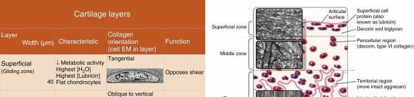
The medial and lateral menisci are crescent-shaped fibrocartilaginous structures that enhance congruity between the femoral condyles and tibial plateaus, distribute axial loads, absorb shock, and contribute to joint lubrication and stability.
Musculature
- Quadriceps Femoris: Primary knee extensor, comprising rectus femoris, vastus lateralis, vastus medialis, and vastus intermedius.
- Hamstrings: Primary knee flexors, comprising semitendinosus, semimembranosus, and biceps femoris.
- Gastrocnemius: Contributes to knee flexion and ankle plantarflexion.
- Popliteus: Initiates knee flexion by internally rotating the tibia, and stabilizes the posterolateral corner.
Biomechanics of the Knee
The biomechanical goals of TKA involve restoring proper alignment, stability, and range of motion.
*
Mechanical Axis:
The primary objective is to restore the mechanical axis of the lower limb, a straight line extending from the center of the femoral head through the center of the knee to the center of the ankle. In a normal knee, this axis passes through the center of the knee, resulting in a neutral alignment. Deviation leads to varus (medial compartment overload) or valgus (lateral compartment overload) deformities. TKA aims to achieve 0-3 degrees of varus or valgus.
*
Joint Kinematics:
The knee exhibits complex motion including rolling and gliding. In TKA, implant design aims to replicate this as closely as possible, influencing stability and patellar tracking.
*
Load Distribution:
The articular surfaces normally distribute weight-bearing forces. In OA, this distribution becomes uneven. TKA aims to create balanced flexion and extension gaps, ensuring even load distribution across the prosthetic components.
*
Soft Tissue Balance:
Crucial for achieving stability throughout the range of motion. Ligamentous releases are often necessary to balance the medial and lateral compartments, ensuring equal tension in flexion and extension.
Indications & Contraindications
Indications for Total Knee Arthroplasty
The primary indication for TKA is debilitating pain and functional impairment due to end-stage arthritis that has failed non-operative management.
*
Primary Osteoarthritis:
Severe, symptomatic degeneration of the articular cartilage.
*
Inflammatory Arthritis:
Rheumatoid arthritis, psoriatic arthritis, ankylosing spondylitis, leading to joint destruction.
*
Post-Traumatic Arthritis:
Articular damage following fractures or ligamentous injuries.
*
Avascular Necrosis (AVN) of the Femoral Condyles:
Collapse of subchondral bone leading to severe pain and joint destruction.
*
Failed Conservative Management:
Persistent pain, stiffness, and loss of function despite an adequate trial of non-surgical interventions.
*
Severe Deformity:
Significant varus, valgus, or flexion contracture impairing function and mobility.
*
Severe Joint Pain at Rest or Nocturnal Pain:
Unresponsive to analgesics.
*
Radiographic Evidence:
Kellgren-Lawrence Grade III or IV osteoarthritis, with joint space narrowing, osteophytes, subchondral sclerosis, and cysts.
Contraindications for Total Knee Arthroplasty
Contraindications can be absolute or relative.
*
Absolute Contraindications:
*
Active Infection:
Especially periprosthetic joint infection (PJI) or active distant infection, which mandates eradication prior to TKA.
*
Extensor Mechanism Insufficiency:
Non-functional quadriceps mechanism (e.g., irreparable quadriceps rupture), making rehabilitation and ambulation impossible.
*
Rapidly Progressive Neuropathic Arthropathy (Charcot Joint):
High risk of implant loosening and failure.
*
Recurrens Deformity due to Quadriceps Paralysis:
Without effective surgical correction.
*
Severe Peripheral Vascular Disease:
Significantly increased risk of wound complications and infection.
*
Patient Unwillingness or Inability to Participate in Rehabilitation:
Essential for successful outcomes.
*
Relative Contraindications:
*
Severe Obesity (BMI > 40):
Increased risk of complications (infection, wound issues, DVT, loosening), though benefits may still outweigh risks in selected patients.
*
Morbidly Obese Patients (BMI > 50):
Often associated with higher rates of complications.
*
Previous Surgical Arthrodesis:
While possible, TKA in this setting is technically demanding and carries higher risks.
*
Neurological Conditions:
Progressive conditions like Parkinson's disease or multiple sclerosis, which may compromise rehabilitation and functional gains.
*
Psoriasis or Other Skin Conditions:
In the operative field, increasing infection risk.
*
Poorly Controlled Diabetes Mellitus (HbA1c > 8%):
Increased risk of infection and wound complications.
*
Smoking:
Increased risk of wound complications, infection, and DVT.
*
Unrealistic Patient Expectations:
Requires thorough preoperative counseling.
Summary of Operative vs. Non-Operative Indications
| Category | Operative Indications (TKA) | Non-Operative Indications (Conservative Management) |
|---|---|---|
| Pain | Severe, debilitating pain at rest, nocturnal pain, or pain with minimal activity | Mild to moderate pain, pain with specific activities, controlled by analgesics |
| Function | Significant functional limitation, inability to perform ADLs, loss of independence | Mild to moderate functional limitation, able to perform most ADLs with adaptations |
| Deformity | Fixed varus/valgus deformity (>10-15 degrees), severe flexion contracture (>20-30 degrees) | Mild to moderate angular deformity, correctable, or mild flexion contracture (<10-15 degrees) |
| Radiographs | Kellgren-Lawrence Grade III-IV OA, significant joint space narrowing, bone-on-bone | Kellgren-Lawrence Grade I-II OA, minimal joint space narrowing, osteophytes, but preserved cartilage space |
| Response to Rx | Failure of an adequate trial (typically >6 months) of non-operative treatments | Symptomatic relief and functional improvement with non-operative modalities |
| Comorbidities | Acceptable medical risk for surgery | Significant medical comorbidities precluding safe surgery, or patient preference to avoid surgery |
| Specific Dx | End-stage primary OA, inflammatory arthritis, severe post-traumatic arthritis, AVN | Early-stage OA, meniscal tears (if amenable to repair/debridement), isolated chondral defects (if amenable to repair) |
| Age | Generally for older adults (>60 years), but increasingly performed in younger, active patients with specific indications. | Any age, particularly younger patients, to preserve native joint function |
Pre-Operative Planning & Patient Positioning
Meticulous preoperative planning is essential to minimize complications and optimize outcomes in TKA.
Clinical Assessment
- History: Detailed medical history, comorbidities, previous surgeries, medications (anticoagulants, NSAIDs), allergies, social history (smoking, alcohol), functional limitations, pain characteristics, and patient expectations.
-
Physical Examination:
- Gait analysis: Observe walking pattern and limb alignment.
- Neurovascular status: Assessment of sensory, motor, and vascular integrity distal to the knee.
- Range of Motion (ROM): Active and passive flexion, extension, and assess for fixed flexion contracture.
- Ligamentous stability: Assessment of MCL, LCL, and cruciate ligaments.
- Deformity: Quantify varus/valgus alignment, rotational deformities, and patellar tracking.
- Skin integrity: Note any lesions, rashes, or signs of infection.
Radiographic Assessment
-
Standard Views:
- Anteroposterior (AP) weight-bearing view: Evaluates joint space narrowing, osteophyte formation, and overall alignment.
- Lateral view: Assesses posterior femoral condylar offset, patellar height (Insall-Salvati or Caton-Deschamps ratio), and fixed flexion contracture.
- Patella skyline view: Evaluates patellar tilt and subluxation, and patellofemoral arthritis.
- Long-leg Alignment View: A crucial full-length standing AP radiograph extending from the femoral head to the ankle, used to determine the mechanical axis of the limb and quantify varus or valgus deformity. This view is indispensable for accurate preoperative templating and surgical planning to restore neutral mechanical alignment.
- Advanced Imaging (as needed): MRI may be used for specific soft tissue pathology or tumor workup; CT for severe bony deformities or previous hardware, aiding in custom jig planning or robotic-assisted surgery.
Templating and Implant Selection
- Implant System Selection: Based on surgeon preference, bone stock, ligamentous stability, and patient factors (e.g., posterior stabilized vs. cruciate-retaining design).
- Size Estimation: Using templates on radiographs to estimate femoral and tibial component sizes, considering options for augmented components for bone defects.
- Component Alignment: Planning the distal femoral cut (typically 5-7 degrees valgus relative to anatomical axis) and proximal tibial cut (90 degrees to mechanical axis, with appropriate posterior slope).
- Bone Defect Management: Planning for bone grafting or modular augmentations if significant bone loss is anticipated.
Medical Optimization
- Cardiovascular and Pulmonary Assessment: Referral to internal medicine or cardiology for high-risk patients.
- Diabetes Control: Optimize HbA1c to <8% to reduce infection risk.
- Nutrition: Address malnutrition.
- Smoking Cessation: Encourage cessation at least 4-6 weeks preoperatively.
- Medication Review: Discontinue anticoagulants as per protocol, manage pain medications.
- DVT Prophylaxis: Plan for appropriate pharmacologic and mechanical prophylaxis.
- Anesthesia Consultation: Evaluation for suitability for regional vs. general anesthesia, and pain management strategies.
Patient Education and Consent
Comprehensive discussion of the procedure, expected outcomes, potential complications, rehabilitation protocol, and realistic expectations. Informed consent is obtained.
Patient Positioning
- Supine Position: The patient is positioned supine on the operating table.
- Leg Holder: The operative limb is typically placed in a padded leg holder that allows the knee to be flexed to 90 degrees or more while maintaining stability.
- Tourniquet: A pneumatic tourniquet is applied to the proximal thigh to provide a bloodless field, typically inflated to 250-350 mmHg or 50-100 mmHg above systolic blood pressure, for a duration generally not exceeding 90-120 minutes.
- Foot Support: The foot is often supported by a sterile footrest or assistant for optimal exposure.
- Sterile Prep and Drape: The entire limb is prepped with an antiseptic solution and draped in a sterile fashion, ensuring adequate exposure for the incision and instrument placement.
Detailed Surgical Approach / Technique (Medial Parapatellar Approach)
The medial parapatellar approach is the most common surgical approach for TKA, offering excellent exposure of the knee joint.
1. Skin Incision
- A straight midline or slightly medial longitudinal incision is made, extending from approximately 3-4 cm proximal to the superior pole of the patella to the level of the tibial tuberosity. The length is typically 15-20 cm, guided by the patient's anatomy and surgeon preference.
- Care is taken to avoid injury to the infrapatellar branch of the saphenous nerve, which typically courses inferomedially.
2. Deep Dissection and Arthrotomy
- Subcutaneous Dissection: The subcutaneous tissue is incised, and full-thickness skin and subcutaneous flaps are created medially and laterally to expose the quadriceps tendon, patella, and patellar tendon.
- Medial Parapatellar Arthrotomy: The incision begins proximally by dividing the quadriceps tendon just medial to its midline insertion, extending distally through the vastus medialis obliquus (VMO), the medial retinaculum, and the joint capsule, passing medial to the patella and patellar tendon, down to the tibial tuberosity. This internervous plane is between the vastus medialis (supplied by femoral nerve) and the sartorius/gracilis/semitendinosus (supplied by femoral and obturator nerves for sartorius, obturator and tibial nerves for gracilis, tibial nerve for semitendinosus).
- Patellar Eversion: The patella is then everted laterally to expose the entire articular surface of the distal femur and proximal tibia. Lateral patellar retinacular release may be necessary in cases of significant patellar tilt or tight lateral structures to facilitate patellar eversion and tracking.
3. Bone Resection - Femur
- Osteophyte Removal: Initial removal of large osteophytes from the distal femur and proximal tibia to improve exposure and accurately reference bony landmarks.
-
Distal Femoral Resection:
- Intramedullary (IM) Guide: A guide rod is inserted into the intramedullary canal of the femur through an entry point in the intercondylar notch. The IM guide is typically set to a valgus angle of 5-7 degrees (relative to the anatomical axis) to reproduce the patient's mechanical axis. This angle varies based on the individual's anatomical femorotibial angle.
- Cutting Block Placement: A distal femoral cutting block is then seated on the femoral condyles, referencing the IM guide.
- Resection: A measured amount of distal femur (typically 9-10 mm, corresponding to the thickness of the femoral component) is resected perpendicular to the mechanical axis of the femur.
-
Femoral Sizing and Rotation:
- Anterior and Posterior Referencing: An anterior referencing guide or posterior referencing guide is used to determine the appropriate size of the femoral component.
- Rotation: Femoral component rotation is critical. The external rotation of the femoral component is typically set to be 3 degrees relative to the surgical epicondylar axis (SEA), or parallel to the transepicondylar axis (TEA), or perpendicular to the Whiteside line (posterior condylar axis) in cases of significant deformity. This ensures proper patellar tracking and balanced flexion-extension gaps.
- Femoral Component Cuts: Anterior, posterior, and chamfer cuts are then performed using the chosen cutting block.
4. Bone Resection - Tibia
-
Proximal Tibial Resection:
- Extramedullary (EM) Guide: An EM guide is typically used for the tibial cut, referencing the ankle joint and the mechanical axis of the tibia. The guide is centered over the tibial tuberosity.
- Slope: The tibial cut is typically made perpendicular to the mechanical axis in the coronal plane. In the sagittal plane, a posterior slope of 3-7 degrees is usually incorporated, referencing the native slope or surgeon preference, to aid in flexion and posterior rollback.
- Resection: A measured amount of proximal tibia (typically 8-10 mm from the least involved side) is resected.
5. Gap Balancing
- Trial Reduction and Assessment: Trial components (femoral, tibial, and patellar) are inserted. The knee is then taken through a full range of motion.
-
Flexion and Extension Gaps:
The goal is to create balanced, rectangular flexion and extension gaps.
- Extension Gap: Assessed at full extension. If the gap is tight medially (varus deformity), the MCL may be released (pie-crusting, sequential release). If tight laterally (valgus deformity), the LCL, popliteus tendon, and posterolateral capsule may be released.
- Flexion Gap: Assessed at 90 degrees of flexion. If the gap is tight, posterior osteophytes may need further removal, or posterior capsular release may be performed. If loose, posterior stabilized (PS) components may be preferred to provide stability.
- Soft Tissue Releases: Sequential releases are performed from superficial to deep until appropriate balancing is achieved. For varus knees, common releases include superficial MCL, semimembranosus posteromedial capsule. For valgus knees, releases may involve the LCL, popliteus tendon, and IT band.
6. Patellar Resurfacing
- Decision: The decision to resurface the patella is based on the degree of patellofemoral arthritis, patellar tracking, surgeon preference, and patient factors.
- Technique (if resurfacing): The patella is resected to a depth that allows for the patellar button's thickness, restoring the original patellar thickness. Multiple drill holes are made for cement interdigitation, and a polyethylene button is cemented in place.
- Patellar Tracking: After resurfacing, the knee is moved through a range of motion to assess patellar tracking. If there is lateral subluxation or tilt, a lateral retinacular release may be performed.
7. Final Implant Implantation
- Component Preparation: The prepared bone surfaces are thoroughly cleaned and dried.
- Cementation: Bone cement (polymethylmethacrylate, PMMA) is applied to the bone surfaces and the back of the definitive components (femoral, tibial, and patellar).
- Implantation: The components are then impacted into place. Excess cement is removed.
- Polyethylene Insert: The definitive polyethylene bearing insert is placed between the femoral and tibial components. The knee is then held in full extension for proper cement curing.
- Final Assessment: After cement curing, the knee is ranged to assess stability, patellar tracking, and range of motion.
8. Wound Closure
- Hemostasis: Thorough hemostasis is achieved.
- Drainage (optional): A drain may be placed, though its routine use is controversial.
- Capsular Closure: The medial parapatellar incision is closed in layers: joint capsule/retinaculum, subcutaneous tissue, and skin (sutures or staples).
- Sterile Dressing: A sterile dressing is applied, often with a compressive bandage.
Complications & Management
Despite high success rates, TKA is associated with potential complications, requiring prompt recognition and management.
Common Complications & Management
| Complication | Incidence | Management Strategy |
|---|---|---|
| Periprosthetic Joint Infection (PJI) | 0.5-2% | Acute (<4-6 weeks): Irrigation and debridement (I&D) with polyethylene exchange, followed by prolonged intravenous antibiotics. Chronic (>4-6 weeks) or failed I&D: Two-stage revision arthroplasty (explant with antibiotic spacer, prolonged antibiotics, followed by reimplantation) or permanent resection arthroplasty (arthrodesis or amputation as last resort). |
| Deep Vein Thrombosis (DVT) / Pulmonary Embolism (PE) | DVT: 5-10% (symptomatic) PE: 0.5-2% (symptomatic) | Prophylaxis: Early mobilization, mechanical compression devices, pharmacologic agents (aspirin, LMWH, DOACs). Treatment: Anticoagulation (IV heparin, LMWH, oral DOACs) for 3-6 months. IVC filter for contraindications to anticoagulation or recurrent PE. |
| Periprosthetic Fracture | 0.5-2% | Intraoperative: Depends on fracture location and stability; typically managed with fixation (screws, wires, plates) or conversion to a revision TKA with stemmed components. Postoperative: Depends on fracture type (supracondylar femur, tibial, patellar), stability, and implant fixation. May require ORIF with plates/screws, retrograde nailing, or revision arthroplasty. Immobilization is often required. |
| Arthrofibrosis / Stiffness | 1-10% | Early: Intensive physical therapy, dynamic splinting, manipulation under anesthesia (MUA) if ROM is limited despite therapy and within 3 months of surgery. Late/Refractory: Arthroscopic or open arthrolysis, potentially revision TKA. |
| Wound Complications | 2-5% | Hematoma: Evacuation, compression. Dehiscence: Wound care, secondary closure, or flap coverage. Necrosis: Debridement, local wound care, possible flap coverage. Superficial Infection: Local wound care, oral antibiotics. |
| Neurovascular Injury | <0.5% | Peroneal Nerve Palsy: Most common. Observation, bracing (foot drop), gabapentin for neuropathic pain. Surgical exploration and neurolysis/repair if no improvement. Popliteal Artery Injury: Emergent vascular surgery consultation, repair (graft or primary). |
| Aseptic Loosening | 1-2% at 10 years | Early: May indicate technical error or early wear. Late: Typically due to osteolysis from polyethylene wear. Revision TKA with bone grafting and/or modular components. |
| Patellar Complications | 2-5% | Patellar Fracture: Non-operative (immobilization) or operative (ORIF, patellectomy, revision patellar component). Patellar Instability/Maltracking: Lateral retinacular release, medial patellofemoral ligament reconstruction, or revision TKA. Patellofemoral Overstuffing: Revision of patellar component. |
| Instability | 1-3% | Early: Often due to inadequate soft tissue balancing. Late: May be due to polyethylene wear or implant loosening. Revision TKA with constrained or hinged components, or addressing soft tissue laxity. |
| Chronic Pain (Non-infectious) | 10-20% | Thorough workup to rule out mechanical causes (loosening, instability, maltracking) and infection. Consider nerve blocks, pain management consultation, psychological assessment, and rarely revision TKA if a treatable mechanical cause is identified. |
Post-Operative Rehabilitation Protocols
Post-operative rehabilitation is a critical component of successful TKA, aiming to restore joint function, strength, and mobility, and mitigate complications. Protocols vary, but generally follow a phased approach.
Phase 1: Acute/Protective Phase (Days 0-2 weeks)
Goals:
* Pain and edema control.
* Initiate early range of motion (ROM).
* Prevent complications (DVT, infection).
* Achieve safe functional mobility (bed mobility, transfers, ambulation).
Interventions:
*
Pain Management:
Multimodal analgesia (opioids, NSAIDs, acetaminophen, nerve blocks, gabapentinoids, regional anesthesia) to facilitate participation in therapy.
*
Early Mobilization:
*
Continuous Passive Motion (CPM) machine:
Often initiated on Day 0 or 1, if available, though evidence for routine use is mixed.
*
Ambulation:
Weight-bearing as tolerated (WBAT) with assistive devices (walker, crutches) on Day 0 or 1, focusing on proper gait mechanics.
*
Transfers:
Bed to chair transfers.
*
Range of Motion (ROM) Exercises:
*
Active-assisted and passive ROM:
Focus on achieving full extension (or as close as possible) and progressively increasing flexion. Target 0-90 degrees flexion by 2 weeks.
*
Quadriceps sets, gluteal sets, ankle pumps:
To maintain muscle tone and promote circulation.
*
Cryotherapy and Compression:
To reduce pain and swelling.
*
Patient Education:
Home exercise program, signs of complications, activity precautions.
Phase 2: Subacute/Restoration Phase (Weeks 2-6)
Goals:
* Progressive increase in ROM (especially flexion).
* Restore muscle strength and endurance.
* Improve balance and gait.
* Transition to independent functional activities.
Interventions:
*
ROM Progression:
Continue with active, active-assisted, and passive ROM exercises. Target 0-110 degrees flexion by 6 weeks. Gentle stretching.
*
Strengthening Exercises:
*
Progressive Resistance Exercises (PREs):
Leg presses, mini-squats, hamstring curls, calf raises, step-ups.
*
Closed-chain exercises:
Preferred to open-chain to minimize patellofemoral stress.
*
Balance Training:
Single-leg stance, tandem walking, unstable surfaces.
*
Gait Training:
Weaning from assistive devices, correcting gait deviations.
*
Stair Climbing:
Practice ascending and descending stairs.
*
Proprioception Exercises:
Re-education of joint position sense.
*
Hydrotherapy:
May be beneficial for pain reduction and early mobility in a low-impact environment.
Phase 3: Return to Activity Phase (Weeks 6-12 and beyond)
Goals:
* Maximize strength, endurance, and functional independence.
* Return to desired low-impact activities.
* Maintain long-term joint health.
Interventions:
*
Advanced Strengthening:
Incorporate higher-level resistance training, functional movements (e.g., lunges, single-leg squats).
*
Endurance Training:
Cycling, swimming, walking.
*
Sport-Specific Training (if applicable):
For patients desiring to return to low-impact sports (e.g., golf, doubles tennis). High-impact sports are generally discouraged.
*
Dynamic Balance and Agility:
Lateral stepping, cutting maneuvers (carefully).
*
Home Exercise Program:
Long-term adherence to maintain gains.
*
Patient Education:
Activity modification, joint protection strategies, weight management.
Expected Outcomes:
* Most patients achieve a pain-free or significantly reduced pain status.
* Functional improvement, allowing participation in daily activities and low-impact recreation.
* Average ROM: 0-110 to 120 degrees of flexion.
* Long-term implant survivorship is excellent, with 10-year survivorship rates exceeding 90-95% for modern implants.
Summary of Key Literature / Guidelines
Total Knee Arthroplasty remains one of the most successful and impactful orthopedic procedures, consistently demonstrating excellent long-term outcomes in terms of pain relief, functional restoration, and improvement in quality of life. The vast body of literature supporting TKA has led to the development of robust guidelines by major orthopedic societies.
Key Guidelines and Consensus Statements
- American Academy of Orthopaedic Surgeons (AAOS): Publishes clinical practice guidelines (CPGs) for TKA, covering topics such as prevention of venous thromboembolism, appropriate indications, pain management, and the use of specific technologies. For instance, the AAOS has consistently emphasized shared decision-making, patient education, and a multidisciplinary approach to TKA. Their guidelines on surgical management of osteoarthritis of the knee provide evidence-based recommendations for both non-operative and operative treatments.
- American Association of Hip and Knee Surgeons (AAHKS): Focuses on advancing hip and knee arthroplasty through education, research, and advocacy. AAHKS provides expert consensus statements on various aspects of TKA, including implant design, management of complications, and value-based care.
- European Federation of National Associations of Orthopaedics and Traumatology (EFFORT): Offers European perspectives and guidelines, often aligning with global consensus on best practices, but sometimes addressing specific regional healthcare contexts.
Evolving Trends and Research Frontiers
- Enhanced Recovery After Surgery (ERAS) Protocols: These multidisciplinary, evidence-based pathways are increasingly adopted to optimize perioperative care, reduce length of hospital stay, minimize complications, and improve patient experience. Key elements include preoperative patient education, prehabilitation, multimodal analgesia, regional anesthesia, early mobilization, and optimized nutrition. Literature consistently demonstrates ERAS protocols lead to faster recovery without increasing readmission rates.
- Robotic-Assisted and Navigated Surgery: The advent of robotic and computer-assisted navigation systems aims to improve the precision of bone resections and implant positioning, potentially leading to more consistent limb alignment and soft tissue balance. While initial studies show improved accuracy in component placement, conclusive evidence for superior long-term functional outcomes or implant survivorship compared to conventional methods is still emerging. These technologies are particularly valuable in complex cases with severe deformity.
- Patient-Specific Instrumentation (PSI): Utilizes preoperative CT or MRI scans to create custom-made cutting guides tailored to the individual patient's anatomy. PSI offers the theoretical advantage of improved accuracy and reduced operative time, though similar to robotics, its impact on long-term clinical outcomes remains a subject of ongoing research.
- Cementless TKA: While cemented TKA remains the gold standard, advancements in implant design and porous coatings have led to a resurgence of cementless components. These aim for biological fixation, potentially reducing the risk of cement-related complications and facilitating revision surgery. Long-term studies are needed to fully assess their efficacy and survivorship, particularly in younger, more active patient populations.
- Pain Management Strategies: Ongoing research into non-opioid pain management strategies, including regional nerve blocks (e.g., adductor canal block, IPACK block), periarticular injections, and systemic non-opioid analgesics, is crucial for reducing opioid consumption and improving post-operative recovery.
- Outcomes Research: Large national joint registries (e.g., National Joint Registry for England, Wales, Northern Ireland and the Isle of Man; Australian Orthopaedic Association National Joint Replacement Registry) continue to provide invaluable data on implant performance, complication rates, and long-term survivorship, informing clinical practice and implant design.
In conclusion, TKA is a highly effective procedure for end-stage knee arthritis. Adherence to established surgical principles, meticulous preoperative planning, precise surgical technique, and comprehensive postoperative rehabilitation, guided by a robust understanding of current literature and guidelines, are paramount for achieving optimal and durable patient outcomes. The field continues to evolve, with technological advancements and refined protocols promising further improvements in care.
You Might Also Like