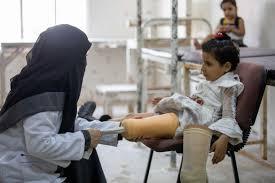
Save Lives: Support Our Orthopedic Program in Yemen
Key Takeaway
Learn more about Save Lives: Support Our Orthopedic Program in Yemen and how to manage it. Our humanitarian program in Yemen delivers essential orthopedic care and rehabilitation services to patients affected by the ongoing crisis. This includes providing life-saving surgeries, prosthetic and orthotic devices, physical therapy, and training for local medical staff. The program targets poor and disabled individuals, aiming to restore mobility and improve quality of life.
Introduction & Epidemiology
The ongoing geopolitical instability and armed conflict in Yemen have precipitated the world's most severe humanitarian crisis, impacting an estimated 24 million individuals, constituting approximately 80% of the nation's populace. The protracted nature of the conflict has led to the systemic degradation of public infrastructure, including healthcare facilities, supply chains, and human resources. This environment has resulted in a critical unmet need for specialized medical services, particularly orthopedic care, which is indispensable for managing the sequelae of high-energy trauma, chronic musculoskeletal conditions exacerbated by displacement, and the long-term disabilities prevalent within the affected population.
Epidemiological data from conflict zones consistently demonstrate a distinct pattern of orthopedic injuries. These primarily include:
*
Blast Injuries:
Polytrauma involving multiple system injuries, often characterized by complex open fractures, traumatic amputations, and soft tissue deficits. The mechanisms involve primary blast wave effects, secondary projectile injuries, and tertiary impact from displacement.
*
Gunshot Wounds (GSW):
Varying in severity from low-velocity penetrating injuries to high-velocity cavitation trauma, resulting in comminuted fractures, extensive soft tissue damage, neurovascular compromise, and high rates of infection.
*
Crush Injuries:
Common in collapsed structures or direct blunt trauma, leading to compartment syndromes, rhabdomyolysis, renal failure, and severe soft tissue and bony destruction.
*
Falls and Indirect Trauma:
Exacerbated by unsafe living conditions, displacement, and impaired mobility due to pre-existing conditions or prior injuries.
The demographic profile of orthopedic trauma patients in Yemen is broad, encompassing combatants and, critically, a significant proportion of civilians, including women and children, often sustaining injuries from explosive remnants of war (ERW) or indiscriminate shelling. The lack of timely access to definitive surgical intervention, combined with poor pre-hospital care and inadequate post-operative rehabilitation, contributes to high rates of complications such as chronic osteomyelitis, non-union, malunion, contractures, and functional limb loss. This situation places an immense burden on an already fragile healthcare system and results in profound long-term disability, significantly impacting quality of life and socioeconomic reintegration.
The challenges are multifaceted:
*
Resource Scarcity:
Severe shortages of surgical equipment, sterile supplies, implants, anesthetics, analgesics, and essential medications (e.g., antibiotics).
*
Infrastructure Collapse:
Damaged hospitals, lack of reliable electricity, clean water, and functional operating theatres.
*
Human Resources:
Depletion of trained orthopedic surgeons, anesthetists, nurses, and rehabilitation specialists due to displacement, conflict-related hazards, and lack of remuneration.
*
Logistical Barriers:
Insecurity, damaged roads, fuel shortages, and bureaucratic hurdles impede patient transport and supply delivery.
*
Public Health Crisis:
Concomitant outbreaks of infectious diseases (e.g., cholera, diphtheria) further compromise patient health and complicate surgical outcomes.
Addressing the orthopedic trauma burden in Yemen necessitates a comprehensive, evidence-based approach that integrates damage control principles, resource-appropriate surgical techniques, and resilient rehabilitation strategies. The sustainability of such initiatives critically relies on robust logistical support and diversified funding mechanisms.
![]()
Surgical Anatomy & Biomechanics
Orthopedic interventions in conflict zones like Yemen frequently involve complex musculoskeletal trauma, necessitating a profound understanding of surgical anatomy and biomechanical principles adapted to austere environments. Common injury patterns include severe open fractures, polytrauma, and traumatic amputations, often compounded by delayed presentation and infection.
Extremity Anatomy: Critical Considerations for Trauma
-
Lower Extremity:
- Femur: High-energy fractures (diaphyseal, supracondylar, intertrochanteric) common. Proximity to femoral artery/vein and sciatic nerve is critical during approaches (e.g., lateral approach for shaft, anterior for supracondylar). Biomechanical stability is paramount given weight-bearing function.
- Tibia/Fibula: Most frequently fractured long bones, often open. Significant risk of compartment syndrome, particularly with high-energy or crush injuries. Anterior compartment is especially vulnerable. Distal tibia (pilon) fractures demand precise articular reduction and stable fixation to prevent post-traumatic arthritis. Medial approaches for tibia are relatively safe; lateral approaches risk peroneal nerve injury.
- Foot & Ankle: Complex anatomy of 26 bones, numerous joints, ligaments, and tendons. Blast injuries often cause devastating destruction, sometimes necessitating primary amputation. Management of calcaneal and talar fractures is challenging, requiring meticulous reduction to restore foot mechanics.
-
Upper Extremity:
- Humerus: Diaphyseal fractures can endanger the radial nerve (spiral groove). Supracondylar fractures in children are common and demand careful neurovascular assessment due to proximity to brachial artery and median nerve. Proximal humeral fractures are often managed non-operatively but require consideration of rotator cuff integrity.
- Radius/Ulna: Forearm fractures often involve both bones. Interosseous membrane integrity is crucial for stability. Monteggia and Galeazzi fracture-dislocations involve both bone and joint instability. Radial nerve (posterior interosseous branch) vulnerability during surgical approaches to the proximal radius is critical.
- Hand & Wrist: Intricate anatomy with critical neurovascular bundles, tendons, and small joints. Blast injuries can cause extensive soft tissue and bony defects. Careful attention to digital nerve and artery preservation is essential for functional recovery. Carpal fractures (scaphoid, lunate) require precise management to prevent avascular necrosis and chronic pain.
Biomechanical Principles in Austere Settings
The principles of fracture fixation aim to restore anatomical alignment, stability, and function while minimizing complications. In resource-limited environments, simpler, robust fixation methods are often preferred due to constraints on equipment, sterilization, and follow-up.
*
External Fixation:
A cornerstone of damage control orthopedics and open fracture management. Provides rigid fixation remote from the injury site, facilitating wound care, debridement, and managing soft tissue compromise. Biomechanically, it offers relative stability, allowing for callus formation. Frame configurations (uniplanar, biplanar, circular) are chosen based on fracture pattern and stability requirements.
*
Internal Fixation (Limited):
When feasible, internal fixation (plates, screws, intramedullary nails) offers absolute stability, enabling early mobilization. However, it demands meticulous surgical technique, sterile environments, and specialized implants, which are often scarce.
*
Intramedullary Nailing:
Preferred for long bone diaphyseal fractures (femur, tibia, humerus) due to load-sharing properties, minimal soft tissue disruption, and high union rates. Requires reaming or unreamed techniques, with reaming offering theoretical biomechanical advantages but higher risk of fat embolism in poly-trauma.
*
Plate Fixation:
Useful for articular fractures, metaphyseal regions, and periarticular fractures. Locking plates provide angular stability, which is advantageous in osteoporotic bone or comminuted fractures. Requires precise contouring and often more extensive soft tissue dissection.
*
Soft Tissue Management:
Crucial for biomechanical integrity and infection prevention. Debridement, negative pressure wound therapy (if available), and delayed primary closure or reconstructive options (local flaps, skin grafts) are critical. The "fix the soft tissues first" principle is often paramount.
*
Amputation Biomechanics:
When limb salvage is not feasible, amputation requires careful consideration of residual limb length, muscle balance, and nerve management to optimize prosthetic fitting and functional outcomes.
Understanding these anatomical relationships and biomechanical principles is critical for safe and effective surgical intervention, especially when faced with the compounded challenges of conflict-related trauma and resource scarcity. The goal is to achieve stable fixation, promote union, and restore maximal function while minimizing infection and other complications under challenging circumstances.
Indications & Contraindications
The decision-making process for orthopedic interventions in a crisis zone like Yemen is complex, balancing established surgical principles with profound logistical and resource constraints. The primary objectives are limb salvage, infection control, pain management, and early mobilization to facilitate patient independence.
Indications for Operative Intervention
General indications are often amplified by the severity and nature of conflict-related trauma.
*
Open Fractures:
All open fractures require urgent surgical debridement and stabilization to prevent infection and promote healing. This is particularly critical for Gustilo-Anderson Type II and III injuries.
*
Compartment Syndrome:
Clinical diagnosis mandates emergency fasciotomy to relieve pressure, prevent muscle necrosis, and preserve neurovascular function.
*
Vascular Injury with Orthopedic Trauma:
Combined vascular and orthopedic injuries necessitate urgent revascularization, typically followed by fracture stabilization (often external fixation) to protect the repair.
*
Nerve Entrapment or Transection:
Acute nerve entrapment (e.g., median nerve in supracondylar humerus fracture) requires emergent decompression. Traumatic transection may indicate primary repair if feasible, though delayed repair is often necessary in contaminated wounds.
*
Unstable Fractures:
Fractures that cannot be adequately reduced or maintained by non-operative means, including:
* Displaced articular fractures (e.g., pilon, tibial plateau, calcaneus) requiring anatomical reduction.
* Unstable long bone diaphyseal fractures (femur, tibia, humerus, forearm) with significant displacement, shortening, or angulation.
* Pelvic and acetabular fractures with instability, hemorrhage, or neurologic compromise.
* Spinal fractures with neurological deficit or biomechanical instability.
*
Traumatic Amputation:
Surgical completion of amputation and wound closure are indicated for devitalized limbs.
*
Infected Non-Union/Malunion:
For patients presenting with chronic osteomyelitis or debilitating deformity, operative debridement, bone transport (if resources allow), or corrective osteotomy may be indicated.
*
Periarticular Fractures:
Displaced fractures involving major joints that lead to significant instability or incongruity, likely resulting in severe post-traumatic arthritis without surgical intervention.
*
Polytrauma:
Damage control orthopedics (DCO) principles dictate early, temporary stabilization of major fractures in hemodynamically unstable patients, followed by definitive fixation once the patient is physiologically optimized.
Contraindications for Operative Intervention
These are often magnified in austere environments due to resource limitations.
*
Systemic Instability:
Hemodynamic instability, uncontrolled sepsis, severe electrolyte imbalances, or other critical medical comorbidities that preclude safe anesthesia and surgery. Damage control principles dictate delay of definitive orthopedic fixation until physiological stability is achieved.
*
Severe Soft Tissue Compromise:
Extensive avulsion, devitalization, or degloving injuries where definitive wound closure or coverage is impossible, predisposing to overwhelming infection or limb loss despite bone stabilization. In such cases, further debridement or amputation may be a more appropriate primary intervention.
*
Irreparable Vascular Injury:
In cases of severe limb ischemia where successful revascularization is deemed impossible or poses an unacceptable risk to the patient's life, primary amputation may be indicated.
*
Lack of Resources:
* Absence of appropriate surgical implants, instruments, or equipment.
* Insufficient blood products for transfusion.
* Lack of sterile operating environment or adequate infection control measures.
* Unavailability of trained surgical, anesthetic, or nursing personnel.
*
Overwhelming Infection:
Established, uncontrolled deep space infection or widespread necrotizing fasciitis that cannot be contained by surgical debridement and antibiotics, potentially requiring amputation as a life-saving measure.
*
Patient Refusal:
Competent patient refusal of surgical treatment, after adequate informed consent regarding risks and benefits.
*
Prognosis for Functional Recovery:
In cases of devastating injuries where functional limb salvage is highly improbable or the patient's overall health status suggests minimal long-term benefit from complex limb reconstruction, considering a primary amputation may be more pragmatic and humane in resource-limited settings.
Operative vs. Non-Operative Indications
| Indication Type | Operative Management | Non-Operative Management |
|---|---|---|
| Fractures | - Open fractures (all types) | - Stable, closed fractures with minimal displacement (e.g., stable distal radius, undisplaced humeral shaft) |
| - Displaced articular fractures (e.g., tibial plateau, pilon, calcaneus) | - Stable avulsion fractures | |
| - Unstable long bone fractures (femur, tibia, humerus, forearm) | - Fractures with established union, no pain or functional deficit | |
| - Unstable pelvic/acetabular fractures (displacement, hemorrhage, neurologic deficit) | - Certain stress fractures (e.g., metatarsal) | |
| - Spinal fractures with instability or neurologic compromise | - Fractures in moribund patients where surgery offers no survival benefit | |
| Soft Tissue | - Compartment syndrome (fasciotomy) | - Minor lacerations/abrasions (wound care) |
| - Significant soft tissue loss requiring debridement/reconstruction | - Contusions, sprains (RICE protocol) | |
| - Vascular injury (repair/bypass) | - Stable hematomas | |
| - Traumatic amputation (completion, wound closure) | ||
| Infection | - Acute osteomyelitis (debridement, antibiotics) | - Cellulitis (antibiotics alone if no deep infection) |
| - Chronic osteomyelitis (radical debridement, reconstruction) | - Abscess drainage if accessible percutaneously | |
| Other | - Nerve entrapment (decompression) | - Minor ligamentous injuries (bracing, physiotherapy) |
| - Severe burns requiring escharotomy/grafting | - Non-specific musculoskeletal pain (analgesia, physiotherapy) | |
| - Deformity (corrective osteotomy) | - Patients with prohibitive surgical risk where non-operative offers equivalent or better outcome |
Pre-Operative Planning & Patient Positioning
Meticulous pre-operative planning and appropriate patient positioning are critical for successful orthopedic surgery, especially in resource-limited environments where surgical latitude is often constrained.
Pre-Operative Planning
-
Patient Assessment and Stabilization:
Adherence to ATLS (Advanced Trauma Life Support) protocols is paramount.
- Primary Survey (ABCDE): Address airway, breathing, circulation, disability, and exposure. Manage life-threatening injuries first.
- Resuscitation: Aggressive fluid resuscitation, hemorrhage control, and blood transfusion (if available). Assess for traumatic brain injury, thoracic, and abdominal injuries.
- Pain Management: Administer appropriate analgesia.
- Antibiotic Prophylaxis: Crucial for all open fractures and high-energy trauma to prevent infection. Broad-spectrum antibiotics (e.g., a cephalosporin with an aminoglycoside) should be initiated as early as possible. Tetanus prophylaxis is also critical.
-
Imaging:
- Plain Radiographs: The primary imaging modality. Obtain two orthogonal views of the injured area, including the joints proximal and distal to the suspected fracture. For long bones, consider imaging the entire length if multiple fracture sites are suspected.
- Limited CT/MRI: Often unavailable. When present, CT can provide detailed information for complex articular fractures (e.g., tibial plateau, pilon, acetabulum) and pelvic fractures. MRI is rarely available for acute trauma.
- Ultrasound: May be utilized for FAST exams in polytrauma, or for vascular assessment in experienced hands.
-
Wound Assessment and Debridement Planning:
- Open Fractures: Meticulous documentation of wound characteristics (size, contamination, soft tissue loss, neurovascular status). Plan for thorough debridement, lavage, and potential staged management.
- Timing: "The Golden Hour" for debridement of open fractures may extend to 6-8 hours, but earlier intervention significantly reduces infection rates. Damage Control Orthopedics (DCO) principles prioritize stabilization and temporary fixation, followed by definitive treatment once the patient is stable.
-
Surgical Strategy:
- Implant Selection: Based on fracture pattern, available inventory, and surgeon experience. Prioritize simple, robust fixation methods (e.g., external fixators for open fractures, intramedullary nails for diaphyseal long bone fractures).
- Approach Selection: Choose an approach that provides adequate exposure with minimal soft tissue stripping, considering the existing trauma wounds.
- Contingency Planning: Always have a backup plan (e.g., if internal fixation is not achievable, be ready for external fixation or amputation).
- Resource Management: Confirm availability of instruments, implants, blood products, and anesthetic agents. Prioritize use of limited resources.
- Informed Consent: Discuss risks (infection, non-union, limb loss, nerve/vascular injury), benefits, and alternatives with the patient or legal guardian, acknowledging the higher complication rates in crisis settings.
Patient Positioning
Proper positioning is essential for surgical access, preventing iatrogenic injury, and maintaining physiological stability.
*
General Principles:
*
Padding:
Protect all pressure points (heels, sacrum, elbows, head, perineum, ulnar nerves) to prevent nerve palsies and pressure ulcers, especially in prolonged cases.
*
Temperature Regulation:
Prevent hypothermia or hyperthermia using blankets or warming devices if available.
*
Neurovascular Monitoring:
Ensure unobstructed access for anesthetic monitoring.
*
Sterile Field:
Ensure adequate sterile draping and preparation of a wide surgical field.
*
Common Positions for Orthopedic Trauma:
*
Supine:
*
Indications:
Upper extremity fractures (clavicle, humerus, forearm, wrist), anterior hip/pelvic approaches, tibial shaft fractures, foot/ankle.
*
Setup:
Arms often abducted on arm boards. For lower extremity, a bump under the ipsilateral hip may improve access. C-arm access is generally good.
*
Lateral Decubitus:
*
Indications:
Humeral shaft fractures (posterior approach), femoral shaft fractures (lateral approach), posterior hip approaches.
*
Setup:
Axillary roll to protect brachial plexus. Pillows between knees. Secure patient with chest and hip straps.
*
Prone:
*
Indications:
Posterior spinal approaches, posterior pelvic approaches, posterior femoral and tibial approaches.
*
Setup:
Chest rolls to allow abdominal respiration. Head support to prevent neck strain. Careful attention to eyes and nose.
*
Semi-Fowler (Beach Chair):
*
Indications:
Shoulder girdle (clavicle, scapula, proximal humerus).
*
Setup:
Torso elevated, legs flexed. Head secured.
*
Fracture Table:
*
Indications:
Femoral shaft fractures (IM nailing), tibial shaft fractures.
*
Setup:
Allows for traction and controlled manipulation. Requires experience to prevent perineal nerve palsy or skin breakdown. Often not available in austere settings.
In all cases, ensure the position allows for necessary C-arm imaging without contamination of the sterile field, and consider the potential for prolonged operative time due to complex injuries or limited resources.
Detailed Surgical Approach / Technique
Given the breadth of orthopedic trauma, a single "detailed surgical approach" is insufficient. Instead, this section will detail principles and common techniques relevant to trauma in austere environments, with a focus on open fractures and long bone stabilization.
Principles of Damage Control Orthopedics (DCO)
In the polytraumatized or hemodynamically unstable patient, DCO is paramount.
1.
Stage 1: Life-Saving Measures:
Address hemorrhage, respiratory failure, etc. Orthopedic intervention is limited to temporary stabilization.
2.
Stage 2: Resuscitation and Optimization:
Intensive care, correction of acidosis, hypothermia, coagulopathy.
3.
Stage 3: Definitive Orthopedic Management:
Once physiologically stable, definitive fixation can proceed.
Management of Open Fractures: A Step-by-Step Approach
Open fractures are a surgical emergency requiring immediate debridement and stabilization.
1.
Pre-Operative:
*
Antibiotics:
Administer broad-spectrum intravenous antibiotics (e.g., Cefazolin + Gentamicin for Gustilo-Anderson Type II/III, or Cefazolin alone for Type I). Tetanus prophylaxis if indicated.
*
Wound Care:
Cover wound with sterile dressing; do not irrigate or explore in the emergency room.
*
Splinting:
Temporarily splint the limb to prevent further soft tissue damage.
*
Photography:
Document the wound pre-debridement.
2.
Surgical Debridement (The Cornerstone):
*
Tourniquet (Optional but Recommended):
Use judiciously to minimize blood loss and enhance visualization during debridement. Inflate only after exanguination, if feasible.
*
Extension of Wound:
Carefully extend the wound (elliptical excision) to gain adequate exposure, respecting skin lines and critical structures.
*
Irrigation:
Copious irrigation with normal saline (e.g., 6-9 liters for Gustilo III fractures, 3-6 liters for Type II, 1-3 liters for Type I). Low-pressure irrigation is generally preferred over high-pressure pulsed lavage to minimize tissue damage and driving contaminants deeper.
*
Excision of Devitalized Tissue (The "Z" Test):
This is the most critical step.
*
Skin:
Excise obviously devitalized, non-bleeding, non-perfused skin edges.
*
Subcutaneous Tissue:
Remove macerated, contaminated fat.
*
Muscle:
Excise muscle until it bleeds, contracts (if stimulated), and has a healthy pink color. This is the "four C's" principle:
C
olor,
C
ontractility,
C
onsistency,
C
apacity to bleed.
*
Fascia:
Excise necrotic fascia.
*
Bone:
Remove clearly contaminated, avascular bone fragments (loose cortical fragments). Preserve large, stable fragments unless grossly contaminated. Leave viable periosteum.
*
Foreign Bodies:
Meticulously remove all foreign bodies (clothing, dirt, projectiles).
*
Neurovascular Assessment:
Re-assess neurovascular status after debridement.
3.
Fracture Stabilization:
*
External Fixation (Primary Choice for Open Fractures in Austere Settings):
*
Principle:
Provides rapid, stable fixation, allowing for repeated wound inspection and debridement without disturbing the fracture.
*
Technique:
Place Schanz pins through small stab incisions, away from the zone of injury, into healthy bone. Typically, two pins proximal and two distal to the fracture. Ensure adequate skin release around pins to prevent tension necrosis. Connect pins with external bars, constructing a stable frame.
*
Frame Types:
Uniplanar frames are simpler; biplanar or circular frames offer more rigidity but require more expertise.
*
Limited Internal Fixation:
In selected clean Gustilo Type I/II wounds, with minimal soft tissue loss and stable conditions, intramedullary nailing or limited plating may be considered after thorough debridement. This is often a secondary procedure in DCO.
4.
Wound Management:
*
Leave Open:
Most open fractures, especially Gustilo Type II/III, are left open after initial debridement.
*
Delayed Primary Closure (DPC):
After 24-72 hours, if the wound appears clean and free of infection, DPC can be performed.
*
Wound Vac/Negative Pressure Wound Therapy (NPWT):
If available, NPWT can be beneficial to manage exudate and promote granulation tissue formation prior to closure or grafting.
*
Repeat Debridement:
Staged debridement every 24-48 hours until the wound is clean is crucial for grossly contaminated injuries.
*
Soft Tissue Coverage:
Once the wound is clean, definitive soft tissue coverage (skin grafts, local/regional flaps) is performed if DPC is not possible.
Example: External Fixation of a Tibial Diaphyseal Fracture
- Patient Positioning: Supine on a radiolucent table. Ensure easy C-arm access.
- Anesthesia: General or regional.
- Surgical Field: Prepare and drape the entire lower leg from mid-thigh to foot, allowing access for pinning.
-
Pin Placement (Proximal Segment):
- Identify the anteromedial surface of the tibia, 2-3 cm distal to the tibial tuberosity.
- Make two small stab incisions (1-2 cm apart) through skin and fascia.
- Carefully insert two Schanz pins (e.g., 5mm diameter) perpendicular to the long axis of the tibia, ensuring bicortical purchase. Aim for the center of the bone in the coronal plane. Cool pins with saline during insertion to prevent thermal necrosis.
-
Pin Placement (Distal Segment):
- Identify the anteromedial surface of the tibia, 2-3 cm proximal to the ankle joint.
- Repeat the process for two pins in the distal segment, similar to the proximal placement. Avoid structures like the anterior tibial artery/nerve by staying medial.
-
Frame Construction:
- Connect the pins using carbon fiber or metal rods/clamps.
- Manually reduce the fracture under fluoroscopic guidance (if available), restoring length, alignment, and rotation.
- Tighten all clamps to secure the frame. Ensure the frame is stable and provides adequate clearance from the skin.
- Wound Management: Address the open fracture wound as described above.
- Post-Procedure: Apply sterile dressings to pin sites.
Amputation Techniques
When limb salvage is impossible or ill-advised due to devastating injury, severe infection, or patient instability.
1.
Principles:
*
Preserve Length:
Maximize residual limb length while ensuring healthy, viable tissue for closure.
*
Muscle Balance:
Create balanced muscle flaps to prevent contractures and facilitate prosthetic fitting. Myodesis (muscle-to-bone attachment) or myoplasty (muscle-to-muscle attachment) is preferred.
*
Nerve Management:
Identify major nerves, pull gently, ligate, and sharply transect at a level that allows retraction into soft tissue to prevent neuroma formation.
*
Vascular Ligation:
Ligate all major arteries and veins.
*
Bone Transection:
Smooth bone edges and bevel if necessary (e.g., tibia anterior edge) to prevent skin breakdown.
2.
Technique (e.g., Transtibial Amputation):
*
Incision:
Design anterior and posterior skin flaps (often posterior flap longer) or circular flap based on viable tissue.
*
Muscle Dissection:
Incise muscles, dissecting down to bone. Identify and ligate vessels.
*
Bone Transection:
Cut tibia and fibula at the desired level. Bevel the anterior aspect of the tibia to avoid pressure on the skin.
*
Nerve Transection:
Identify sciatic nerve branches (tibial, common peroneal), pull gently, ligate, and sharply transect.
*
Closure:
Bring posterior muscle flap anteriorly and suture to anterior compartment muscles (myoplasty). Close skin with interrupted sutures. Drain may be placed.
Complications & Management
Orthopedic complications in crisis zones are often more frequent and severe due to delayed presentation, contamination, limited resources, and suboptimal post-operative care. Proactive management and early recognition are paramount.
Common Complications and Salvage Strategies
| Complication | Incidence (Conflict/Austere) | Clinical Presentation | Management / Salvage Strategy |
|
Infection:
| - 20-50% in open fractures | - Redness, warmth, swelling, pain, purulent discharge, fever, elevated inflammatory markers. | -
Acute:
Immediate surgical debridement (re-debridement may be needed), copious irrigation, appropriate IV antibiotics guided by culture results. Stable fixation (often external fixation).
-
Chronic (Osteomyelitis):
Radical surgical debridement of dead bone/soft tissue (saucerization, sequestrectomy), culture-directed long-term IV antibiotics, often followed by oral. May require complex reconstructive procedures (bone transport with Ilizarov, free tissue transfer) if resources allow. Amputation in intractable cases. |
|
Non-Union:
| - 10-30% in open fractures, especially tibia | - Persistent pain, motion at fracture site after 6-9 months, lack of radiographic progression. | -
Biologically Active:
Bone graft (autograft if available, allograft/synthetic if not), bone marrow aspirate concentrate.
-
Biologically Inactive/Atrophic:
Requires radical debridement of fibrous tissue, often shortening, then stable fixation and bone graft.
-
Hypertrophic:
Stable fixation with dynamization or exchange nailing.
-
Infected Non-Union:
Aggressive debridement of infection first, then stable fixation and bone graft once infection is controlled. Amputation as last resort. |
|
Malunion:
| - Variable, higher with non-operative/suboptimal fixation | - Deformity (angulation, rotation, shortening) leading to pain, joint dysfunction, gait abnormalities. | -
Minor Deformity:
Non-operative management (physiotherapy, bracing) for functional adaptation.
-
Significant Deformity:
Corrective osteotomy (wedge, dome), internal fixation (plate/screws), with or without bone graft. Gradual correction using external fixator (Ilizarov) for severe deformities. |
|
VCompartment Syndrome:
| - Variable, higher with crush/high-energy trauma | - Disproportionate pain, severe pain with passive stretch, tense skin, paresthesias, weakness. Absent pulse is a LATE sign. | -
Emergency Fasciotomy:
Urgent surgical release of all compartments. Skin often left open for delayed primary closure. Monitor for systemic effects of reperfusion injury (renal failure due to rhabdomyolysis). |
|
Nerve Palsy/Injury:
| - Variable, depending on trauma mechanism | - Sensory deficit, motor weakness/paralysis in distribution of affected nerve. | -
Observation:
For neurapraxia or axonotmesis. Majority resolve spontaneously over weeks/months.
-
Exploration:
For neurotmesis (complete transection) or persistent entrapment/compression, especially if progression of deficit occurs. Consider primary repair or grafting if identified acutely.
-
Neurolysis:
For nerve compression.
-
Tendon Transfer:
For irreversible nerve palsy. |
|
Vascular Injury:
| - High incidence with blast/GSW | - Absent or diminished pulses, pallor, coldness, paresthesias, pain, paralysis (the 5 Ps). Expanding hematoma. | -
Emergent Revascularization:
Surgical repair (primary repair, interposition graft using saphenous vein) or bypass. Often requires temporary external fixation of associated fracture to protect vascular repair. Amputation if limb is unsalvageable or prolonged ischemia leads to irreversible damage. |
|
Heterotopic Ossification:
| - 10-20% after severe trauma, particularly head injury/burns | - Pain, swelling, restricted joint motion, typically develops weeks-months post-injury. | -
Prophylaxis:
NSAIDs (Indomethacin) or radiation therapy in high-risk patients.
-
Management:
Physiotherapy to maintain range of motion. Surgical excision after maturation (often 6-12 months post-onset) if functionally limiting. |
|
Pressure Ulcers / Pin Tract Infection:
| - High in patients with prolonged immobility, poor nutrition, external fixators | -
Ulcer:
Localized skin breakdown over bony prominences.
-
Pin Site:
Redness, swelling, pain, discharge around pins. | -
Ulcer:
Frequent turning, meticulous wound care, debridement of necrotic tissue, advanced dressings.
-
Pin Site:
Meticulous pin care (cleaning), oral antibiotics for superficial infection. IV antibiotics and pin removal/replacement for deep infection/osteomyelitis around pin. |
|
Amputation Stump Complications:
| - Variable, including infection, phantom pain, neuroma, skin breakdown, poor prosthetic fit | - Localized pain, swelling, discharge, hypersensitivity, limb length discrepancy. | -
Infection:
Debridement, antibiotics.
-
Phantom Pain:
Multimodal analgesia, neuropathic pain medications.
-
Neuroma:
Surgical excision and careful nerve retraction.
-
Skin Breakdown:
Wound care, revision surgery.
-
Poor Fit:
Prosthetic adjustment, stump revision. |
|
Psychological Trauma:
| - Extremely high, especially in conflict zones | - PTSD, depression, anxiety, grief, difficulty with reintegration. | - Psychosocial support, counseling, peer support groups. Collaboration with mental health professionals. Integration into rehabilitation. |
|
Vascular Injury (cont.):
| - | -
Ischemia Time:
>6 hrs often leads to poor outcome; critical for urgent intervention. | -
Reconstruction Priority:
Restore arterial flow first, then venous. Follow with rigid fracture fixation to protect the vascular repair. |
|
V | | | |
|
Vascular Injury (cont.):
| - | -
Compartment Syndrome:
Common sequela, needs immediate fasciotomy. |
Pre-op:
Resuscitation, urgent fluid balance, antibiotic therapy.
Intra-op:
Consider fasciotomy as prophylactic measure.
Post-op:
Close monitoring. |
|
Early Death:
| - High in severe trauma, often due to hemorrhage or shock | - Hemodynamic instability, uncontrolled bleeding, organ failure. | - Aggressive resuscitation, damage control surgery, early definitive hemorrhage control. Rapid stabilization and transfer to definitive care if possible. |
|
Psychosocial Impact:** | - Near 100% in survivors of conflict trauma | - PTSD, depression, anxiety, social isolation, economic hardship. | - Early identification, psychological first aid, ongoing counseling, community integration support, vocational training. Multidisciplinary approach essential. |
General Management Principles
- Fluid and Electrolyte Management: Aggressive resuscitation in trauma patients; careful monitoring in prolonged procedures or those with crush injuries (risk of hyperkalemia and renal failure).
- Nutrition: Early initiation of enteral feeding, if possible, to support wound healing and immune function.
- Pain Management: Multimodal approach including opioids, NSAIDs, regional blocks (if expertise and supplies allow), and psychological support.
- Physiotherapy and Occupational Therapy: Crucial for regaining function, preventing stiffness, and preparing for activities of daily living. Often limited by resources and safety concerns.
- Prosthetic Provision: A major challenge for amputees. Requires collaboration with NGOs and international organizations for fitting and maintenance.
- Infection Control: Strict adherence to sterile technique, regular wound inspection, and timely antibiotic administration are vital.
- Monitoring: Close post-operative monitoring for signs of infection, compartment syndrome, vascular compromise, and pain control.
Post-Operative Rehabilitation Protocols
Post-operative rehabilitation is a critical, yet often neglected, component of comprehensive orthopedic care in crisis zones like Yemen. The aim is to restore maximum possible function, minimize long-term disability, and facilitate psychosocial reintegration, often with significant resource limitations. Protocols must be pragmatic, adaptable, and patient-centered.
Phases of Rehabilitation
-
Acute Phase (In-Hospital):
- Pain Management: Optimal control using available analgesics to facilitate early movement and cooperation with therapy.
- Wound Care: Meticulous dressing changes, infection surveillance, and management of pin sites for external fixators.
-
Early Mobilization (as tolerated and permitted by fixation):
- Bed Mobility: Teaching patients to change positions, transfer in/out of bed.
- Range of Motion (ROM): Passive, active-assisted, or active ROM exercises for uninvolved joints and, where appropriate, for the injured limb itself (e.g., knee flexion for femoral IM nails).
- Weight-Bearing: Initiate partial or full weight-bearing as dictated by fracture stability, fixation method, and surgeon's protocol (e.g., touchdown weight-bearing for lower extremity fractures, protected weight-bearing for joint injuries).
- Strengthening: Isometric exercises for muscles around the injured area, and progressive resistive exercises for uninvolved limbs.
- Respiratory Hygiene: Deep breathing exercises, incentive spirometry (if available) to prevent pulmonary complications.
- Splinting/Bracing: Custom or off-the-shelf orthoses to support the limb, prevent contractures, or provide controlled motion.
- Patient Education: Instruct patients on their injury, wound care, safe transfers, and activity restrictions.
- Amputees: Early stump care (compression, shaping), desensitization, and contralateral limb strengthening.
-
Subacute Phase (Transition to Outpatient/Community):
- Progressive ROM and Strengthening: Gradually increase intensity and range of exercises. Focus on restoring functional movement patterns.
- Gait Training: For lower extremity injuries, progress from non-weight-bearing (crutches/walker) to partial, then full weight-bearing. Emphasize proper technique to prevent compensatory patterns.
- Balance and Proprioception: Exercises to improve stability, particularly important after lower extremity and spinal injuries.
- Activities of Daily Living (ADLs): Training for self-care activities (dressing, bathing, eating) to promote independence.
- Scar Management: Massage, silicone sheeting (if available) to minimize hypertrophic scarring and contractures, especially important in burn or blast injuries.
- External Fixator Care: Ongoing pin site hygiene, monitoring for loosening or infection. Potential for fixator adjustment or removal.
- Psychosocial Support: Continue counseling, address post-traumatic stress, anxiety, and depression. Facilitate peer support groups.
- Prosthetic/Orthotic Fitting (Amputees): Assessment for definitive prosthetic or orthotic needs. In resource-limited settings, simple, robust prosthetics are often the only option. Training in prosthetic use and maintenance.
-
Chronic/Long-Term Phase (Community Integration):
- Functional Reintegration: Focus on return to work, school, or community roles. May require vocational training or adaptation of tasks.
- Advanced Strengthening and Endurance: Progressive exercise programs to build strength, power, and cardiovascular fitness.
- Sport/Recreational Activities: Encourage participation in adaptive sports or recreational activities to enhance quality of life.
- Management of Chronic Pain: Non-pharmacological (e.g., physiotherapy, psychological techniques) and pharmacological approaches.
- Monitoring for Complications: Regular follow-up for issues such as non-union, malunion, osteoarthritis, pressure sores, and psychological well-being.
- Adaptation and Assistive Devices: Provision of crutches, wheelchairs, walkers, or other assistive devices.
Challenges in Yemen
- Limited Rehabilitation Personnel: Scarcity of physiotherapists, occupational therapists, prosthetists, and mental health professionals.
- Lack of Equipment: Basic gym equipment, assistive devices, and prosthetic components are often unavailable.
- Infrastructure: Damaged or non-existent rehabilitation centers.
- Security Concerns: Difficulty for patients to attend follow-up appointments.
- Nutrition and Hygiene: Poor nutritional status impedes healing; inadequate hygiene increases infection risk.
- Socioeconomic Factors: Poverty, displacement, and lack of social support hinder long-term recovery.
Protocols must be adapted to these realities, emphasizing home-based exercises, community health worker involvement, and basic, durable assistive devices. Training of local personnel in basic rehabilitation techniques is paramount.
Summary of Key Literature / Guidelines
Orthopedic practice in humanitarian crises is informed by established trauma principles adapted for austere environments, with a strong emphasis on evidence-based approaches from international organizations.
Key Guidelines and Recommendations:
-
World Health Organization (WHO):
- Emergency Trauma Care Guidelines: Provide a framework for initial assessment, resuscitation, and management of trauma in low-resource settings. Emphasize ATLS principles, basic surgical skills, and public health interventions.
- Guidelines for Trauma Care in Complex Emergencies: Focus on establishing trauma systems, training of personnel, and essential equipment lists. Advocate for simple, robust fixation methods over complex reconstructive procedures unless advanced resources are sustainable.
- Surgical Care at the District Hospital: A foundational text for general surgeons, applicable to orthopedic residents, detailing common procedures, equipment lists, and peri-operative care in settings without specialized infrastructure.
-
International Committee of the Red Cross (ICRC):
-
War Surgery:
The ICRC Manual on War Surgery is a cornerstone for orthopedic surgeons working in conflict zones. It provides pragmatic, experience-based guidance on:
- Open Fracture Management: Emphasizes aggressive debridement, copious irrigation, leaving wounds open, and the use of external fixation as the primary stabilization method.
- Ammunition Wounds: Detailed classification and management principles for various types of ballistic injuries.
- Amputation: Guidelines for appropriate indications, levels, and surgical techniques for conflict-related traumatic amputations, optimizing for prosthetic fitting.
- Infection Control: Strict protocols for preventing and managing wound infection and osteomyelitis, recognizing the high risk in contaminated war wounds.
- Guidelines on Physical Rehabilitation: Focus on comprehensive care for individuals with physical disabilities, including early rehabilitation, prosthetic and orthotic provision, and psychosocial support.
-
War Surgery:
The ICRC Manual on War Surgery is a cornerstone for orthopedic surgeons working in conflict zones. It provides pragmatic, experience-based guidance on:
-
Doctors Without Borders (Médecins Sans Frontières - MSF):
- Clinical Guidelines for Humanitarian Action: While comprehensive, these include specific sections on trauma surgery, focusing on practical approaches in resource-constrained environments. They underscore the importance of standardized protocols, basic surgical skills, and infection prevention.
- Damage Control Orthopedics in Austere Environments: MSF often employs DCO principles, with primary external fixation for unstable polytrauma patients, allowing for staged definitive care once patient physiology is optimized.
-
Orthopedic Trauma Association (OTA) and American Academy of Orthopaedic Surgeons (AAOS):
- Clinical Practice Guidelines (CPGs): While developed for high-resource settings, the fundamental principles of fracture management (e.g., timing of fixation, infection prophylaxis, anatomical reduction of articular fractures) remain relevant. They serve as ideal benchmarks, which practitioners in austere environments strive to emulate within available constraints.
- Principles of Fracture Care: Foundational textbooks and courses (e.g., AO Principles of Fracture Management) provide the biomechanical and surgical basis for fracture fixation. Adaptation for limited resources involves prioritizing simpler, more robust techniques.
Key Literature Themes:
- Epidemiology of Conflict Trauma: Studies consistently report a high prevalence of open fractures, blast injuries, and GSWs, with significant rates of infection, non-union, and limb loss. The tibia and femur are frequently involved.
- Role of External Fixation: Widely regarded as the preferred method for initial stabilization of open and highly contaminated fractures in war zones, allowing for staged debridement and management of soft tissue envelope.
- Infection Management: Aggressive surgical debridement and appropriate antibiotic regimens are critical. Rates of chronic osteomyelitis remain high despite best efforts.
- Amputation Rates: Higher in conflict-related trauma due to severe limb destruction, vascular injuries, or overwhelming infection. Focus on optimizing residual limb for prosthetic fitting.
- Rehabilitation Challenges: A recurring theme is the significant gap in rehabilitation services, leading to poorer functional outcomes and increased societal burden. Innovative, community-based approaches are advocated.
- Humanitarian Surgical Training: Emphasis on training local healthcare providers and deploying experienced, adaptable international surgical teams.
In summary, orthopedic care in Yemen requires a profound understanding of trauma surgery principles, rigorous application of damage control orthopedics, a strong emphasis on infection prevention and control, and pragmatic adaptation of surgical techniques to severe resource constraints. The literature and guidelines from leading humanitarian organizations provide the essential framework for delivering effective, life-saving orthopedic care in such challenging environments.
You Might Also Like