Acromioclavicular (AC) Joint Dislocation: Epidemiology, Anatomy, Biomechanics, and Clinical Evaluation
Key Takeaway
Acromioclavicular (AC) joint dislocation is a common traumatic shoulder injury characterized by disruption of the AC and coracoclavicular (CC) ligaments, leading to clavicular displacement. Typically resulting from direct shoulder impact, injuries are categorized using the Rockwood classification (Type I-VI) based on ligamentous integrity and clavicular displacement, crucial for diagnosis.
Acromioclavicular Dislocation: Your Guide to AC Joint Care

Introduction & Epidemiology
Acromioclavicular (AC) joint dislocation represents a common traumatic injury to the shoulder girdle, frequently encountered in orthopedic practice. The injury involves disruption of the ligamentous stabilizers of the AC joint, leading to varying degrees of clavicular displacement relative to the acromion. These dislocations are observed across all age groups but exhibit a bimodal distribution, with peaks in young, active males and an older population due to falls.
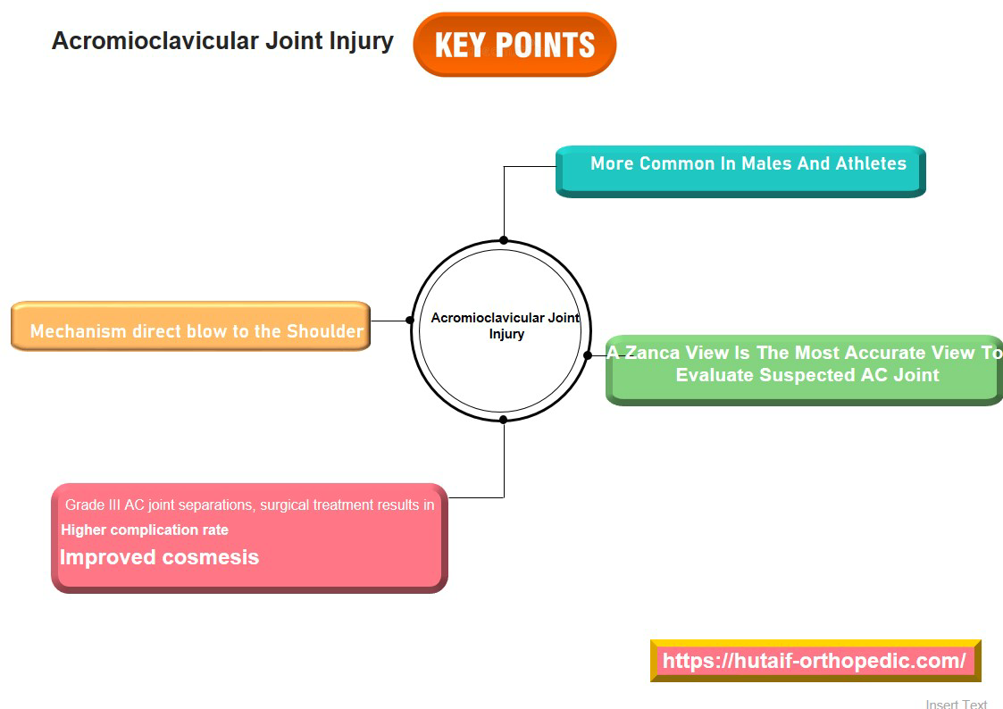
Epidemiologically, AC joint dislocations constitute approximately 9% of all injuries to the shoulder girdle and 4% of all upper extremity injuries. The incidence is notably higher in males, particularly those participating in contact sports, cycling, or other activities associated with high-energy trauma. The mechanism typically involves a direct force applied to the superior aspect of the shoulder, often with the upper extremity adducted, driving the acromion inferiorly relative to the clavicle. Less commonly, an indirect mechanism, such as a fall onto an outstretched arm, can also result in AC joint disruption. The spectrum of injury ranges from mild sprains (Rockwood Type I) to complete disruption of both the AC and coracoclavicular (CC) ligaments with significant clavicular displacement (Rockwood Type III-VI). Precise diagnosis and appropriate classification are paramount in guiding management, balancing the potential benefits of operative stabilization with the morbidity associated with surgical intervention.
Surgical Anatomy & Biomechanics
Joint Structure
The AC joint is a diarthrodial articulation between the distal clavicle and the medial aspect of the acromion. Both articular surfaces are typically covered by fibrocartilage, and an intra-articular disc or meniscus is present in approximately 30-80% of individuals, though its functional significance diminishes with age due to degeneration. The joint line orientation is variable, ranging from nearly vertical to approximately 50° medially inclined, a factor that can influence injury patterns and surgical planning. The AC joint allows for subtle gliding and rotation, contributing to the overall motion of the scapulothoracic articulation.
Ligamentous Stabilizers
Stability of the AC joint is conferred by a complex of intrinsic and extrinsic ligaments:
-
Acromioclavicular Ligaments: These ligaments directly reinforce the thin joint capsule, forming superior, inferior, anterior, and posterior bands. The superior AC ligament is the strongest and most consistently defined, blending intimately with the overlying deltotrapezial fascia. These ligaments primarily provide anteroposterior (AP) stability to the joint, resisting translation of the clavicle relative to the acromion in the horizontal plane.
-
Coracoclavicular (CC) Ligaments: These are the primary vertical stabilizers of the AC joint, connecting the undersurface of the distal clavicle to the coracoid process of the scapula. They consist of two distinct bundles:
- Trapezoid Ligament: Positioned more laterally and anterolaterally. It originates from the superior aspect of the coracoid process, running superolaterally to insert onto the trapezoid line on the conoid tubercle of the distal clavicle. Its fibers run almost vertically, providing significant resistance to vertical displacement and posterior rotation of the clavicle.
-
Conoid Ligament:
Positioned more medially and posteromedially. It originates from the base of the coracoid process, running almost vertically to insert onto the conoid tubercle of the clavicle. Its fibers are more fan-shaped and resist superior translation and anterior rotation of the clavicle.
The average distance between the conoid and trapezoid ligaments at their clavicular attachments is approximately 2 cm, and the average coracoclavicular distance is 1.1-1.3 cm in neutral radiographs.
-
Deltotrapezial Fascia: The fibers of the deltoid and trapezius muscles blend with the AC ligaments and invest the AC joint, forming a critical fascial envelope. This fascia acts as a secondary stabilizer, particularly in resisting superior displacement of the distal clavicle and providing horizontal stability. Its integrity is crucial for successful non-operative and operative outcomes.
Biomechanics
Normal AC joint motion includes rotation, protraction/retraction, and elevation/depression, synchronized with scapulothoracic movement. The CC ligaments act as a suspensory sling, holding the clavicle close to the scapula, while the AC ligaments prevent excessive AP shear.
Upon trauma, the sequence of ligamentous failure typically begins with the AC ligaments, followed by progressive disruption of the CC ligaments. Complete disruption of the AC ligaments and variable tearing of the deltotrapezial fascia, without complete CC ligament disruption, characterizes Type II injuries. Type III injuries involve complete disruption of both AC and CC ligaments, leading to significant superior displacement of the clavicle. More severe injuries (Types IV-VI) involve additional disruption of the deltotrapezial fascia and displacement of the clavicle into specific anatomical locations.

Indications & Contraindications
Clinical Evaluation
Patients typically present with localized pain over the AC joint, swelling, and often an observable deformity ("step-off" sign). Abrasions over the distal clavicle are common with direct impact mechanisms. Physical examination should be performed with the patient seated and the arm in a dependent position to accentuate any clavicular prominence.
- Inspection: Assess for ecchymosis, swelling, and asymmetry compared to the contralateral shoulder. A palpable "step-off" may be evident.
- Palpation: Tenderness directly over the AC joint is universally present. Palpate the distal clavicle, acromion, coracoid process, and surrounding structures to identify associated injuries (e.g., clavicle fracture, scapula fracture, rotator cuff pathology).
- Range of Motion: Painful active and passive shoulder elevation and cross-body adduction are typical.
-
Stability Tests:
- AC Shear Test: Compression of the anterior clavicle and posterior acromion.
- Cross-body Adduction Test: Pain elicited with forced adduction across the chest.
- Neurovascular Status: A thorough neurovascular examination of the ipsilateral upper extremity is critical to rule out brachial plexus injury or subclavian vessel compromise, particularly with high-grade dislocations (Types IV-VI).
Imaging
Initial imaging is essential for accurate diagnosis and classification.
-
Radiographs:
- AP View: Standard anteroposterior projection, typically 10-15° cephalic tilt, centered on the AC joint.
- Axillary Lateral View: Helps assess AP displacement and rule out posterior dislocation of the clavicle (Type IV).
- Zanca View: AP view with 10-15° cephalic tilt, reducing exposure by 50%. This projects the AC joint clearer by angling perpendicular to the joint space.
- Stress Views: Bilateral AP views with 10-15 lb weights held by the patient's wrists can accentuate CC ligament disruption and quantify superior displacement by measuring the coracoclavicular distance (from the superior aspect of the coracoid to the inferior cortex of the clavicle). A difference of >5 mm compared to the contralateral side, or an absolute CC distance >13 mm, is indicative of significant CC ligament injury. However, the utility of stress views for Type III injuries is debated, and they are less critical for overt higher-grade injuries.
- Computed Tomography (CT) Scan: Useful for detecting subtle clavicular or acromial fractures, especially Type IV (posterior displacement) or Type VI (subcoracoid) dislocations, and for pre-operative planning. 3D reconstructions can precisely define osseous relationships.
- Magnetic Resonance Imaging (MRI): Provides detailed soft tissue assessment, confirming AC and CC ligament disruption, deltotrapezial fascia integrity, and identifying concomitant injuries (e.g., rotator cuff tears, labral pathology, brachial plexus edema). MRI is typically not indicated for routine diagnosis of acute AC dislocations but may be considered in equivocal cases or when associated soft tissue injuries are suspected.

Classification
The Rockwood classification system is the most widely accepted method for categorizing AC joint injuries based on the degree and direction of clavicular displacement and the extent of ligamentous disruption.
- Type I: AC joint sprain. Mild stretching of AC ligaments, no clavicular displacement. CC ligaments intact.
- Type II: Subluxation. Complete tear of AC ligaments. CC ligaments sprained but intact. Slight superior subluxation of the distal clavicle (<50% of clavicular height).
- Type III: Dislocation. Complete tear of AC and CC ligaments. Disruption of deltotrapezial fascia. Superior displacement of distal clavicle (25-100% of clavicular height, >5mm CC distance compared to contralateral side). Clavicle not completely dislocated over acromion but significantly elevated.
- Type IV: Dislocation with posterior displacement. Complete tear of AC and CC ligaments. Disruption of deltotrapezial fascia. Distal clavicle displaced posteriorly into or through the trapezius muscle. Best visualized on axillary lateral view.
- Type V: Severe Dislocation. Complete tear of AC and CC ligaments. Gross disruption of deltotrapezial fascia and muscle attachments. Marked superior displacement of distal clavicle (>100% of clavicular height, >25mm CC distance). The skin may be tented by the clavicle.
- Type VI: Inferior Dislocation. Rare. Complete tear of AC and CC ligaments. Distal clavicle displaced inferiorly, beneath the coracoid process or acromion. Requires significant force and often associated with other severe injuries.

Indications for Operative vs. Non-Operative Management
The decision to proceed with operative or non-operative management is multifactorial, considering the Rockwood classification, patient activity level, hand dominance, associated injuries, and chronicity of presentation.
| Feature | Non-Operative Indications | Operative Indications |
|---|---|---|
| Rockwood Type | Type I & II: Standard of care. | Type IV, V, VI: Absolute indication for acute surgical stabilization due to gross instability and risk of skin compromise/neurovascular injury (Type V) or specific anatomical displacement patterns. |
| Type III: Controversial. Often managed non-operatively, particularly in sedentary patients, older individuals, or those with low functional demands. |
Type III:
Relative indication.
Considered for:
- High-demand overhead athletes or laborers. - Young, active patients. - Patients with persistent pain or dysfunction after a trial of non-operative management (chronic instability). - Open injuries or associated fractures (e.g., coracoid fracture). |
|
| Patient Factors | Low functional demands, sedentary lifestyle, older age, significant medical comorbidities contraindicating surgery. | High-demand athlete, manual laborer, young active individual, desire for maximal functional recovery, cosmesis concerns in certain professions. |
| Associated Injuries | Isolated AC injury. | Brachial plexus injury, subclavian vessel injury, open fracture/dislocation, ipsilateral scapula or proximal humerus fracture, glenohumeral instability requiring concomitant management. |
| Chronicity | Acute (<3 weeks) Type I/II, select Type III. | Acute Type IV-VI. Chronic dislocations (>3 weeks) of Type III-VI with persistent pain, instability, or significant functional impairment, typically requiring reconstruction rather than repair. |
| Functional Outcome | Acceptable function with potential for residual deformity, mild pain, and endurance deficits. | Aims for anatomical reduction and restoration of stability, potentially leading to better cosmetic outcome and higher functional return, but with surgical risks. |
Contraindications for Operative Management
Absolute contraindications are rare and typically relate to the patient's overall medical status (e.g., severe cardiopulmonary disease precluding general anesthesia) or active local infection. Relative contraindications include advanced age with low functional demands, non-compliance with post-operative rehabilitation, or poorly controlled systemic diseases.
Pre-Operative Planning & Patient Positioning
Thorough pre-operative planning is critical to ensure a successful outcome and minimize complications.
Pre-operative Workup
- History and Physical: Reconfirm diagnosis, assess patient expectations, identify comorbidities, and ascertain activity level.
- Imaging Review: Review all radiographs (AP, axillary lateral, Zanca, stress views if taken), CT, and MRI scans to fully understand the injury pattern, rule out associated fractures, and plan the surgical approach. Pay close attention to the morphology of the coracoid process and distal clavicle for tunnel placement.
- Informed Consent: Discuss the risks (infection, nerve injury, hardware failure, loss of reduction, chronic pain, stiffness, re-operation) and benefits of the chosen procedure. Detail rehabilitation expectations.
- Antibiotic Prophylaxis: Administer intravenous broad-spectrum antibiotics (e.g., Cefazolin) within one hour prior to incision.
- Anesthesia: General anesthesia is standard. A regional interscalene block can provide excellent post-operative pain control but must be carefully considered given its potential to mask neurological deficits.
Patient Positioning
The
beach chair position
is generally preferred for AC joint surgery.
*
Setup:
The patient is placed supine on the operating table, then the head and back are elevated to a semi-recumbent position (30-60° of torso elevation). The head is supported by a doughnut headrest.
*
Arm Positioning:
The ipsilateral arm is typically free-draped, allowing for intraoperative manipulation to assess reduction and range of motion. It should be supported by an arm board or assistant in a neutral position to prevent traction on the brachial plexus.
*
Padding:
Adequate padding of all pressure points (sacrum, heels, contralateral arm, ulnar nerve at the elbow) is crucial to prevent pressure neuropathies.
*
Fluoroscopy:
Ensure a C-arm can be brought into the field to obtain adequate AP and lateral views for real-time assessment of reduction and hardware placement. The image intensifier should be positioned to allow unobstructed views of the clavicle, AC joint, and coracoid process.
*
Sterile Prep and Drape:
The surgical field extends from the ipsilateral neck to the mid-humerus, including the entire shoulder girdle, and medially across the sternum to the contralateral clavicle.
Alternatively, the
lateral decubitus position
can be used, particularly if a concomitant arthroscopic glenohumeral procedure is planned.
*
Setup:
Patient is placed on the contralateral side, supported by a bean bag. The ipsilateral arm is often suspended in traction.
*
Advantages:
May facilitate visualization of the coracoid if extensive subcoracoid work is anticipated.
*
Disadvantages:
Can make external rotation of the arm for posterior hardware assessment more challenging.

Detailed Surgical Approach / Technique
The goal of surgical management for AC joint dislocation is anatomical reduction of the distal clavicle to the acromion and coracoid, with restoration of both vertical and horizontal stability, while promoting biological healing of the disrupted ligaments. Numerous techniques exist, broadly categorized into temporary fixation, direct AC ligament repair, CC ligament repair/reconstruction, and distal clavicle excision. Modern approaches often combine CC ligament reconstruction/augmentation with repair of the deltotrapezial fascia.
General Principles
- Reduction: Achieve and maintain anatomical reduction of the clavicle relative to the coracoid and acromion.
- Stabilization: Provide stable fixation that allows for early motion while protecting the healing ligaments.
- Restoration: Address both vertical (CC ligaments) and horizontal (AC ligaments, deltotrapezial fascia) stability.
Common Surgical Techniques
1. Coracoclavicular Suture Button Fixation (e.g., Dog Bone, TightRope)
This is a popular contemporary technique, often performed open or arthroscopically-assisted, for acute and chronic AC joint instabilities. It provides strong, flexible fixation, allowing dynamic stability.
-
Incision and Exposure:
- A transverse or saber-cut incision (3-5 cm) is made centered over the distal clavicle and AC joint.
- Dissection proceeds through the subcutaneous tissue. The deltotrapezial fascia, typically torn in Type III-VI injuries, is identified. Careful separation of the deltoid and trapezius muscle fibers is performed to expose the AC joint and the superior surface of the distal clavicle.
- The torn AC joint capsule and ligaments are débrided or prepared for direct repair if possible.
- The coracoid process is then identified. This can be challenging. A useful landmark is to follow the lateral border of the clavicle inferiorly, palpating the tip and base of the coracoid. Dissection should proceed cautiously along the anterior aspect of the coracoid, just medial to the conjoined tendon origin, protecting the neurovascular structures located medially.
-
Reduction of the AC joint is achieved by downward pressure on the clavicle and upward pressure on the elbow. Provisional reduction can be held with a provisional K-wire from the clavicle into the acromion, or manually.

-
Tunnel Creation:
- Clavicular Tunnels: Two parallel guide pins are drilled vertically from the superior surface of the clavicle, exiting on its inferior cortex. These are typically placed approximately 2 cm medial to the lateral border of the clavicle, straddling the anatomical footprint of the conoid and trapezoid ligaments. The distance between the tunnels should correspond to the specific suture button system. The clavicular holes are then reamed to the appropriate diameter (e.g., 3.0-4.0 mm).
- Coracoid Tunnel: A single guide pin is drilled through the base of the coracoid process, perpendicular to its superior cortex. This tunnel should be placed just medial to the trapezoid ligament's insertion on the coracoid, aiming for a robust bone stock. The coracoid tunnel is then reamed.
-
Important Consideration:
Ensuring the clavicular tunnels are not too lateral (risk of distal clavicle fracture) and the coracoid tunnel is not too medial (risk of neurovascular injury) is paramount. Careful C-arm fluoroscopy verification of guide pin placement is essential.

-
Suture Button Passage and Tensioning:
- The suture button device (e.g., two cortical buttons connected by high-strength sutures) is prepared. One button is passed through the clavicular tunnels. The other button is passed through the coracoid tunnel.
- The sutures are then carefully pulled, reducing the clavicle onto the coracoid. This is performed under direct visualization and fluoroscopic guidance to ensure anatomical reduction. The sutures are then tensioned and secured, locking the buttons to maintain reduction.
-
Dynamic tensioning by cycling the shoulder through a range of motion can help ensure optimal tension.

-
Augmentation and Closure:
- The torn deltotrapezial fascia is meticulously repaired using strong, non-absorbable sutures to provide additional horizontal stability and protect the primary fixation.
- The subcutaneous tissues and skin are closed in layers.
2. Hook Plate Fixation
This temporary fixation method involves a plate with a hook that engages the undersurface of the acromion, providing strong superior-inferior stability. It is typically used for acute Type III-V injuries.
- Incision and Exposure: Similar to suture button technique.
- Plate Application: The hook plate is contoured and applied to the superior surface of the distal clavicle. The hook is carefully passed beneath the acromion, ensuring proper engagement and minimal impingement.
- Fixation: The plate is secured to the clavicle with screws.
- Deltotrapezial Repair: Crucial for horizontal stability and preventing redislocation after plate removal.
- Removal: The hook plate is a temporary device and requires a second surgery for removal, typically at 3-6 months. Complications include subacromial erosion, pain, and fracture upon removal.
3. Coracoclavicular Ligament Reconstruction (Weaver-Dunn with Graft Augmentation)
Primarily used for chronic AC joint dislocations or acute injuries requiring robust biological reconstruction.
-
Weaver-Dunn Procedure (modified):
- Distal clavicle resection (1-2 cm) to address potential impingement and AC joint arthritis.
- Transfer of the coracoacromial (CA) ligament from the acromion to the distal clavicle. The CA ligament provides a biological soft tissue sling for the distal clavicle.
-
Graft Augmentation:
Autograft (e.g., semitendinosus, gracilis) or allograft (e.g., Achilles tendon, tibialis anterior) is typically used to reconstruct the CC ligaments.
- Graft is passed through a drill hole in the clavicle (or two holes for a double-bundle reconstruction) and around the base of the coracoid process.
- Fixation of the graft is achieved using interference screws, suture buttons, or staples.
- The CA ligament transfer is typically performed in conjunction with graft reconstruction for enhanced stability.
- Deltotrapezial Fascia Repair: Essential for providing further stability and protecting the reconstruction.
4. Arthroscopic-Assisted Techniques
Many of the open techniques, particularly suture button fixation, can be performed with arthroscopic assistance.
- Advantages: Minimally invasive, smaller incisions, potentially less soft tissue dissection, better visualization of the coracoid undersurface.
- Approach: Standard posterior portal for glenohumeral evaluation if needed. An anterolateral portal is often used for visualization of the AC joint and coracoid. A superolateral portal may be used for instrument passage and drilling.
- Visualization and Reduction: Arthroscopic visualization confirms glenohumeral health and assists in identifying the coracoid. A small incision over the clavicle is still required for drilling and button placement.
- Caveat: Arthroscopic techniques require specialized training and equipment.
Intraoperative Pearls
- Anatomical Reduction: Aim for true anatomical reduction rather than just reduction to the level of the superior acromial surface. Fluoroscopy in both AP and lateral planes is crucial.
- Neurovascular Protection: Meticulous dissection around the coracoid process is essential to avoid injury to the brachial plexus and subclavian vessels. Stay close to the bone.
- Deltotrapezial Repair: Do not underestimate the importance of meticulous repair of the deltotrapezial fascia; it is a critical dynamic stabilizer.
- Fixation Strength: Ensure adequate tensioning of the suture button systems or secure plating to prevent loss of reduction.
- Concomitant Pathology: Address any concomitant intra-articular pathology (e.g., rotator cuff tear) identified pre-operatively or intra-operatively.

Complications & Management
Despite advancements in surgical techniques, AC joint stabilization procedures are associated with a range of potential complications. Recognition and appropriate management are crucial for salvage and patient outcomes.
Common Complications and Salvage Strategies
| Complication | Incidence | Management Strategies |
|---|---|---|
| Loss of Reduction/Recurrence of Instability | 5-20% |
Early:
Revision surgery with a stronger construct (e.g., supplemental graft, revision suture button with additional points of fixation).
Late/Chronic: If symptomatic, consider a formal CC ligament reconstruction with autograft/allograft and distal clavicle excision if AC joint arthritis is present. |
| Infection | 1-5% |
Superficial:
Oral antibiotics, wound care.
Deep: Surgical irrigation and débridement, culture-guided intravenous antibiotics, potential hardware removal if non-salvageable. Early identification is key. |
| Hardware-Related Complications | ||
| * Hardware prominent/painful | Up to 30-50% (Hook Plate) | Symptomatic hardware typically requires removal after appropriate healing time (e.g., 3-6 months for hook plates, 6-12 months for suture buttons if causing impingement). Suture buttons are often left in situ if asymptomatic. |
| * Hardware migration/failure | 2-10% (all types) |
Migration:
Excision of migrated hardware. If reduction is lost, consider revision.
Failure (e.g., suture breakage): If symptomatic and unstable, revision surgery with a different or augmented fixation technique. |
| * Clavicle/Coracoid Fracture | <5% |
Intraoperative:
Requires immediate stabilization of the fracture (e.g., plate and screws) and potentially a different AC joint fixation method.
Postoperative: Non-displaced fractures may heal with immobilization. Displaced fractures may require open reduction and internal fixation (ORIF). |
| AC Joint Arthritis | Up to 20-30% |
Often related to inadequate reduction, persistent instability, or articular cartilage damage.
Management: Conservative measures (NSAIDs, injections). If persistent and symptomatic, consider distal clavicle excision (Mumford procedure) with or without continued CC ligament stabilization if instability is also a factor. |
| Stiffness/Adhesive Capsulitis | 5-15% |
Prevention:
Early, controlled range of motion (ROM) as dictated by rehabilitation protocol.
Management: Intensive physical therapy, NSAIDs, glenohumeral corticosteroid injections. If refractory, consider arthroscopic capsular release. |
| Neurovascular Injury | Rare (<1%) |
Prevention:
Meticulous surgical technique, careful identification and protection of structures (brachial plexus, subclavian vessels) during coracoid drilling and dissection.
Management: Intraoperative repair if identified. Postoperative: Neurology consultation, nerve exploration/repair, vascular repair. |
| Heterotopic Ossification (HO) | 5-10% |
Prevention:
Prophylaxis (NSAIDs, radiation therapy) in high-risk patients (e.g., trauma patients with head injury, ankylosing spondylitis).
Management: If symptomatic and mature, surgical excision. |
| Residual Pain | Highly variable (10-40%) |
Often multifactorial. Investigate causes (instability, arthritis, impingement, nerve irritation).
Management: Targeted physical therapy, injections, pain management, revision surgery for specific identifiable pathology. |
| Deltoid/Trapezius Dehiscence | <5% |
Prevention:
Meticulous repair of the deltotrapezial fascia.
Management: If symptomatic and significant, surgical re-repair. |
Specific Considerations
- Hook Plate Removal: Routine removal of hook plates is necessary, usually between 3-6 months, to prevent complications such as acromial osteolysis, pain, and fracture, making it a two-stage procedure.
- Suture Button Systems: While generally well-tolerated, button prominence, suture breakage, and osteolysis around the tunnels can occur. Often, the hardware is left in place unless symptomatic.
- Chronic Instability: Management of chronic AC dislocations often requires CC ligament reconstruction with autograft or allograft, frequently combined with distal clavicle excision due to the high incidence of post-traumatic AC joint arthritis.


Post-Operative Rehabilitation Protocols
Post-operative rehabilitation is paramount for optimizing outcomes after AC joint stabilization. Protocols vary based on the surgical technique, the integrity of the deltotrapezial fascia repair, and surgeon preference, but generally follow a phased approach focused on protecting the repair, restoring range of motion, and regaining strength.
General Principles
- Protection: Guard against stress to the healing ligaments and fixation construct.
- Gradual Progression: Slowly increase mobility and load to allow for tissue healing and adaptation.
- Pain Management: Address pain to facilitate participation in therapy.
- Scapular Control: Emphasize scapular stabilization throughout all phases.
Phase I: Protection & Early Motion (Weeks 0-6)
- Goals: Protect surgical repair, minimize pain and swelling, initiate early passive range of motion (PROM).
-
Immobilization:
- Sling immobilization is typically used for 4-6 weeks to protect the deltotrapezial repair and reduce tension on the CC reconstruction. The arm should be removed from the sling for exercises.
-
Exercises:
- Passive Range of Motion (PROM): Pendulum exercises (gentle), passive flexion to 90-120°, passive external rotation to 30-45° (shoulder adducted). Avoid active shoulder elevation, abduction, or cross-body adduction during this phase.
- Elbow, Wrist, Hand ROM: Active exercises for the elbow, wrist, and hand are encouraged to prevent stiffness.
- Scapular Mobility: Gentle scapular retraction and protraction exercises.
- Core Stability: Maintain core strength.
-
Precautions:
- NO active shoulder elevation or abduction.
- NO lifting with the operative arm.
- NO pulling or pushing.
- Maintain good posture.

Phase II: Gradual Motion & Early Strengthening (Weeks 6-12)
- Goals: Restore full passive ROM, initiate active assisted range of motion (AAROM) and active range of motion (AROM), begin light strengthening.
-
Immobilization:
- Discontinue sling as tolerated, typically around 6 weeks.
-
Exercises:
- AAROM/AROM: Progress from AAROM to AROM for shoulder flexion, abduction, and external/internal rotation within pain-free limits.
- Isometric Strengthening: Begin with isometric exercises for rotator cuff (internal/external rotation), deltoid, and scapular stabilizers.
- Light Resistance: Introduce light resistance exercises using resistance bands or very light weights for rotator cuff and deltoid.
- Scapular Strengthening: Wall slides, rows, prone extension.
-
Precautions:
- Avoid heavy lifting or sudden movements.
- No overhead lifting of weights.
- Limit activities that place direct stress on the AC joint.
Phase III: Advanced Strengthening & Return to Activity (Weeks 12-24)
- Goals: Achieve full functional strength, endurance, and power; gradually return to sport-specific or work-specific activities.
-
Exercises:
- Progressive Resistance: Advance strengthening exercises with increasing resistance and weight for all shoulder muscles (rotator cuff, deltoid, pectorals, latissimus, biceps, triceps).
- Functional Exercises: Incorporate exercises that mimic daily activities or sport-specific movements.
- Plyometrics: For athletes, introduce plyometric exercises as appropriate.
- Proprioception: Balance and proprioceptive training.
- Endurance: Light cardio and progressive endurance exercises for the upper extremity.
-
Return to Activity:
- Return to light sports or work activities may begin around 4-6 months, depending on the individual's progress and the demands of the activity.
- Return to contact sports or overhead activities typically requires 6-9 months, contingent upon achieving full strength, stability, and surgeon clearance.
-
Precautions:
- Listen to the body; avoid pain.
- Continue a maintenance exercise program.
Long-Term Considerations
Full recovery can take 6-12 months. Patients should be counseled on the potential for residual hardware sensation, stiffness, or low-grade discomfort. Compliance with rehabilitation is the strongest predictor of a successful outcome.

Summary of Key Literature / Guidelines
The management of AC joint dislocations, particularly Type III injuries, remains an area of ongoing debate and evolving evidence. Current literature supports tailored approaches based on injury severity, patient factors, and desired outcomes.
Evidence-Based Recommendations
- Type I & II: Consensus strongly supports non-operative management. A recent systematic review confirmed excellent outcomes for these types with conservative treatment, including early mobilization and symptomatic relief.
-
Type III:
This is the most controversial classification.
- Historically, many Type III injuries were treated non-operatively, with meta-analyses showing similar functional outcomes (e.g., DASH scores, pain levels) between operative and non-operative groups at long-term follow-up. Non-operative management typically results in an earlier return to activity but may leave a residual cosmetic deformity and potentially some fatigue endurance deficits.
- However, a subset of patients with Type III injuries, particularly young, active individuals, overhead athletes, or laborers, may benefit from surgical stabilization. Some studies suggest superior strength and endurance in operatively treated Type III injuries, especially with CC ligament reconstruction. The primary goal of surgery in these cases is to restore anatomical alignment and maximize shoulder function, potentially reducing the risk of chronic pain or instability.
- The decision for Type III must be individualized, considering patient expectations and activity demands.
- Type IV, V, VI: Strong consensus for acute surgical stabilization. These high-grade injuries are associated with severe instability, gross deformity, and potential neurovascular compromise, necessitating anatomical reduction and robust fixation. Delays in surgical intervention for these types can make reduction and soft tissue repair significantly more challenging.
Key Literature Trends and Controversies
-
Optimal Surgical Technique:
- Hook Plates: While providing rigid fixation, their temporary nature and high rates of hardware-related complications (up to 50% requiring removal) have led to a decline in their primary use. They may still be considered in select cases for specific patient populations or as a salvage option.
- Coracoclavicular Suture Button Fixation: Techniques like TightRope (Arthrex) or Dog Bone (Arthrex) have gained popularity due to their minimally invasive nature (often arthroscopically-assisted), strong biomechanical properties, and preservation of biological structures. Meta-analyses suggest favorable outcomes and lower reoperation rates compared to hook plates.
- Graft Reconstruction: For chronic instability or situations where primary repair is not feasible, autograft or allograft reconstruction of the CC ligaments, often combined with distal clavicle excision and CA ligament transfer (modified Weaver-Dunn), demonstrates good results.
- Combined AC and CC Ligament Reconstruction: Current trends favor techniques that address both vertical and horizontal stability, often combining CC ligament reconstruction with robust deltotrapezial fascia repair.
-
Acute vs. Chronic Repair:
- Acute repair (<3 weeks post-injury) generally allows for primary repair of the torn ligaments and deltotrapezial fascia, yielding more predictable results.
- Chronic injuries (>3 weeks) typically require reconstruction with tissue grafts due to retraction and degeneration of native ligaments.
-
Distal Clavicle Excision:
- Routine distal clavicle excision (DCE) as part of acute AC joint reconstruction is debated. While it prevents post-traumatic AC joint arthritis, it can compromise horizontal stability.
- DCE is generally reserved for patients with symptomatic AC joint arthritis, particularly in chronic cases or failures of previous stabilization.
-
Role of Biological Augmentation:
- The use of allografts or autografts to augment suture button constructs or as standalone CC ligament reconstructions is increasingly common, especially for chronic injuries or in high-demand athletes, to provide biological stability.
- Research into tissue engineering and growth factors to enhance ligament healing is ongoing.
Future Directions
Future research is focused on refining existing techniques, developing novel biodegradable fixation devices, and exploring biological augmentation strategies to enhance native ligament healing. Personalized treatment algorithms, potentially incorporating advanced imaging and biomechanical modeling, will further optimize patient selection and surgical outcomes. Understanding the long-term implications of various surgical constructs on surrounding joint mechanics and the development of arthritis will also remain a critical area of study. The ongoing evolution underscores the importance for orthopedic surgeons to remain current with the scientific literature and evidence-based guidelines in managing AC joint dislocations.
Clinical & Radiographic Imaging


You Might Also Like