The Role of Magnetic Resonance Imaging in Operative Orthopaedics: A Masterclass in Surgical Planning
Key Takeaway
Magnetic Resonance Imaging (MRI) has revolutionized operative orthopaedics by providing unparalleled soft-tissue resolution and multiplanar capabilities. This comprehensive guide details the critical role of MRI in surgical planning, detailing specialized coil applications, optimal imaging protocols, and region-specific diagnostic criteria. By bridging the gap between radiological findings and surgical execution, orthopaedic surgeons can optimize approaches, anticipate intraoperative challenges, and improve postoperative outcomes across knee, hip, spine, shoulder, and foot and ankle pathologies.
Introduction to Advanced Orthopaedic Magnetic Resonance Imaging
Magnetic Resonance Imaging (MRI) has fundamentally transformed the landscape of operative orthopaedics. Moving beyond the limitations of plain radiography and computed tomography (CT), MRI provides unparalleled soft-tissue contrast, multiplanar imaging capabilities, and the ability to detect occult osseous pathologies. The advent of smaller, highly specialized surface coils has allowed for dramatically higher spatial resolution and smaller fields of view (FOV), all while maintaining—and often enhancing—satisfactory image signal-to-noise ratios (SNR).
Continued improvements in both hardware (e.g., 3.0 Tesla and 7.0 Tesla magnets) and software (e.g., fast spin-echo sequences, isotropic 3D imaging) have shortened imaging times and reduced motion artifacts. However, optimal image quality is not solely a product of technology; it requires meticulous attention to imaging technique by the radiologist and MRI technologist. Furthermore, a synergistic interaction between the operating orthopaedic surgeon and the musculoskeletal radiologist is paramount to ensure that imaging protocols are tailored to solve specific clinical and surgical problems.
Clinical Pearl: Never treat the MRI; treat the patient. While MRI is highly sensitive, its specificity can vary. Incidental findings, such as asymptomatic meniscal degeneration or mild disc bulging, are common in the general population. Surgical indications must always be dictated by a rigorous clinical examination that correlates with the advanced imaging findings.
General Principles of MRI in Surgical Planning
Before delving into region-specific pathologies, the orthopaedic surgeon must understand the fundamental sequences that dictate surgical decision-making:
* T1-Weighted Sequences: Excellent for defining regional anatomy and evaluating bone marrow. Fat appears bright, making it ideal for detecting occult fractures or avascular necrosis (AVN).
* T2-Weighted Sequences: Highly sensitive to fluid (water appears bright). Essential for identifying edema, effusions, cysts, and acute ligamentous or tendinous tears.
* STIR (Short Tau Inversion Recovery) / Fat-Suppressed T2: Suppresses the bright signal of fat to exquisitely highlight bone marrow edema, stress fractures, and acute inflammatory processes.
* Proton Density (PD): Provides a balance between T1 and T2, offering exceptional anatomical detail of articular cartilage, menisci, and ligaments.
* MR Arthrography (MRA): Involves the intra-articular injection of dilute gadolinium. The capsular distension separates intra-articular structures, making it the gold standard for evaluating labral tears in the shoulder and hip.
The Knee: Meniscal, Ligamentous, and Chondral Pathologies
The knee is the most frequently imaged joint in orthopaedics. MRI has largely replaced diagnostic arthroscopy, serving as the definitive preoperative roadmap.
Meniscal Injuries
MRI is highly accurate in detecting meniscal tears, utilizing fast spin-echo PD sequences. Tears are categorized by their configuration (e.g., horizontal, longitudinal, radial, root, or bucket-handle), which directly dictates the surgical approach (meniscectomy vs. repair).
* Grading: Grade 1 and 2 signals are confined within the meniscus substance (myxoid degeneration) and are treated conservatively. Grade 3 signal extends to an articular surface, representing a true tear.
* Surgical Implication: A "double PCL sign" or an absent "bow-tie sign" on sagittal images indicates a displaced bucket-handle tear. This necessitates prompt surgical intervention. The surgeon must prepare for a potential inside-out or all-inside meniscal repair, positioning the patient appropriately with a lateral post or leg holder to allow for valgus/varus stress during arthroscopy.
Anterior Cruciate Ligament (ACL) Tears
Acute complete tears of the ACL are diagnosed with near 100% accuracy on MRI.
* Biomechanics & Bone Bruises: MRI frequently reveals characteristic "kissing" bone marrow edema patterns in the lateral femoral condyle and the posterolateral tibial plateau. This pathognomonic finding is a footprint of the pivot-shift mechanism of injury.
* Postoperative Evaluation: MRI is invaluable for evaluating ACL reconstructions. It can assess graft incorporation, impingement (e.g., roof impingement due to anterior tibial tunnel placement), and the integrity of the hardware.
Surgical Warning: When evaluating a postoperative meniscus for a re-tear, standard MRI is notoriously difficult to interpret due to persistent altered signal from the prior healing process. MR arthrography or evaluating for fluid tracking into the tear on T2 sequences is required to confirm a true re-tear.
The Hip: Avascular Necrosis, Impingement, and Occult Fractures
MRI of the hip requires a large FOV for bilateral comparison (e.g., for AVN) followed by a small FOV for detailed articular evaluation.
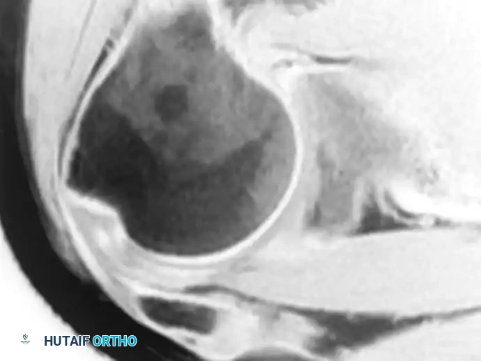
Avascular Necrosis (AVN) of the Femoral Head
MRI is the most sensitive modality for early detection of AVN, often identifying lesions long before plain radiographic changes (Ficat Stage I).
* Imaging Characteristics: The classic "band-like" lesion on T1 or the "double-line sign" on T2 represents the reactive interface between necrotic and viable bone.
* Surgical Planning: Volumetric analysis of the necrotic segment using MRI is critical. Lesions involving less than 30% of the weight-bearing surface may be amenable to joint-preserving procedures like core decompression or vascularized fibular grafting. Larger lesions with subchondral collapse (crescent sign) typically require Total Hip Arthroplasty (THA).
Femoroacetabular Impingement (FAI) and Labral Tears
MR arthrography is the modality of choice for evaluating the acetabular labrum and articular cartilage.
* Cam and Pincer Morphology: MRI allows for precise measurement of the alpha angle (Cam impingement) and acetabular version (Pincer impingement).
* Surgical Approach: Preoperative MRI mapping dictates the extent of femoral osteochondroplasty or acetabular rim trimming required during hip arthroscopy. The patient is positioned supine or in the lateral decubitus position on a traction table, and the MRI guides the exact location for portal placement to address the specific labral pathology.
Occult Fractures
In elderly patients with hip pain following a fall and negative plain radiographs, MRI is the definitive diagnostic tool. It can rapidly differentiate between an occult femoral neck fracture (requiring urgent surgical fixation to prevent displacement) and an isolated greater trochanteric fracture or pelvic ring injury (often managed conservatively).
The Shoulder: Instability and Rotator Cuff Pathology
Shoulder MRI requires specialized surface coils and specific patient positioning to optimize diagnostic yield.
Glenohumeral Instability
For patients with recurrent anterior shoulder instability, MR arthrography is essential to evaluate the capsulolabral complex.
* Positioning: The ABER (Abduction and External Rotation) position is a specialized MRI technique. By placing the arm in ABER, the anterior band of the inferior glenohumeral ligament (IGHL) is placed under tension. This exquisitely demonstrates anteroinferior labral tears (Bankart lesions) and articular-sided partial rotator cuff tears (PASTA lesions).
* Surgical Implication: Identification of significant glenoid bone loss or an engaging Hill-Sachs lesion on MRI will pivot the surgical plan from an arthroscopic Bankart repair to an open Latarjet procedure.
Rotator Cuff Tears
MRI accurately determines the size, retraction, and chronicity of rotator cuff tears.
* Muscle Atrophy and Fatty Infiltration: The Goutallier classification, adapted for MRI, assesses fatty infiltration of the rotator cuff muscle bellies (particularly the supraspinatus and infraspinatus). High-grade fatty infiltration (Grade 3 or 4) is generally irreversible and portends a high failure rate for primary repair, often steering the surgeon toward superior capsular reconstruction (SCR) or Reverse Total Shoulder Arthroplasty (RTSA).
Pitfall: Suprascapular nerve entrapment can mimic a massive rotator cuff tear clinically. Always evaluate the spinoglenoid and suprascapular notches on sagittal MRI for paralabral cysts, which require arthroscopic decompression rather than primary tendon repair.
The Spine: Degeneration, Infection, and Trauma
Spinal MRI is the cornerstone of modern spine surgery, dictating whether a patient requires decompression, fusion, or conservative care.
Disc Degeneration and Herniation
MRI clearly delineates the relationship between herniated nuclear material and the traversing or exiting nerve roots.
* Postoperative Spine: Differentiating between recurrent disc herniation and postoperative epidural fibrosis (scar tissue) is a common clinical dilemma.
* Protocol: Intravenous Gadolinium contrast is mandatory in this scenario. Epidural scar tissue is highly vascular and will enhance uniformly with gadolinium, whereas a recurrent avascular disc fragment will not enhance (though it may show a thin rim of peripheral enhancement).
Spinal Infections
MRI is highly sensitive for detecting discitis and vertebral osteomyelitis.
* Imaging Characteristics: Decreased signal on T1 and increased signal on T2/STIR within the disc space and adjacent vertebral endplates are classic. Contrast enhancement helps define epidural abscesses.
* Surgical Approach: The presence of an epidural abscess causing neurologic deficit or significant spinal instability dictates urgent surgical decompression and debridement, often requiring a combined anterior-posterior approach depending on the extent of ventral destruction.
Vertebral Compression Fractures
Differentiating benign osteoporotic compression fractures from malignant pathological fractures is critical. Benign fractures typically show preserved normal marrow signal in the pedicles and a retropulsed bone fragment, whereas malignant fractures exhibit complete marrow replacement, pedicle involvement, and an epidural soft-tissue mass.
The Foot and Ankle: Complex Soft Tissue and Osteochondral Lesions
The complex, compact anatomy of the foot and ankle necessitates high-resolution MRI with small FOVs.
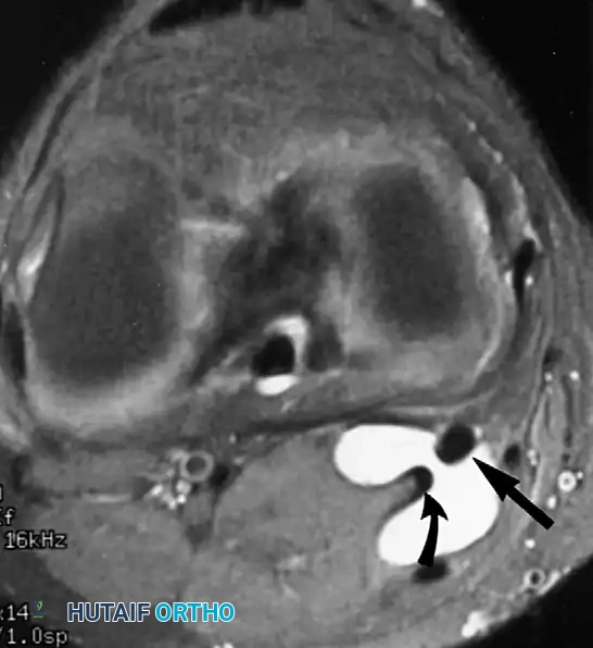
Osteochondral Lesions of the Talus (OCLT)
MRI is superior to helical CT for assessing the stability of osteochondral lesions.
* Stability Criteria: High T2 signal fluid tracking completely behind the osteochondral fragment indicates instability.
* Surgical Planning: Unstable lesions or those failing conservative management require surgery. Anterior lesions are typically addressed via standard anterior arthroscopic portals. Posterior lesions may require a posterolateral approach or even a medial malleolar osteotomy if the lesion is large, deep, and centrally located.
Tendon Dysfunction
Posterior tibial tendon dysfunction (PTTD) is a primary cause of adult acquired flatfoot deformity.
* MRI Findings: MRI can stage the tendinopathy, showing tenosynovitis, partial tearing, or complete rupture.
* Magic Angle Artifact: The radiologist and surgeon must be aware of the "magic angle" phenomenon. When a tendon (like the PTT as it curves around the medial malleolus) is oriented at 55 degrees to the main magnetic field, it can artificially show increased signal on short TE sequences (T1, PD), mimicking a tear. Evaluating the tendon on T2 sequences (long TE) resolves this artifact.
Diabetic Foot Osteomyelitis
In diabetic patients with foot ulcers, differentiating osteomyelitis from neuropathic (Charcot) arthropathy is notoriously difficult. MRI is the modality of choice. Confluent bone marrow edema directly adjacent to a skin ulcer or sinus tract is highly predictive of osteomyelitis, guiding the surgeon in determining the necessary level of amputation or bone resection.
Conclusion and Future Directions
The integration of Magnetic Resonance Imaging into operative orthopaedics has elevated the precision of surgical interventions to unprecedented levels. By providing a detailed preoperative roadmap, MRI allows the orthopaedic surgeon to anticipate intraoperative challenges, select the optimal surgical approach, and counsel patients accurately regarding their prognosis.
As technology advances, the future of orthopaedic MRI lies in ultra-high-field imaging (7.0 Tesla) and compositional cartilage mapping (such as dGEMRIC, T2 mapping, and T1rho). These techniques will allow surgeons to evaluate the biochemical integrity of articular cartilage before macroscopic morphological changes occur, ushering in a new era of truly preventative and joint-preserving orthopaedic surgery. Ultimately, the greatest outcomes are achieved when meticulous radiological interpretation is seamlessly married to masterful surgical execution.
You Might Also Like