Comprehensive Guide to Hip Arthroscopy: Principles, Anatomy, and Patient Selection
Key Takeaway
Hip arthroscopy is a minimally invasive surgical technique addressing intra-articular and peritrochanteric hip pathologies, primarily FAI and labral tears. It aims to preserve the native hip joint, reduce pain, and restore function. Success hinges on a deep understanding of surgical anatomy, biomechanics, neurovascular structures, and careful patient selection for optimal outcomes.
Principles of Hip Arthroscopy: Essential Steps for Success
Introduction & Epidemiology
Hip arthroscopy has evolved significantly over the past two decades, transitioning from a niche diagnostic tool to a widely adopted, minimally invasive surgical technique for addressing a broad spectrum of intra-articular and peritrochanteric pathologies. Its development parallels advancements in instrumentation, imaging, and understanding of hip joint kinematics and pathomechanics. The primary impetus for the growth of hip arthroscopy has been the recognition of femoroacetabular impingement (FAI) and labral pathology as significant precursors to hip osteoarthritis, particularly in young, active adults. By addressing these structural abnormalities early, hip arthroscopy aims to preserve the native hip joint, alleviate pain, restore function, and potentially delay or prevent the need for total hip arthroplasty.
Epidemiologically, conditions such as FAI and labral tears are prevalent, with studies indicating a high incidence of morphologic variations associated with FAI in asymptomatic individuals, underscoring the importance of symptomatic presentation for intervention. The demographic profile of patients undergoing hip arthroscopy typically includes younger adults (20-50 years old) involved in sports or physically demanding occupations, although its utility extends to select adolescent and older adult populations. The procedure is increasingly recognized as a cornerstone of hip preservation surgery, demonstrating favorable outcomes in pain reduction and functional improvement when applied to appropriate indications.
Surgical Anatomy & Biomechanics
A thorough understanding of hip anatomy is paramount for safe and effective hip arthroscopy. The hip is a complex ball-and-socket synovial joint, characterized by its inherent stability due to deep osseous congruity, a robust capsule, and strong ligamentous support.
Bony Anatomy
The primary bony structures include the acetabulum (comprising ilium, ischium, and pubis) and the femoral head. Key landmarks for portal placement and orientation include the anterior superior iliac spine (ASIS), pubic tubercle, greater trochanter, and the inferior pubic ramus. The relationship between the femoral head-neck junction and the acetabular rim is critical in understanding FAI. Cam-type impingement involves an aspherical femoral head (pistol grip deformity), while pincer-type impingement involves excessive acetabular overcoverage.
Articular Cartilage & Labrum
The acetabulum is lined by hyaline cartilage in a horseshoe shape, leaving the acetabular fossa bare. The acetabular labrum, a fibrocartilaginous ring, deepens the socket, enhances joint stability by increasing the surface area of contact, creates a suction seal, and distributes stress across the articular cartilage. Tears of the labrum, often associated with FAI, can compromise this suction seal and lead to pain and progressive cartilage damage.
Joint Capsule & Ligaments
The hip joint capsule is strong and fibrous, attaching proximally to the acetabular rim and transverse acetabular ligament, and distally to the intertrochanteric line anteriorly and the base of the femoral neck posteriorly. Three primary capsular ligaments reinforce the capsule:
*
Iliofemoral ligament (Y-ligament of Bigelow):
The strongest ligament, preventing hyperextension. Its anterior fibers are relevant during capsulotomy for potential instability.
*
Pubofemoral ligament:
Prevents excessive abduction and external rotation.
*
Ischiofemoral ligament:
Prevents excessive internal rotation and extension.
The
ligamentum teres
, an intra-articular ligament originating from the acetabular fossa and inserting into the fovea of the femoral head, contains small vessels supplying the femoral head (especially in childhood). While its role in adult hip stability is debated, its pathology (tears, hypertrophy) can cause pain.
Neurovascular Structures at Risk
Careful attention to neurovascular anatomy is crucial to minimize iatrogenic injury.
*
Lateral Femoral Cutaneous Nerve (LFCN):
Courses inferior to the ASIS and is at risk with anterolateral portals, particularly during skin incision and fascial dissection.
*
Femoral Nerve, Artery, and Vein:
Located anterior to the hip joint, medial to the ASIS. They are vulnerable during placement of anteromedial or straight anterior portals. A safe zone for anterior portals is typically lateral to these structures.
*
Sciatic Nerve:
Runs posterior to the hip joint, inferior to the piriformis. At risk with posterolateral portals, especially with excessive traction or deep cannula placement.
*
Pudendal Nerve:
Courses along the medial aspect of the ischial tuberosity, susceptible to compression injury from the perineal post during prolonged traction.
*
Peroneal Nerve:
The superficial peroneal nerve is vulnerable to compression from traction boots if not properly padded or if traction is excessive. The seed content mentions the placement of TED stockings 2 inches below the fibular head to avoid peroneal nerve compression, highlighting its importance.
Biomechanics
Normal hip kinematics involve coupled motions of flexion-extension, abduction-adduction, and internal-external rotation. FAI alters these normal mechanics by causing premature contact between the femoral head-neck junction and the acetabular rim, particularly during flexion and internal rotation. This repetitive microtrauma leads to labral tears and chondral delamination, initiating a cascade of degenerative changes. The suction seal created by the labrum is vital for joint lubrication and stability; its disruption can lead to increased joint friction and further cartilage damage.
Indications & Contraindications
Careful patient selection is paramount for successful outcomes in hip arthroscopy.
Indications
| Operative Indications (Primary) | Operative Indications (Secondary/Adjunct) |
|---|---|
| Femoroacetabular Impingement (FAI): | Capsular Laxity/Instability: |
| - Cam impingement (femoral head-neck deformity) | - Post-traumatic or atraumatic microinstability |
| - Pincer impingement (acetabular overcoverage) | - Capsular plication/repair |
| - Mixed impingement | Peritrochanteric Conditions: |
| Labral Tears: | - Gluteus medius/minimus tears (tendinopathy/repair) |
| - Symptomatic tears, often associated with FAI | - External snapping hip (IT band release) |
| Loose Bodies: | - Trochanteric bursitis (bursectomy) |
| - Intra-articular osteochondral fragments | Avascular Necrosis (AVN): |
| Chondral Lesions: | - Early stages (Stage I/II) for diagnostic or microfracture |
| - Focal chondral defects (debridement, microfracture) | Septic Arthritis: |
| Paralabral Cysts: | - Diagnostic aspiration and arthroscopic washout |
| - Decompression and management of underlying labral pathology | Diagnostic Arthroscopy: |
| Ligamentum Teres Pathology: | - Unexplained hip pain with equivocal imaging |
| - Tears, impingement, or hypertrophy | |
| Internal Snapping Hip: | |
| - Iliopsoas impingement/tendinopathy (release) |
Contraindications
| Absolute Contraindications | Relative Contraindications |
|---|---|
| Advanced Osteoarthritis: | Severe Obesity: |
| - Tönnis grade ≥ 3 | - Technical difficulty, increased risk of complications |
| - Joint space narrowing ≤ 2mm | Advanced Dysplasia: |
| Active Systemic or Local Infection: | - LCEA < 18 degrees (arthroscopic treatment may worsen instability) |
| - Unless performed for infection washout | Prior Open Hip Surgery: |
| Unreconstructable Acetabular Bone Loss: | - Adhesions, altered anatomy, increased difficulty |
| Severe Neurological Deficit: | Severe Capsular Laxity (without reconstructive plan): |
| - Precluding post-operative rehabilitation | - May exacerbate instability if not addressed |
| Poor General Health: | Ankylosed Hip: |
| - Unfit for anesthesia or surgery | - Inability to distract or create working space |
| Diffuse Inflammatory Arthropathy: | Limited Distraction Capacity: |
| - Rheumatoid arthritis, ankylosing spondylitis (unless for diagnostic biopsy) | - Due to severe adhesions or bone-on-bone contact |
Pre-Operative Planning & Patient Positioning
Meticulous preoperative planning and precise patient positioning are fundamental to the safety and success of hip arthroscopy.
Preoperative Preparation
- Patient Education and Consent: Comprehensive discussion with the patient regarding the diagnosis, surgical plan, potential benefits, risks (including specific nerve injuries, fluid extravasation, infection, and the possibility of conversion to open surgery), and expected recovery.
-
Imaging Review:
All preoperative imaging studies (plain radiographs, CT, and/or MRI with arthrogram) must be meticulously reviewed and available for viewing in the operating room.
- Radiographs: AP pelvis, lateral, Dunn 45°, false profile views are essential to assess FAI morphology (alpha angle, LCEA, crossover sign), joint space, and Tönnis grade for osteoarthritis.
- CT Scan: Provides detailed 3D bony morphology, crucial for planning osteoplasty in complex FAI or re-orienting acetabular rim trimming.
- MRI with Arthrogram: Best for evaluating labral tears, chondral lesions, paralabral cysts, and capsular integrity.
- Templating: Consider templating key measurements or specific resection angles based on 3D CT reconstructions.
-

- Thromboembolic Prophylaxis: As per institutional guidelines. Mechanical prophylaxis with thromboembolic deterrent (TED) stockings or sequential compression devices (SCDs) is standard. TED stockings are carefully placed on both lower extremities, ensuring the superior margin is at least 2 inches below the level of the fibular head to avoid compression of the peroneal nerve.
- Hair Removal: Hair on the operative extremity is trimmed, not shaved, to reduce the risk of surgical site infection. The typical area extends medially to 1 inch medial to the anterior superior iliac spine (ASIS), posterolaterally to the midbuttock, proximally to 2 inches proximal to the inguinal crease, and distally to 6-8 inches distal to the inguinal crease.
-

Anesthesia
- Anesthesia Plan Discussion: A detailed plan is discussed with the anesthesia provider before surgery, emphasizing the need for muscle relaxation, controlled hypotension, and awareness of traction-related neuropraxia.
- Endotracheal Intubation: Rather than a laryngeal mask airway, endotracheal intubation is recommended to ensure a secure airway, facilitate muscle paralysis, and manage potential fluid extravasation.
- Muscular Paralysis: Induced and maintained during critical portions of the procedure (e.g., initial distraction, portal placement) to optimize distraction and minimize muscle spasms.
- Controlled Hypotension: The goal is typically a systolic blood pressure of approximately 100 mm Hg, or a mean arterial pressure (MAP) between 60-70 mm Hg, to minimize intra-articular bleeding and improve visualization. This requires close communication with the anesthesia team.
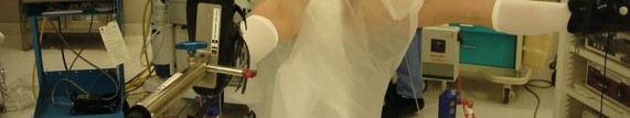
Patient Positioning, Fluoroscopy, Traction, and Draping
Hip arthroscopy can be performed with the patient in either a supine or a lateral decubitus position. Each position has its advantages and disadvantages regarding access, traction efficacy, and specific pathologies. Our preference is supine positioning, which offers advantages in terms of fluoroscopic imaging, ease of conversion to open surgery if needed, and familiar anatomical orientation.
Supine Positioning
- Table Setup: The patient is positioned supine on a commercially available fracture table or a standard operating table equipped with an extension and traction mechanism.
-
Perineal Post:
A well-padded perineal post is strategically placed against the contralateral (non-operative) perineum. Proper padding is critical to prevent pudendal nerve compression or skin breakdown.
-

-
-
Contralateral Extremity Padding:
Large pads are placed on the heel and dorsal and plantar aspects of the contralateral foot to protect pressure points. The hip and knee are often slightly flexed.
-

-
-
Operative Extremity Preparation:
- A single protective pad is placed on the dorsal aspect of the operative foot.
- The foot and ankle are then wrapped meticulously with a self-adherent wrap (e.g., Coban, 3M, St. Paul, MN) to prevent slippage within the traction boot. This also helps distribute pressure evenly.
-
 - Both feet are placed securely in the traction boots. The operative leg is positioned to allow free range of motion, particularly for internal rotation and hyperextension/flexion during traction.
-

-
C-arm Positioning:
The C-arm is brought into the sterile field from the contralateral side of the operative hip, positioned to allow unobstructed AP, lateral, and oblique fluoroscopic views throughout the procedure. Initial images confirm patient alignment.
-

-
-
Traction Application:
- Traction is applied gradually and continuously until adequate distraction of the femoral head from the acetabulum is achieved. This is verified under fluoroscopy, aiming for 8-12 mm of joint space distraction. The amount of traction required varies by patient body habitus, muscle relaxation, and chronicity of joint pathology.
- Excessive traction should be avoided, and traction time limited, typically not exceeding 2 hours in a single application, to mitigate neurological complications. Intermittent release of traction may be considered.
-

- Draping: After establishing adequate traction, the operative limb is prepared and draped in a sterile fashion. A split sheet or specialized hip arthroscopy drape is used to allow manipulation of the limb while maintaining a sterile field. The perineal post, traction apparatus, and distal limb are often excluded from the sterile field.
Lateral Decubitus Positioning
- Advantages: May offer easier access to the posterior compartment, and potentially less risk of pudendal nerve neuropraxia due to the absence of a perineal post.
- Disadvantages: More challenging fluoroscopic views, potential for difficulties in maintaining stable patient position, and more complex C-arm maneuvering. Requires a beanbag or specialized hip positioner to stabilize the pelvis. The operative limb is suspended, and counter-traction is applied via the perineal region with a padded post or strap.
Detailed Surgical Approach / Technique
The surgical technique in hip arthroscopy follows a systematic approach, typically involving portal placement, capsulotomy, systematic inspection of compartments, addressing pathology, and closure.
1. Distraction and Initial Setup
-
Verification of Distraction:
Under fluoroscopy, confirm adequate joint distraction (8-12 mm of articular space between the femoral head and acetabulum). A neutral rotation of the operative limb is generally maintained for initial portal placement.
-

-
-
Bony Landmarks and Neurovascular Structures:
Palpate and mark the ASIS, greater trochanter, pubic tubercle, and course of the femoral neurovascular bundle and LFCN. These surface markings guide safe portal placement.
-

-
2. Portal Placement
Portal placement is critical for safe access and effective visualization. The sequence and specific portals may vary based on surgeon preference and pathology.
-
Anterolateral (AL) Portal (Primary Viewing Portal):
- Located approximately 1 cm anterior to the tip of the greater trochanter and 1 cm distal to a line extending from the ASIS to the greater trochanter.
- It offers a direct line of sight to the central compartment structures.
- Technique: After skin incision, a blunt trocar is directed towards the femoral head, aiming parallel to the femoral neck axis. A "hip poke" maneuver (slight internal rotation/flexion/adduction) can aid entry into the joint capsule.
- Fluid extravasation into the soft tissues can obscure this entry.
-
Mid-Anterior Portal (MAP) / Anteromedial Portal (AM) (Working Portal):
- Located 1-2 cm lateral to the femoral artery pulse at the level of the ASIS, or more commonly, established under direct arthroscopic visualization through the AL portal.
- Allows access to the anterior labrum, acetabular rim, and femoral head-neck junction.
- Risk: Femoral neurovascular bundle. Careful cannulation from lateral to medial, under vision, is paramount.
-
Distal Anterolateral (DAL) Portal (Accessory Working Portal):
- Located 2-3 cm distal and slightly anterior to the AL portal.
- Provides a more inferior angle of approach, useful for cam resection, specific labral repairs, or removing loose bodies from the inferior aspect of the joint.
-
Posterolateral Portal (PL) (For Posterior Compartment):
- Typically located posterior to the greater trochanter, at the level of its tip.
- Requires the patient to be slightly turned or positioned laterally. Primarily used for pathologies in the posterior compartment (e.g., posterior FAI, loose bodies, posterior capsular repair).
- Risk: Sciatic nerve. Careful blunt dissection and awareness of anatomical depth are essential.
-
Modified Anterior Portal (MAP) / Direct Anterior Portal (DAP):
- Located approximately 1 cm lateral to the ASIS and 1 cm distal. This portal is often created using an outside-in technique under fluoroscopic guidance, directing a guidewire towards the acetabular notch. A flexible guide wire is advanced, followed by a cannulated obturator.
-
 -

3. Capsulotomy
Once portals are established and arthroscopic visualization is achieved, a capsulotomy is performed to gain access to the central compartment.
-
Interportal Capsulotomy:
The most common approach, connecting the AL and AM portals. The extent of capsulotomy can vary:
- T-capsulotomy: Involves a longitudinal incision connected to a transverse limb, allowing for wider exposure.
- Limited Capsulotomy: For simple procedures, a smaller incision may suffice.
- Capsular Closure: The trend is towards capsular repair or plication at the end of the case, especially in cases of hip instability or large capsulotomies, to help restore biomechanics and prevent iatrogenic instability.
4. Systematic Intra-articular Inspection (Central Compartment)
With adequate distraction and capsular access, a systematic inspection of the central compartment is performed using the arthroscope through one portal and instruments through the others.
- Labrum: Assess for tears (radial, longitudinal, avulsion), degeneration, and delamination. Note tear morphology, extent, and stability.
- Acetabular Rim: Evaluate for pincer impingement (overcoverage) and cartilage damage at the transition zone.
- Articular Cartilage: Inspect the acetabular and femoral head cartilage for delamination, fibrillation, chondromalacia, and defects (Outerbridge classification).
- Ligamentum Teres: Examine for tears, hypertrophy, or impingement.
- Acetabular Fossa: Look for loose bodies or synovitis.
5. Management of Pathology (Central Compartment)
-
Labral Repair/Debridement:
- Repair: Preferred for unstable labral tears, especially avulsions from the acetabular rim. Anchors are placed into the acetabular rim, and sutures are passed through the labrum to re-approximate it to the bone.
- Debridement: Resection of unstable, severely degenerated, or irreparable labral tissue. The goal is to create a stable rim without sacrificing too much tissue.
-
Pincer Osteoplasty:
- Performed for acetabular overcoverage. Using a burr, the prominent acetabular rim is resected to restore a normal spherical relationship with the femoral head, typically from the 12 o'clock to 3 o'clock position (right hip). Fluoroscopy guides the extent of resection.
- Caution: Avoid excessive resection, which can lead to iatrogenic instability or dysplasia.
-
Chondral Lesions:
- Debridement: Unstable flaps or delaminated cartilage are debrided to a stable rim.
- Microfracture: For contained full-thickness chondral defects (typically < 2-3 cm²), microfracture can stimulate fibrocartilage formation.
- Loose Body Removal: Retrieved using graspers or suction.
6. Peripheral Compartment Access and Management
After addressing central compartment pathology, traction is released, and the hip is placed in extension and external rotation, which creates space in the peripheral compartment.
- Systematic Inspection: Inspect the femoral head-neck junction, capsule, synovium, and gluteal tendons.
-
Cam Osteoplasty:
- For cam-type FAI, the prominent bone at the anterior-superior femoral head-neck junction is resected using a burr. The goal is to restore the normal concave contour of the femoral neck, increasing the femoral head-neck offset and preventing impingement.
- Dynamic fluoroscopy (flexion/internal rotation) is used to confirm adequate resection and freedom from impingement.
-

- Synovectomy/Capsulectomy: For diffuse synovitis or hypertrophic capsule.
- Iliopsoas Release: For internal snapping hip, the iliopsoas tendon is released at its insertion or a portion of the tendon is released where it crosses the anterior acetabular rim, typically with electrocautery or a shaver.
7. Closure
- Dynamic Impingement Test: Perform a final dynamic impingement test under fluoroscopy with the hip in flexion and internal rotation to ensure complete resolution of FAI.
- Capsular Repair/Plication: If a significant capsulotomy was performed, particularly in patients with pre-existing or iatrogenic instability, the capsule is repaired or plicated using absorbable sutures to restore capsular integrity and enhance stability.
- Portal Closure: After ensuring hemostasis and removing all instruments, portals are closed with sutures or sterile strips.
-
Dressing:
A sterile dressing is applied.
-

-
Complications & Management
While hip arthroscopy is generally safe, potential complications exist. Proactive measures and prompt recognition are key to effective management.
| Complication | Incidence (%) | Mechanism | Salvage Strategies / Management |
|---|---|---|---|
| Neuropraxia: | |||
| - Pudendal Nerve | 0.5-2.0 | Compression by perineal post during traction | Adjust post, limit traction time, padding, typically resolves spontaneously within weeks/months. |
| - Lateral Femoral Cutaneous Nerve | 1.0-5.0 | Traction, direct injury during AL/AM portal placement | Minimize traction, careful portal technique, resolve spontaneously, consider nerve block for persistent symptoms. |
| - Sciatic Nerve | < 0.1 | Traction, deep/posterior portal placement | Minimize traction, careful PL portal, immediate traction release, neurological consultation. |
| - Peroneal Nerve | < 0.1 | Traction boot compression at fibular head | Proper padding, avoid compression at fibular head, typically resolves spontaneously. |
| Iatrogenic Chondral/Labral Injury: | 1.0-3.0 | Instrument scuffing, reaming error, guide wire malposition | Careful technique, visualization, protective cannulae, microfracture for full-thickness defects. |
| Fluid Extravasation: | 1.0-4.0 | Infiltration into soft tissues, retroperitoneum | Maintain low intra-articular pressure (40-60 mmHg), monitor fluid output, discontinue irrigation, abdominal binder for compartment syndrome. |
| Infection (Septic Arthritis): | 0.05-0.5 | Contamination | Strict asepsis, systemic antibiotics, aspiration, arthroscopic irrigation and debridement. |
| Deep Vein Thrombosis (DVT)/Pulmonary Embolism (PE): | < 0.1-0.5 | Immobilization, hypercoagulable state, traction | Pre-operative risk assessment, mechanical prophylaxis (TEDs, SCDs), chemical prophylaxis in high-risk patients, early mobilization. |
| Heterotopic Ossification (HO): | 0.5-10.0 (variable) | Periosteal injury, genetic predisposition | Indomethacin or celecoxib post-operatively, especially after extensive bony resection or in high-risk patients. |
| Inadequate Impingement Correction: | 1.0-5.0 | Incomplete cam/pincer resection | Re-evaluation with imaging, revision arthroscopy for residual impingement. |
| Continued Pain/Residual Symptoms: | 5.0-20.0 | Incomplete pathology address, unrecognized pathology, poor rehab, psychosocial factors | Re-evaluation, imaging, physical therapy, pain management, revision surgery. |
| Capsular Instability/Laxity: | Rare (increasing recognition) | Excessive capsulotomy without repair | Capsular repair/plication, physical therapy, revision arthroscopic capsular plication or open stabilization. |
| Vascular Injury: | Extremely rare | Direct injury from instruments, guidewires | Immediate pressure, vascular surgery consult, angiography, surgical repair. |
| Fracture (Femoral Neck/Subtrochanteric): | Rare | Aggressive cam osteoplasty, osteopenic bone | Conservative management if stable, internal fixation if unstable. |
Management Principles for Complications:
- Prevention: The best management is prevention through meticulous preoperative planning, precise surgical technique, controlled traction, and appropriate intra-operative monitoring.
- Early Recognition: Vigilance for signs and symptoms of complications, both intraoperatively and postoperatively.
- Prompt Intervention: Rapid assessment and intervention are crucial for minimizing long-term sequelae. This may involve stopping the procedure, releasing traction, administering medication, or surgical revision.
- Multidisciplinary Approach: Collaboration with anesthesia, neurology, vascular surgery, and physical therapy is often necessary for complex complications.
Post-Operative Rehabilitation Protocols
Post-operative rehabilitation is a critical component for optimizing outcomes after hip arthroscopy. Protocols vary based on the specific procedures performed (e.g., labral repair vs. debridement, capsular repair, extent of bony resection) and surgeon preference. However, general principles apply.
Phase I: Immediate Post-operative (Weeks 0-6)
- Goals: Protect repaired structures, control pain and swelling, restore basic range of motion (ROM) without impingement, initiate muscle activation.
-
Weight-Bearing (WB):
- Labral Repair/Microfracture/Capsular Repair: Often touch-down weight-bearing (TDWB) or protected partial weight-bearing (PWB) (20-50% body weight) with crutches for 2-6 weeks. The use of a brace (e.g., hip abduction brace) is often controversial but may be used in cases of extensive capsular repair or instability.
- Isolated Debridement/Cam Osteoplasty: May allow PWB to WBAT (weight-bearing as tolerated) more quickly, often within 1-2 weeks, transitioning to crutches for comfort.
-
Range of Motion (ROM):
- Restrictions: Avoid extreme hip flexion (>90 degrees), adduction past midline, and internal/external rotation into impingement positions, especially for the first 2-4 weeks post-FAI correction. For capsular repair, specific external rotation limits may be imposed.
- Initiation: Gentle passive ROM (PROM) within protected arcs, often using a continuous passive motion (CPM) machine for 4-6 hours daily for the first 2-4 weeks to promote cartilage health and prevent adhesions.
- Exercises: Hip flexion/extension, abduction/adduction in supine. Gentle isometric gluteal sets, quadriceps sets.
- Pain & Swelling Management: Ice, NSAIDs (if no contraindication), analgesics, and activity modification.
Phase II: Intermediate (Weeks 6-12)
- Goals: Restore full, pain-free ROM, improve muscular strength and endurance, normalize gait, advance proprioception.
- Weight-Bearing: Progress from protected PWB to full weight-bearing (FWB) as pain allows and muscle control improves. Discontinue crutches once a stable, pain-free gait is achieved.
- Range of Motion: Progress active-assisted ROM (AAROM) to active ROM (AROM). Focus on regaining full physiological ROM without impingement. Stretching, especially for hip flexors and piriformis, can be initiated cautiously.
-
Strengthening:
- Progressive resistance exercises (PREs) for hip abductors, adductors, flexors, extensors, and core musculature. Examples: clamshells, side-lying leg lifts, glute bridges, wall slides, light resistance bands.
- Start proprioceptive exercises (e.g., single-leg stance, balance board).
- Functional Activities: Begin light stationary cycling without resistance, elliptical.
Phase III: Advanced (Weeks 12-24)
- Goals: Maximize strength, power, endurance, sport-specific mechanics, and prepare for return to activity/sport.
- Strengthening: Advanced PREs, plyometrics, functional training. Focus on eccentric control and dynamic stability.
- Sport-Specific Training: Gradually introduce agility drills, sport-specific movements (e.g., cutting, jumping), and progressive running programs (if applicable for athletes).
- Return to Activity: Gradual return to recreational activities and sports, typically after 4-6 months, provided the patient demonstrates full ROM, strength, and confidence, and passes functional tests. High-impact sports may require 6-12 months.
Important Considerations:
- Individualization: Protocols must be tailored to the individual patient's progress, pain levels, and specific surgical findings.
- Physical Therapy: Close collaboration with an experienced physical therapist specializing in hip rehabilitation is essential.
- Avoidance of Impingement Positions: Emphasize strict avoidance of positions that reproduce impingement symptoms, especially in the early phases.
- Patience: Full recovery can take 6-12 months, and patients must be counselled on the importance of adhering to the protocol to prevent re-injury or sub-optimal outcomes.
Summary of Key Literature / Guidelines
Hip arthroscopy literature has rapidly expanded, providing a growing evidence base for its efficacy and indications. Key themes and consensus guidelines include:
- Efficacy in FAI and Labral Tears: Numerous systematic reviews and randomized controlled trials (RCTs) support the effectiveness of hip arthroscopy for symptomatic FAI and associated labral tears. For example, the FIRST (Femoroacetabular Impingement Randomised Surgical Trial) and FAIT (Femoroacetabular Impingement Trial) studies have provided Level I evidence demonstrating superior outcomes with arthroscopic FAI correction compared to conservative management in carefully selected patient populations.
- Joint Preservation: The primary goal of hip arthroscopy is joint preservation. Studies show that successful FAI correction and labral repair can improve patient-reported outcomes (PROs) and activity levels, potentially delaying or preventing the progression to osteoarthritis and the need for total hip arthroplasty (THA). However, long-term data (beyond 10 years) on the prevention of OA remain an active area of research.
- Capsular Management: There is an evolving understanding and increasing consensus regarding the importance of capsular repair or plication, especially after extensive capsulotomy, to prevent iatrogenic microinstability. Several studies demonstrate improved outcomes and reduced revision rates when the capsule is repaired.
- Predictors of Outcome: Factors associated with less favorable outcomes include advanced osteoarthritis (Tönnis grade >2, joint space narrowing <2 mm), severe dysplasia, obesity, older age, and duration of symptoms. Conversely, younger age, lower Tönnis grade, and absence of significant cartilage damage predict better results.
- Role of Physical Therapy: Rehabilitation is universally recognized as integral to surgical success. Structured, progressive physical therapy protocols are critical for regaining strength, motion, and function.
- International Society for Hip Arthroscopy (ISHA): This society plays a crucial role in establishing consensus guidelines for indications, surgical techniques, and rehabilitation protocols. Their published guidelines and consensus statements serve as valuable resources for practitioners.
- Comparison to Open Surgery: While open surgical dislocation remains a viable option for complex FAI cases, hip arthroscopy has largely superseded it for most intra-articular pathologies due to its minimally invasive nature, lower morbidity, and comparable outcomes in appropriate settings.
- Future Directions: Research continues into optimal treatment strategies for severe chondral damage, biological augmentation techniques (e.g., PRP, stem cells), and the long-term impact on preventing OA progression. The development of advanced instrumentation and navigation systems further refines precision and safety.
In conclusion, hip arthroscopy is a sophisticated and effective procedure that demands a comprehensive understanding of anatomy, meticulous surgical technique, and a disciplined approach to rehabilitation. Adherence to established principles and guidelines, coupled with continuous learning, is essential for achieving successful patient outcomes in hip preservation.
Clinical & Radiographic Imaging


You Might Also Like