Unstable Intertrochanteric Femur Fracture: Case Study, Diagnosis & Clinical Management
Key Takeaway
An unstable intertrochanteric femur fracture is diagnosed by severe hip pain and limb deformity after a fall. Radiographs confirm comminution, displacement, and specific patterns like AO/OTA 31-A2.2. Clinical examination, detailed imaging, and lab results are crucial for accurate classification and guiding the complex surgical management of this common geriatric orthopedic injury.
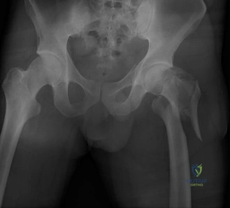
You are presented with this 78-year-old male who has sustained a ground-level fall. Look at the provided radiographs. How do you classify this fracture, and what is your primary concern regarding the stability of this injury?
Candidate: This is an intertrochanteric hip fracture. Based on the radiographs, it's comminuted and involves the lesser trochanter. I would classify it as an AO/OTA 31-A2.2 fracture. My main concern is that the posteromedial wall is gone, which makes it unstable.
The candidate focuses only on the posteromedial cortex. They fail to mention the lateral wall, which is the most critical anatomical structure for choosing between an SHS (Sliding Hip Screw) and a Cephalomedullary Nail (CMN) in the modern FRCS exam.
The candidate defines this as an AO/OTA 31-A2.2 unstable intertrochanteric fracture. They highlight three points: 1) The posteromedial void (loss of calcar support) renders the fracture inherently unstable. 2) The lateral wall integrity—measuring less than 20.5mm—is the key determinant for implant selection. 3) They state that because the lateral wall is compromised, an extramedullary device like an SHS is contraindicated due to the high risk of catastrophic lateral wall blowout during impaction.
The patient is on Apixaban. How does this impact your decision-making for surgical timing, and what are the key risks if you proceed prematurely?
Candidate: I would stop the Apixaban and wait 48 hours before surgery. If I operate too early, the patient might bleed significantly or have an issue if I use spinal anesthesia.
Failing to mention the patient's CKD Stage 3a (eGFR 52). The clearance of DOACs is renal-dependent; a "blanket" 48-hour rule is dangerous in patients with significant renal impairment, as the drug half-life is extended.
The candidate provides a structured approach: 1) Multidisciplinary Input: Involve hematology/geriatric medicine. 2) Renal Consideration: Acknowledge that the patient's eGFR (52 mL/min) dictates a longer clearance time for Apixaban. 3) Anesthesia: Note that neuraxial anesthesia is contraindicated while anticoagulated due to the risk of epidural hematoma. 4) Resuscitation: Emphasize that we do not use reversal agents (like Andexanet alfa) routinely to "speed up" surgery due to their prothrombotic risk, but rather wait for the drug to clear naturally unless there is a life-threatening hemorrhage.
During the procedure, you have inserted your cephalomedullary nail. Explain the concept of the Tip-Apex Distance (TAD) and why it is critical for this patient.
Candidate: TAD is the distance from the tip of the screw to the apex of the femoral head. It needs to be under 25mm to prevent the screw from cutting out of the bone.
Providing a vague definition without mentioning the correction for magnification or the specific radiographic views required (AP and lateral). Examiners want to hear the mechanics of why it works.
The candidate explains: 1) Definition: TAD is the sum of the distance from the lag screw tip to the femoral head apex on both AP and lateral views, corrected for magnification. 2) Clinical Threshold: A TAD > 25mm is the strongest independent predictor of lag screw cut-out. 3) Optimizing Strength: They add that the screw should ideally be placed in the inferior quadrant on the AP view and central on the lateral view to engage the dense subchondral bone of the calcar, thereby maximizing pull-out strength in this osteoporotic patient.
