Sciatic Nerve Injuries: Surgical Anatomy, Approaches, and Management
Key Takeaway
The sciatic nerve is the most critical neural structure in the lower extremity, analogous to the brachial plexus in the upper limb. Injuries typically arise from high-energy trauma, posterior hip dislocations, penetrating wounds, or iatrogenic causes during arthroplasty. Management requires a profound understanding of its complex surgical anatomy, precise clinical evaluation, and timely surgical exploration. This guide details the indications, operative approaches, and neurolysis techniques essential for optimizing functional recovery in sciatic nerve lesions.
INTRODUCTION TO SCIATIC NERVE PATHOLOGY
The sciatic nerve is analogous in its importance in the lower extremity to the brachial plexus in the upper extremity. As the largest single nerve in the human body, it provides critical motor innervation to the posterior compartment of the thigh and all compartments of the leg and foot, alongside extensive sensory coverage.
Sciatic nerve injuries present a profound challenge to the orthopaedic surgeon. They are most frequently injured by high-energy trauma, such as gunshot wounds to the thigh or buttock. Less often, but of equal clinical significance, the nerve is compromised by posterior dislocations and fracture-dislocations of the hip, by misplaced intramuscular injections into the gluteal region, or iatrogenically during complex reconstructive surgery around the hip joint. Furthermore, delayed neuropathies can occur; compression of the sciatic nerve by wear debris, metallosis, or pseudotumors from long-standing total hip arthroplasty (THA) represents a rare but increasingly recognized etiology.
This comprehensive guide delineates the surgical anatomy, clinical evaluation, indications for exploration, and operative management of sciatic nerve lesions, providing a strictly evidence-based framework for the practicing orthopaedic surgeon.
SURGICAL ANATOMY AND BIOMECHANICS
A masterful grasp of the sciatic nerve's topographical and internal anatomy is non-negotiable for safe surgical exploration and repair.
Origin and Course
The sciatic nerve arises from the lumbosacral plexus, specifically the ventral rami of L4 through S3. It is composed of two distinct nerve trunks—the tibial nerve (medial, derived from anterior divisions) and the common peroneal nerve (lateral, derived from posterior divisions)—which are enveloped in a common epineural sheath from their origin down to the distal third of the thigh.
The nerve exits the pelvis through the greater sciatic foramen, typically emerging inferior to the piriformis muscle. However, anatomical variants are well-documented, including the common peroneal division piercing or exiting superior to the piriformis.
As it descends through the gluteal region, it lies deep to the gluteus maximus, resting on the posterior surface of the ischium, the obturator internus, the gemelli, and the quadratus femoris. In the posterior thigh, it is crossed obliquely by the long head of the biceps femoris and rests on the adductor magnus.
Branching and Distribution
The sciatic nerve supplies the hamstring muscles (semitendinosus, semimembranosus, long head of biceps femoris via the tibial division; short head of biceps femoris via the common peroneal division) and the ischial head of the adductor magnus.
Surgical Warning: The blood supply to the proximal sciatic nerve is primarily derived from the inferior gluteal artery and the medial circumflex femoral artery. Aggressive circumferential mobilization of the nerve over long distances can devascularize the extrinsic blood supply, compromising nerve regeneration.
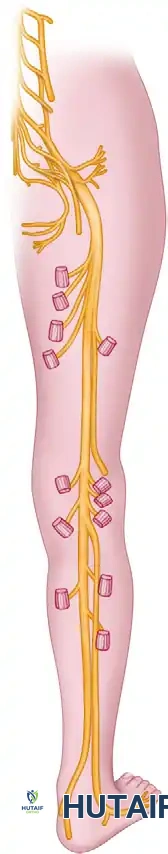
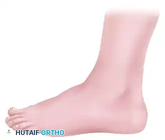
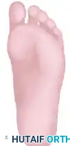
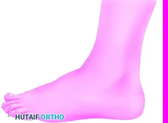
Dermatomal and Autonomous Sensory Zones
Evaluating the autonomous sensory zones of the foot is critical for localizing the specific division of the sciatic nerve that has been injured. The terminal branches of the sciatic nerve (via the tibial and common peroneal nerves) provide distinct sensory territories.
The following diagrams illustrate the autonomous zones of the sciatic nerve and its terminal branches, which must be meticulously mapped during the preoperative clinical examination:


ETIOLOGY AND MECHANISMS OF INJURY
Understanding the mechanism of injury dictates the timing and necessity of surgical intervention.
Traumatic Injuries
- Penetrating Trauma: Gunshot wounds and lacerations to the buttock or posterior thigh are primary causes of direct sciatic nerve transection or severe contusion.
- Fracture-Dislocations: Posterior hip dislocations (with or without posterior wall acetabular fractures) frequently stretch or compress the nerve. The common peroneal division is disproportionately affected due to its lateral position, larger fascicles, and tighter tethering at the fibular head.
- Femoral Shaft Fractures: High-energy fractures of the femur, particularly those with significant posterior displacement, can impale or stretch the nerve.
Iatrogenic and Compressive Injuries
- Total Hip Arthroplasty (THA): The nerve is at risk during posterior approaches (due to retractor placement or direct trauma) and during significant limb lengthening (stretch injury).
- Wear Debris and Pseudotumors: In long-standing THA, particularly metal-on-metal articulations, the accumulation of wear debris can lead to massive pseudotumor formation, causing delayed, progressive sciatic nerve compression.
- Intramuscular Injections: Misplaced injections outside the upper outer quadrant of the buttock can cause direct needle trauma or severe chemical neurotoxicity.
- External Compression: Prolonged external compression, such as from a poorly fitted cast, prolonged surgical positioning, or unusual postures (e.g., prolonged crossing of the legs or coma), can induce severe neurapraxia or axonotmesis.
CLINICAL EVALUATION AND DIAGNOSIS
A meticulous neurological examination is paramount. Motor testing must isolate the tibial division (plantar flexion, toe flexion, foot inversion) and the common peroneal division (dorsiflexion, toe extension, foot eversion).
The Role of Percussion (Tinel's Sign)
In patients with multiple wounds or complex trauma, percussing along the anatomical course of the nerve to the point where tingling (Tinel's sign) is most pronounced is a highly accurate method of locating the exact zone of injury.
Clinical Pearl: Exact knowledge of the point of emergence of the various nerve branches is helpful; however, if a surgeon attempts to locate an injury by this anatomical knowledge alone, they are more likely to err than when using percussion. This is because a branch may be injured after it emerges from the main nerve trunk, making the percussion point the most reliable clinical indicator of the lesion's exact location.
Electrodiagnostic Studies
Electromyography (EMG) and Nerve Conduction Studies (NCS) are essential adjuncts. Baseline studies are typically obtained at 3 to 4 weeks post-injury to allow for Wallerian degeneration to become electrically apparent. Serial EMGs help differentiate between neurapraxia (which will recover) and axonotmesis/neurotmesis (which may require surgery).
INDICATIONS FOR SURGICAL INTERVENTION
The decision to explore the sciatic nerve requires a nuanced understanding of the injury mechanism and the timeline of recovery.
Indications for Early Exploration (Acute)
- Penetrating Trauma: If a sciatic nerve lesion is caused by a penetrating injury (e.g., knife wound, glass laceration), especially if the wound is proximal in the buttock, early exploration and repair are highly worthwhile. Early repair ensures that the distal structures are denervated for the shortest possible time, maximizing the potential for motor endplate reinnervation.
- Complete Division with Fracture: If a complete division of the sciatic nerve complicates a dislocation or fracture around the hip, early exploration of the nerve assists in determining the extent of the injury, removing bony impingement, and assessing whether primary repair is possible.
- Iatrogenic Transection: Known or highly suspected transection during arthroplasty or tumor resection.
Indications for Delayed Exploration (3 to 6 Months)
- Closed Trauma/Stretch Injuries: If complete division of the nerve complicates a femoral shaft fracture or fracture-dislocation around the knee, and no clinical or EMG signs of recovery are apparent by 3 to 4 months, exploration is justified.
- External Compression: If an injury to a branch of the nerve has been caused by external compression (e.g., poorly fitted cast, unusual posture), the primary cause must be corrected immediately. If the compression has been of long duration and fails to improve after several months, exploration and neurolysis may be warranted.
- Prognostic Warning: The prognosis for long-duration compressive lesions, even after meticulous neurolysis, remains extremely guarded due to intraneural fibrosis.
SURGICAL APPROACHES TO THE SCIATIC NERVE
Surgical exposure of the sciatic nerve must be extensile, allowing for proximal and distal control before entering the zone of injury.
Patient Positioning
The patient is placed in the prone position on a radiolucent Jackson table. All bony prominences are heavily padded. The entire lower extremity, from the iliac crest to the toes, is prepped and draped free to allow for intraoperative manipulation of the hip and knee, which is crucial for assessing nerve tension during repair. A sterile tourniquet may be applied to the proximal thigh if only the distal nerve is being explored, but it is generally avoided for proximal gluteal exposures.
The Gluteal and Subgluteal Approach
- Incision: A "question mark" incision is utilized. Begin at the posterior superior iliac spine (PSIS), curve laterally and distally along the fibers of the gluteus maximus toward the greater trochanter, and then extend vertically down the posterior midline of the thigh.
- Superficial Dissection: Incise the fascia lata in the thigh and the gluteal fascia proximally. The gluteus maximus can be split in line with its fibers or, for massive exposures, detached from its femoral insertion and reflected medially.
- Deep Dissection: Identify the sciatic nerve as it emerges beneath the piriformis muscle, located midway between the ischial tuberosity and the greater trochanter.
- Vascular Control: Meticulous hemostasis is required, particularly when managing the inferior gluteal artery and the cruciate anastomosis, which lie in close proximity to the nerve.
The Posterior Thigh Approach
- Incision: A longitudinal incision is made down the posterior midline of the thigh, extending from the gluteal fold to the popliteal fossa.
- Fascial Incision: The deep fascia is incised in line with the skin incision.
- Muscle Retraction: The long head of the biceps femoris is identified. The nerve is found deep to this muscle. Retracting the biceps laterally and the semitendinosus medially exposes the nerve resting on the adductor magnus.
- Distal Tracing: The nerve is traced distally to its bifurcation into the tibial and common peroneal nerves at the superior apex of the popliteal fossa.
OPERATIVE TECHNIQUES: NEUROLYSIS AND REPAIR
Once the nerve is exposed, the surgeon must utilize intraoperative nerve stimulation and surgical microscopy to dictate the next steps.
External and Internal Neurolysis
If the nerve is in continuity but encased in scar tissue (e.g., from a previous fracture or pseudotumor), an external neurolysis is performed. The epineurium is freed from the surrounding fibrotic bed.
If the nerve feels indurated and intraoperative stimulation yields no distal motor response, an epineurotomy (internal neurolysis) may be indicated.
Surgical Pitfall: Internal neurolysis must be performed under high magnification. Overzealous interfascicular dissection can easily destroy the delicate intraneural microcirculation, converting a neuropraxic lesion into an ischemic neurotmesis. Proceed with extreme caution.
Primary End-to-End Repair
If the nerve is transected, primary repair is the gold standard, provided it can be achieved without tension.
1. Preparation of Nerve Ends: The neuromatous proximal stump and gliomatous distal stump are serially resected using a fresh scalpel blade until healthy, pouting fascicles ("mushrooming") are visualized.
2. Mobilization: The nerve is mobilized proximally and distally. Flexing the knee to 90 degrees and extending the hip can provide several centimeters of length.
3. Neurorrhaphy: Using 8-0 or 9-0 non-absorbable monofilament suture under microscopic magnification, an epineural repair is performed. Alignment of the longitudinal epineural vessels and fascicular topography (keeping the peroneal division lateral and tibial division medial) is critical to prevent cross-innervation.
Nerve Grafting
If a tension-free primary repair is impossible after maximal mobilization and joint positioning, nerve grafting is mandatory. Tension across a nerve repair guarantees failure due to ischemia.
1. Graft Harvest: The sural nerve is the standard autograft donor. It can provide up to 30-40 cm of graft material.
2. Cable Grafting: Because the sciatic nerve is massive, multiple strands of sural nerve (cable grafts) are required to bridge the defect.
3. Fibrin Glue: Fibrin tissue adhesive is often used to augment the suture lines and secure the cable grafts in place.
POSTOPERATIVE PROTOCOL AND REHABILITATION
The success of a sciatic nerve repair relies heavily on strict postoperative management to protect the anastomosis.
Immobilization
If the repair was performed under tension requiring joint positioning, the patient is placed in a custom hip-knee-ankle-foot orthosis (HKAFO) or a spica cast with the hip extended and the knee flexed (typically 45 to 90 degrees). This position is maintained for 3 to 4 weeks.
Progressive Mobilization
At 4 weeks, the immobilization is gradually adjusted. The knee is extended by 10 to 15 degrees per week to slowly stretch the nerve without disrupting the repair site. Full extension should be achieved by 6 to 8 weeks postoperatively.
Rehabilitation and Orthotics
- Ankle-Foot Orthosis (AFO): Because foot drop is immediate and recovery takes months to years, a rigid or dynamic AFO is prescribed immediately to prevent equinus contractures and facilitate a normal heel-strike during gait.
- Physical Therapy: Aggressive passive range of motion of the ankle and toes is instituted to prevent joint stiffness. Once reinnervation begins, active-assisted and strengthening exercises are initiated.
- Sensory Re-education: As sensory axons reach the foot, patients must be educated on daily skin inspections to prevent neuropathic ulcerations in the autonomous zones.
PROGNOSIS AND OUTCOMES
The prognosis following sciatic nerve injury depends heavily on the level of the lesion, the mechanism of injury, and the age of the patient.
Proximal lesions (gluteal level) have a poorer prognosis for distal motor recovery (intrinsic foot muscles) due to the sheer distance regenerating axons must travel (approximately 1 mm per day). However, recovery of proximal hamstring function and protective sensation to the sole of the foot is frequently achievable.
The tibial division consistently demonstrates superior recovery rates compared to the common peroneal division. If common peroneal recovery fails after 18 to 24 months, tendon transfer surgery (e.g., posterior tibial tendon transfer to the dorsum of the foot) remains a highly effective salvage procedure to restore active dorsiflexion and eliminate the need for an AFO.
You Might Also Like